MembraneDamaging Toxins 1 Hemolysins These toxins cause lysis
 Hemolysins. These toxins cause lysis of red blood cells which leading")
Membrane-Damaging Toxins: (1) Hemolysins. These toxins cause lysis of red blood cells which leading to anemia, There are many different types of hemolysins but their net action is the same. (2) Phospholipase. This enzyme attacks any cell with phospholipids in its membrane. The result is widespread cell lyses. (3) Lecithinase : it is an enzyme which breaks down lecithin in the human cell plasma membrane, resulting in cell lyses. It is active on red blood cells. (4) Pore-forming toxins: These toxins disrupt the selective influx and efflux of ions across the plasma membrane by inserting a transmembrane pore.

Non membrane damging toxin 1 -Hyaluronidase. This is an enzyme that catalyzes the breakdown of hyaluronic acid, the substance that cements human cells together. This allows the bacterial cells to spread through tissue, causing cellulitis. 2 - Fibrinolysin. This toxin catalyzes the conversion of plasminogen to the fibrinolytic enzyme plasmin. 3 - Lipase. Production of excessive amounts of lipase allows bacteria to penetrate fatty tissue with the consequent formation of abscesses. (4) Collagenase. This enzyme catalyzes the degradation of collagen, a scleroprotein found in tendons, nails, and hair.

The nature and common character of toxin There are common characters for all toxins soluble in water , they responded to reactions characteristic to protein – substances. Toxins are precipitated by the action of ammonium sulphate, zinc chloride , alcohol. C)Toxins are destroyed at 60˚C ( 95% of molecules are destroyed at 56˚C ) also destroyed by acid reaction of PH 5. 5
 Protein toxin molecules consistes of two parts :")
Subunit arrangement in toxin molecules (A+B) Protein toxin molecules consistes of two parts : Sub- unit A (toxophore) which responsple for toxin activety Sub-unit B (Haptophore ) which resposiple for binding of toxin to specific receptors in host tissue Isolated A subunit: are enzymatically active but lack binding and cell entry capability. . Subunit B: Concerned with binding to a specific receptor on the host cell membrane and transferring the enzyme cross the membrane.

Attachment and entry of toxin There are two mechanisms of toxin entry into target cells: 1 -direct entry : the B subunit of the native (A+B) toxin binds to a specific receptor on the target cell and induces the formation of a pore in the membrane through which the A subunit is transferred into the cell cytoplasm. 2 -receptor-mediated endocytosis (RME). : -the native toxin binds to the target cell and the A+B structure is taken into the cell. The toxin is internalized in the cell in a membrane-enclosed vesicle called an endosome.
: is")
Determination of the potency of toxins Minimal lethal dose (M. L. D. ): is the smallest definite dose of toxin which kill an animal(definite weight) in a definite time which varies from different toxins to others No observed adverse effect level of toxin (NOAEL) Low observed advers effect level(LOAEL) estimated dose that cause 50% mortality of tested organism LD 50%

Toxoid properties Toxoids are detoxified toxins, used for artificial immunization against diseases caused by pathogens, was first discovered by Ehrlich who coined the term toxoids for this product. The formation of toxoids can be accelerated by treating exotoxin to be(inactive form) by using chemical treatment, with a variety of reagents including formalin, iodine, pepsin, ascorbic acid, ketones. for using in vaccines and is no longer damage to tissue. A very small dose of toxins will stimulate body cells to produce specific antibodies or antitoxins, which neutralize the toxins in definite proportions.

1 -Toxins that interfere with cellular metabolism a-Diphtheriae toxin The best known and studied bacterial toxin is the diphtheria toxin, produced by Corynebacterium diphtheriae. Diphtheria toxin is a bacterial exotoxin of the A/B prototype. It is produced as single polypeptide chain with a molecular weight of 60, 000 Daltons. 1 -The toxin firstly binds to a receptor on the host cell membrane and then enters to the cell by an endocytic process. 2 - The toxin blocks protein synthesis by inactivating elongation factor 2(EF-2), which an enzyme necessary for the growth of the polypeptide chain in translation process.

Cornybacterium diphtheria toxin is a bacterial exotoxin of the A/B prototype. It is produced as single polypeptide chain with a molecular weight of 60, 000 Daltons. The toxin is distinguish into two parts: -subunit A, with a Mol. wt. of 21, 000 Daltons, contains the enzymatic activity for inhibition of elongation factor-2 involved in host protein synthesis subunit B, with a Mol. wt. of 39, 000 Daltons, is responsible for binding to the membrane of a susceptible host cell. The B subunit possesses a region T (translocation) domain which inserts into the endosome membrane thus curing the release of the enzymatic component into the cytoplasm component.

Diphtheria diseases causatative agent : toxigenic strain of corynebacterium diphtheria , Gram +ve bacilli , non acid fast, non motile have club shap sweeling at the end resemble of Chinese letter reservoir : Man , Age: > 15 years Incubation period: 1 -7 days Mode of transmission: contact with direct droplet infection. Usually recovery (2 weeks) or less & seldom more than 4 weeks effective antibiotic therapy terminate shedding. The rare chronic carrier may shed the organism for 6 months

Toxoid Preparation of toxoids : It can be converted into toxoid (toxin that has lost toxicity , not antigenecity) by 1 – prolonged storage at 37˚C. 2 – incubation at 37˚C for 4 to 6 weeks. 3 – 0. 2 to 4% formalin. Toxin production is also influenced by the concentration of iron in the medium. 0. 1 mg of iron per ml is the optimum concentration in the medium for toxin production 0. 5 mg per ml of iron in the medium inhibits toxin production.

Mechanism of action for toxin. Toxin consists of 2 factors A and B. A is a lethal factor and B is a spreading factor. Fragment A inhibits polypeptide chain elongation in the presence of nicotinamide adenine dinucleotide (NAD). -by inactivating the elongation of EF-2. , toxin fragment A inactive the EF-2 by catalyzing the reactions that yields free nicotinamide and an inactive adenosine diphosphate ribose (ADR) complex , that due to depression of protein synthesis and for necrotizing and neurotoxic effects of diphtheria toxin - The bacilli remain localize to site of entry where they multiply and form toxin. This toxin produces area

B- altering cellular metabolism : Cholerae toxin 1 -the causative agent is Vibrio cholera, they are water-borne microorganism , are Gram –ve , in pairs rods arranged in S_forms or comma shape grow only in alkaline environment (TCBS) produce yellow colony in 8 hours, with short generation time , secreted an choleraenterotoxin that alters the regulatory control in a cell and directly damaging it.
 consistes of 5 B binding subunits and A active")
Virulance factors 1 -Enterotoxin (choleragen) consistes of 5 B binding subunits and A active subunit made up of 2 peptides (A 1 and A 2) , the B subunit specific receptor on the intestinal epithelial cells, so it facillate entrance of subunit A and activate adenylate cyclase enzyme causing rise up in c. AMP production leading to massive secretion of elecrolytes. 2 -Toxin-coregulated pili it is adhesion factorfor intestinal colonization 3 -Mucinase enzyme dissolves the glycoprotein over the intestinal cells. 4 -flagella allow the organism to move through the intestinal mucosa

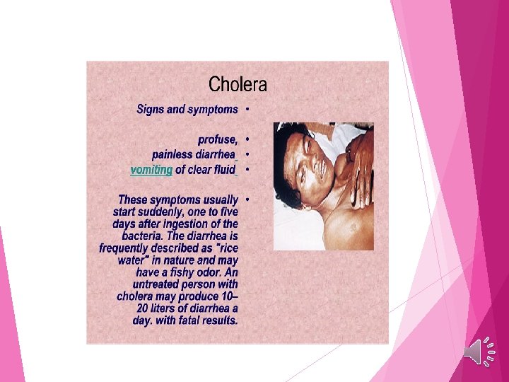
Mechanism of action 1 -Cholera toxin binds specifically to intestinal epithelial cells. 2 - when enters the cell , causes increased and uncontrolled production of cyclic AMP. (which is the mediator of a number of regulatory systems in cells) 3 -The increased levels of cyclic AMP cause unregulated secretion of chloride and bicarbonate ions from the epithelial cells lining the intestine. 4 - This changed in ionic balance resulting a massive outflow of water from the cells into the lumen of the intestine, ending by a profuse diarrhea. The great loss of water that cholera victims may die from extreme dehydration if their fluids and electrolytes are not replaced
- Slides: 16