Membrane Structure and Transport Processes A B C

Membrane Structure and Transport Processes A. B. C. D. The Phospholipid Bilayer Membrane Proteins and Carbohydrates Diffusion and Active Transport Electrical Properties of Membranes Last Updated: Jan 2008

A. The Phospholipid Bilayer 1. 2. 3. Lipid Structure Experimental systems Properties of Lipid Bilayers

A. 1. Lipid Structure Glycerol-based Phospholipids – Glycerol Molecule – Two Fatty Acid Chains – Polar Head Group, attached via phosphate – The fatty acid chains may be saturated or unsaturated – Double bonds in unsaturated fatty acids may be cis or trans

A. 1. Lipid Structure

A. 1. Lipid Structure Sphingosine-based Lipids – Sphingomyelin – Galactocerebroside – Gangliosides Cholesterol

A. 2. Experimental Systems Liposomes – Artificial vesicles made by mixing pure phospholipids in water

A. 2. Experimental Systems Black Membranes – Artificial lipid bilayer formed between two chambers containing aqueous solutions Erythrocyte Membranes

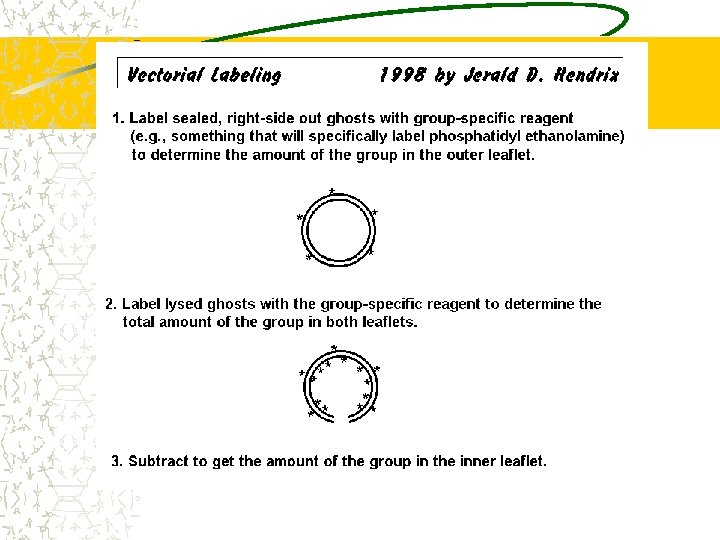
A. 2. Experimental Systems Erythrocyte Membranes – Separate erythrocytes from blood plasma by centrifugation – Suspend the erythrocytes in a hypotonic buffer – The erythrocytes swell and burst to produce erythrocyte “ghosts” – By adjusting buffer conditions, sealed ghosts (either normal or inverted) or leaky ghosts can be formed.

A. 3. Properties of Lipid Bilayers Overall Structure Fluidity – Lateral Diffusion of phospholipids: ~10 -8 cm 2/sec – Rotation – Flexion – Transverse Diffusion of phospholipids: Almost nonexistent. Movement of PL from one leaflet to the other requires enzymes called phospholipid translocators (flippases and scramblases)
")
A. 3. Properties of Lipid Bilayers Measurements of Fluidity – Differential Scanning Calorimetry (DSC) • Determination of the “melting point” at which a bilayer undergoes phase transition – Electron Spin Resonance (ESR) spectroscopy • Attaching an ESR label such as a nitroxyl group to the fatty acid chain; can detect motility and other interactions within the lipid core of the bilayer – Tagged lipid / real time video microscopy • Attach gold particle or fluorescent group to individual lipid molecules and track movement via digitally enhanced imaging – Laser photobleaching experiments

A. 3. Properties of Lipid Bilayers Factors Affecting Fluidity – Length of the fatty acid side chains – Presence of double bonds in the fatty acid chains (degree of saturation) – Size of the polar head groups – Presence of cholesterol – http: //www. nyu. edu/pages/mathmol/library/lipids/

A. 3. Properties. . .

A. 3. Properties. . .

A. 3. Properties. . .

A. 3. Properties of Lipid Bilayers Compositional Symmetry – Refers to the percentages of each phospholipid in the inner and outer leaflets of the bilayer – In artificial liposomes: Leaflets have exactly the same composition – In membranes: The compositions are different (Asymmetric) – Determined by vectorial labeling experiments
")
A. 3. Properties of Lipid Bilayers Erythrocyte Membrane (numbers are % of total lipid) Outside Inside Sphingomyelin 20% 5% P. choline 25% 5% P. ethanolamine 5% 25% P. serine 0% 5%

A. 3. Properties of Lipid Bilayers Lipid rafts – Areas in a bilayer where specific lipids are more concentrated – In a liposome consisting of 1: 1: 1 phosphatidylcholine: sphingomyelin: cholesterol, the sphingomyelin and cholesterol will form patches that may be similar to lipid rafts – There is evidence that some integral membrane proteins may require specific lipid molecules for activity

A. 3. Properties of Lipid Bilayers Role of compositional asymmetry – Charge differences between outer & inner surfaces – see difference in distribution of PC and PS in the erythrocyte membrane – Binding and activation of cell signaling proteins • Protein kinase C binds to negative cytosolic face • Phosphatidylinositol can be modified to create specific binding sites for signaling

A. 3. Properties of Lipid Bilayers Role of compositional asymmetry – Compositional asymmetry is used in mammals to detect cells that have undergone apoptosis (programmed cell death) • When the cell dies, the phospholipid translocator that moves PS to the cytosolic leaflet is inactivated • A scramblase that moves phospholipids across the membrane nonspecifically in both directions is activated • PS rapidly moves from the cytosolic leaflet to become equally distributed between the cytosolic leaflet and the exterior leaflet

B. 1. 2. 3. Membrane Proteins and Carbohydrates Membrane Proteins Mobility of Membrane Proteins Membrane Carbohydrates

B. 1. Membrane Proteins Peripheral, Integral, and Lipid. Anchored Proteins Solubilization experiments – Peripheral protein: Can be removed by high ionic strength wash – Integral protein: Requires detergent treatment to be solubilized – Lipid-Anchored Protein: Solubilized by detergent or by enzymatic lysis from lipid anchor

B. 1. Membrane Proteins It was predicted that transmembrane domains would be rich in hydrophobic amino acids This was confirmed by amino acid sequencing, then later by determination of integral 3 -D structures The two major transmembrane configurations are α-helical segments and β-pleated sheets that form beta barrel structures

B. 1. Membrane Proteins Examples of Membrane Proteins – Erythrocyte Membrane Proteins http: //www. unipv. it/bioscipv/sds-page. htm • Integral Proteins – Band 3 – Glycophorin A • Peripheral Proteins – “Membrane Skeleton” – – Spectrin Ankyrin Band 4. 1 Actin

B. 1. Membrane Proteins – Bacteriorhodopsin • The first integral membrane protein in which the 3 -D structure was determined • This is a light-driven hydrogen ion pump found in the plasma membrane of the archaen Halobacterium salinarum • Located in specialized regions called “purple patches” where it is found in crystalline-like arrays • This unique structure allowed it’s 3 -D structure to be determined by electron diffraction analysis • Its transmembrane domain consists of seven αhelical segments

B. 1. Membrane Proteins Bacterial porins – Found in the outer membrane of gramnegative bacteria – Transmembrane domain is a “beta barrel” structure consisting of a ring of β-pleated sheets that form a large channel

B. 2. Mobility of Membrane Proteins Membrane proteins often exhibit lateral mobility, but not transverse mobility (flip-flop) Demonstrations of lateral mobility – “Patching-and-capping” of immunoglobulins on B-lymphocytes http: //www 3. niaid. nih. gov/labs/aboutlabs/lig/lymphocyte. Activation. Section/ – Cell fusion experiments – Photobleaching experiments (FRAP) – Single particle tracking experiments http: //www. censsis. neu. edu/public_docs/13 b-07. pdf

B. 2. Mobility of Membrane Proteins Lateral mobility of integral proteins may be limited by interactions with peripheral proteins or other components Membrane protein mobility and distribution may be restricted to specific “domains” (regions) of the cell’s plasma membrane – Example: • Intestinal epithelial cell • Mammalian sperm cells

VI. B. 3. Membrane Carbohydrates Glycocalyx – Many integral membrane proteins have carbohydrate groups attached to their exterior domains – This carbohydrate, together with carbohydrate attached to phospholipid molecules, forms the glycocalyx Functions – Cell adhesion – Cell recognition

C. Diffusion and Active Transport Simple Diffusion – Movement of substances directly across a phospholipid bilayer, with no need for a transport protein – Movement from high low concentration – No energy expenditure (e. g. ATP) from cell

C. Diffusion and Active Transport Facilitated Diffusion – Movement of substances across a membrane with the assistance of a transport protein – Movement from high low concentration – No energy expenditure (e. g. ATP) from cell – Two mechanisms: Channel & Carrier Proteins – Carrier proteins may be uniporters, symporters, or antiporters

C. Diffusion and Active Transport – Movement of substances across a membrane with the assistance of a transport protein – Movement from low high concentration – Energy expenditure (e. g. ATP or ion gradients) from cell – Active transport pumps are usually carrier proteins – Examples: • Na+ K+ ATPase pump
 – Active transport ATPase pumps are divided")
C. Diffusion and Active Transport (cont. ) – Active transport ATPase pumps are divided into three types: • P-type pumps phosphorylate themselves during their cycle and include the plasma membrane Na+K+ pump, the sarcoplasmic reticulum Ca 2+ pump, and many other ion pumps • ABC transporters primarily pump small molecules rather than ions; these are the largest family of membrane transport proteins. Each member of this family contains two highly conserved ATP binding sites
 • F-type pumps are H+ driven ATP")
C. Diffusion and Active Transport (cont. ) • F-type pumps are H+ driven ATP synthases in mitochondria and chloroplasts; also are similar to V-type pumps that use ATP to pump H+ into organelles such as lysosomes

D. Electrical Properties of Membranes Gated Ion Channels: – Voltage-gated, mechanically-gated, or ligand-gated • Example: Voltage-gated K+ channel – Ion channels are usually selective; the best understood is the bacterial K+ channel

D. Electrical Properties of Membranes Events in a Nerve Impulse – The resting potential of a nerve cell: The exterior of the cell is positively charged, due to the gradients of Na+ and K+. (these are maintained by K+ leak channels and Na+-K+ ATPase) – In the resting state, the voltage-gated Na+ channels are closed. – A “nerve impulse” is a wave of depolarization along the neuron, caused by the Na+ channels opening and Na+ rushing
 – Within")
D. Electrical Properties of Membranes Events in a Nerve Impulse (cont. ) – Within a millisecond after a nerve impulse passes a section in a neuron, the Na+ channel goes into an “inactive” state until the membrane is repolarized, when it returns to the “closed” configuration – A voltage-gated, delayed K+ channel opens and lets K+ rush outside the cell. This, together with the action of the Na+-K+ pump, returns the region to its resting
 – When")
D. Electrical Properties of Membranes Events in a Nerve Impulse (cont. ) – When a nerve impulse reaches the end of a neuron, it triggers the release of neurotransmitter molecules (e. g. , acetylcholine or glutamate) from vesicles within the cell. – The neurotransmitter diffuses across the synapse, activates ligand-gated Na+ channels in the next neuron to start the wave of depolarization in the next neuron.
- Slides: 38