Membrane Structure and Function Cell Membrane A Plasma

Membrane Structure and Function

Cell Membrane A. Plasma membrane is selectively permeable Allows some substances to cross more easily than others B. Fluid Mosaic Model Fluid: membrane held together by weak interactions Mosaic: phospholipids, proteins, carbs
 Davson/Danielli – Sandwich model phospholipid bilayer between 2 protein layers")
Early membrane model (1935) Davson/Danielli – Sandwich model phospholipid bilayer between 2 protein layers Problems: varying chemical composition of membrane, hydrophobic protein parts

Fluid Mosaic Model
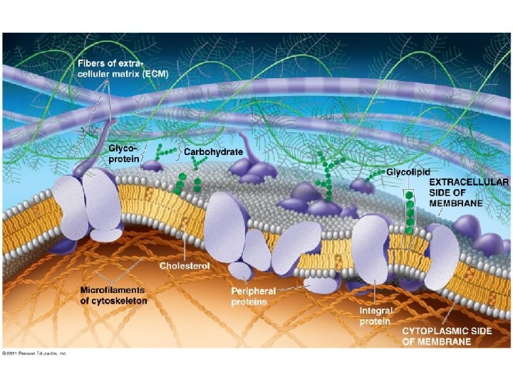

Phospholipids Bilayer Amphipathic = hydrophilic head, hydrophobic tail Hydrophobic barrier: keeps hydrophilic molecules out
 Cholesterol resists changes")
Membrane fluidity Low temps: phospholipids w/unsaturated tails (kinks prevent close packing) Cholesterol resists changes by: limit fluidity at high temps hinder close packing at low temps Adaptations: bacteria in hot springs (unusual lipids); winter wheat ( unsaturated phospholipids)

Evolution of Differences in Membrane Lipid Composition Variations in lipid composition of cell membranes of many species appear to be adaptations to specific environmental conditions Ability to change the lipid compositions in response to temperature changes has evolved in organisms that live where temperatures vary © 2011 Pearson Education, Inc.

Membrane Proteins and Their Functions A membrane is a collage of different proteins, often grouped together, embedded in the fluid matrix of the lipid bilayer Proteins determine most of the membrane’s specific functions © 2011 Pearson Education, Inc.

Peripheral proteins are bound to the surface of the membrane Integral proteins penetrate the hydrophobic core Integral proteins that span the membrane are called transmembrane proteins The hydrophobic regions of an integral protein consist of one or more stretches of nonpolar amino acids, often coiled into alpha helices © 2011 Pearson Education, Inc.

Integral & Peripheral proteins

Some functions of membrane proteins

The Role of Membrane Carbohydrates in Cell-Cell Recognition • Cells recognize each other by binding to surface molecules, often containing carbohydrates, on the extracellular surface of the plasma membrane Membrane carbohydrates may be covalently bonded to lipids (forming glycolipids) or more commonly to proteins (forming glycoproteins) Carbohydrates on the external side of the plasma membrane vary among species, individuals, and even cell types in an individual © 2011 Pearson Education, Inc.

Carbohydrates Function: cell-cell recognition; developing organisms Glycolipids, glycoproteins Eg. blood transfusions are type-specific

Synthesis and sidedness of membranes

Synthesis and Sidedness of Membranes have distinct inside and outside faces The asymmetrical distribution of proteins, lipids, and associated carbohydrates in the plasma membrane is determined when the membrane is built by the ER and Golgi apparatus © 2011 Pearson Education, Inc.
 cross easily (hydrocarbons, hydrophobic molecules, CO 2,")
Selective Permeability Small molecules (polar or nonpolar) cross easily (hydrocarbons, hydrophobic molecules, CO 2, O 2) Hydrophobic core prevents passage of ions, large polar molecules

Transport Proteins Transport proteins allow passage of hydrophilic substances across the membrane Some transport proteins, called channel proteins, have a hydrophilic channel that certain molecules or ions can use as a tunnel Channel proteins called aquaporins facilitate the passage of water. Other transport proteins, called carrier proteins, bind to molecules and change shape to shuttle them across the membrane A transport protein is specific for the substance it moves. . © 2011 Pearson Education, Inc.

Passive transport is diffusion of a substance across a membrane with no energy investment Diffusion is the tendency for molecules to spread out evenly into the available space Although each molecule moves randomly, diffusion of a population of molecules may be directional At dynamic equilibrium, as many molecules cross the membrane in one direction as in the other © 2011 Pearson Education, Inc.
 WATER Net diffusion (a)")
Figure 7. 13 a Molecules of dye Membrane (cross section) WATER Net diffusion (a) Diffusion of one solute Net diffusion Equilibrium
 Diffusion of two solutes")
Figure 7. 13 b Net diffusion Equilibrium (b) Diffusion of two solutes

Substances diffuse down their concentration gradient, the region along which the density of a chemical substance increases or decreases No work must be done to move substances down the concentration gradient The diffusion of a substance across a biological membrane is passive transport because no energy is expended by the cell to make it happen © 2011 Pearson Education, Inc.

Effects of Osmosis on Water Balance Osmosis is the diffusion of water across a selectively permeable membrane Water diffuses across a membrane from the region of lower solute concentration to the region of higher solute concentration until the solute concentration is equal on both sides © 2011 Pearson Education, Inc.
 Higher concentration of solute Sugar molecule")
Figure 7. 14 Lower concentration of solute (sugar) Higher concentration of solute Sugar molecule H 2 O Selectively permeable membrane Osmosis Same concentration of solute

Water Balance of Cells Without Walls Tonicity is the ability of a surrounding solution to cause a cell to gain or lose water Isotonic solution: Solute concentration is the same as that inside the cell; no net water movement across the plasma membrane Hypertonic solution: Solute concentration is greater than that inside the cell; cell loses water Hypotonic solution: Solute concentration is less than that inside the cell; cell gains water © 2011 Pearson Education, Inc.
 Animal cell Hypotonic solution H 2 O Lysed (b) Plant")
Figure 7. 15 (a) Animal cell Hypotonic solution H 2 O Lysed (b) Plant cell H 2 O Cell wall Turgid (normal) Isotonic solution H 2 O Normal H 2 O Flaccid Osmosis H 2 O Hypertonic solution H 2 O Shriveled H 2 O Plasmolyzed

Water Balance of Cells with Walls Cell walls help maintain water balance A plant cell in a hypotonic solution swells until the wall opposes uptake; the cell is now turgid (firm). If a plant cell and its surroundings are isotonic, there is no net movement of water into the cell; the cell becomes flaccid (limp), and the plant may wilt. In a hypertonic environment, plant cells lose water; eventually, the membrane pulls away from the wall, a usually lethal effect called plasmolysis © 2011 Pearson Education, Inc.

Hypertonic or hypotonic environments create osmotic problems for organisms Osmoregulation, the control of solute concentrations and water balance, is a necessary adaptation for life in such environments The protist Paramecium, which is hypertonic to its pond water environment, has a contractile vacuole that acts as a pump © 2011 Pearson Education, Inc.

Figure 7. 16 Contractile vacuole 50 m

Facilitated Diffusion: Passive Transport Aided by Proteins In facilitated diffusion, transport proteins speed the passive movement of molecules across the plasma membrane Channel proteins provide corridors that allow a specific molecule or ion to cross the membrane Channel proteins include Aquaporins, for facilitated diffusion of water Ion channels that open or close in response to a stimulus (gated channels) Carrier proteins undergo a subtle change in shape that translocates the solute-binding site across the membrane © 2011 Pearson Education, Inc.
 A channel protein Channel protein Solute CYTOPLASM Carrier")
Figure 7. 17 EXTRACELLULAR FLUID (a) A channel protein Channel protein Solute CYTOPLASM Carrier protein (b) A carrier protein Solute

Active transport uses energy to move solutes against their gradients Facilitated diffusion is still passive because the solute moves down its concentration gradient, and the transport requires no energy Some transport proteins, however, can move solutes against their concentration gradients The Need for Energy in Active Transport Active transport moves substances against their concentration gradients Active transport requires energy, usually in the form of ATP Active transport is performed by specific proteins embedded in the membranes © 2011 Pearson Education, Inc.
![Figure 7. 18 -6 EXTRACELLULAR [Na ] high FLUID [K ] low Na Na](http://slidetodoc.com/presentation_image_h2/d23995a7cce6f24a5a533f34faf2edc0/image-33.jpg "Figure 7. 18 -6 EXTRACELLULAR [Na ] high FLUID [K ] low Na Na")
Figure 7. 18 -6 EXTRACELLULAR [Na ] high FLUID [K ] low Na Na CYTOPLASM Na 1 [Na ] low [K ] high P ADP 2 ATP P 3 K K K 6 K K K 5 4 P Pi

Figure 7. 19 Diffusion Passive transport Facilitated diffusion Active transport ATP

Figure 7. 20 ATP Proton pump H CYTOPLASM EXTRACELLULAR FLUID H H H H H

Concept 7. 5: Bulk transport across the plasma membrane occurs by exocytosis and endocytosis Small molecules and water enter or leave the cell through the lipid bilayer or via transport proteins Large molecules, such as polysaccharides and proteins, cross the membrane in bulk via vesicles Bulk transport requires energy In exocytosis, transport vesicles migrate to the membrane, fuse with it, and release their contents Many secretory cells use exocytosis to export their products. © 2011 Pearson Education, Inc.

In phagocytosis a cell engulfs a particle in a vacuole. The vacuole fuses with a lysosome to digest the particle In pinocytosis, molecules are taken up when extracellular fluid is “gulped” into tiny vesicles In receptor-mediated endocytosis, binding of ligands to receptors triggers vesicle formation A ligand is any molecule that binds specifically to a receptor site of another molecule © 2011 Pearson Education, Inc.

Figure 7. 22 a Phagocytosis EXTRACELLULAR FLUID Solutes Pseudopodium of amoeba 1 m Bacterium Food vacuole Pseudopodium “Food” or other particle An amoeba engulfing a bacterium via phagocytosis (TEM). Food vacuole CYTOPLASM

Figure 7. 22 b 0. 5 m Pinocytosis Plasma membrane Pinocytosis vesicles forming in a cell lining a small blood vessel (TEM). Vesicle

Figure 7. 22 c Receptor-Mediated Endocytosis Plasma membrane Receptor Ligand Coat proteins 0. 25 m Coat proteins Top: A coated pit. Bottom: A coated vesicle forming during receptor-mediated endocytosis (TEMs). Coated pit Coated vesicle
- Slides: 40