Membrane potential Th e plasma membranes of all

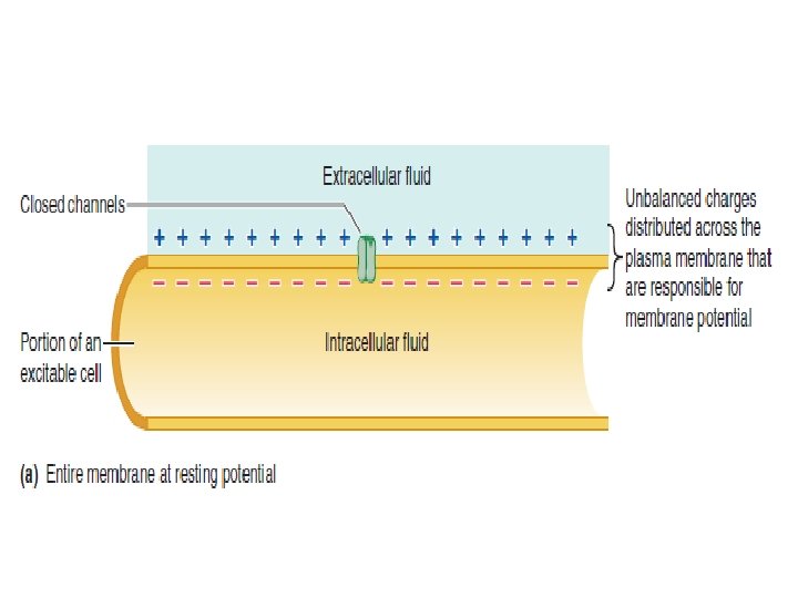
Membrane potential Th e plasma membranes of all living cells have a membrane potential, or are polarized electrically. Th e term membrane potential refers to a separation of opposite charges across the membrane or to a difference in the relative number of cations and anions in the ICF and ECF.

The cells of excitable tissues—namely, nerve cells and muscle cells—have the ability to produce rapid, transient changes in their membrane potential when excited.

These brief fluctuations in potential serve as electrical signals. The constant membrane potential present in the cells of nonexcitable tissues and those of excitable tissues when they are at rest—that is, when they are not producing electrical signals—is known as the resting membrane potential.

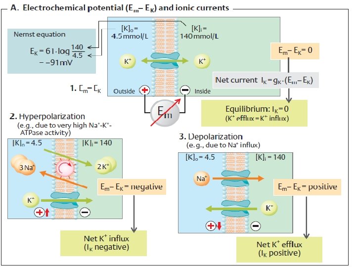
EFFECT OF THE SODIUM–POTASSIUM PUMP ON MEMBRANE POTENTIAL The Na+–K+ pump transports three Na+ out for every two K+ it transports in. Because Na+ and K+ are both positive ions, this unequal transport separates charges across the membrane, with the outside becoming relatively more positive and the inside becoming relatively more negative as more positive ions are transported out than in.

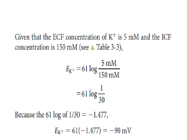
EFFECT OF THE MOVEMENT OF POTASSIUM ALONE ON MEMBRANE POTENTIAL: EQUILIBRIUM POTENTIAL FOR K+

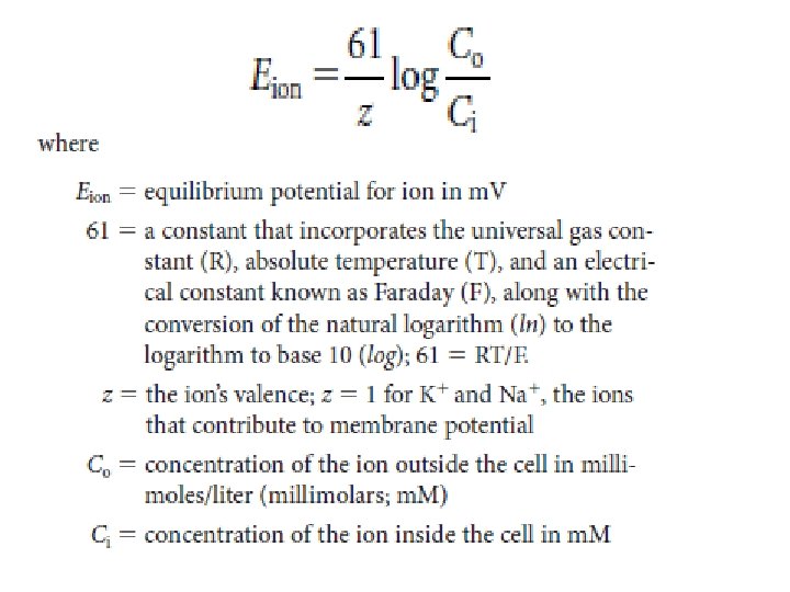
The equilibrium potential for a given ion with differing concentrations across a membrane can be calculated by means of the Nernst equation as follows:

EFFECT OF MOVEMENT OF SODIUM ALONE ON MEMBRANE POTENTIAL: EQUILIBRIUM POTENTIAL FOR Na +
 as calculated by the")
the concentrations for Na, the equilibrium potential for Na+ (ENa+) as calculated by the Nernst equation would be 61 m. V. Given that the ECF concentration of Na is 150 m. M and the ICF concentration is 15 m. M,

CONCURRENT POTASSIUM AND SODIUM EFFECTS ON MEMBRANE POTENTIAL

Nerve and muscle are excitable tissue The constant membrane potential present when a cell is electrically at rest, that is, not producing electrical signals, is referred to as the resting membrane potential

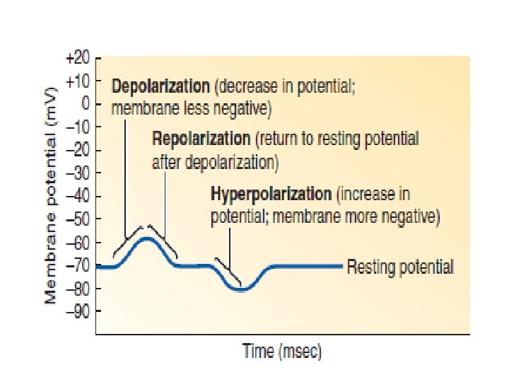
Membrane potential becomes less negative during depolarization and more negative during hyperpolarization. 1. Polarization: Charges are separated across the plasma membrane, so the membrane has potential. Any time the value of the membrane potential is other than 0 m. V, in either the positive or negative direction, the membrane is in a state of polarization.

magnitude of the potential is directly proportional to the number of positive and negative charges separated by the membrane. 2. Depolarization: The membrane becomes less polarized; the inside becomes less negative than at resting potential, with the potential moving closer to 0 m. V.

3. Repolarization: The membrane returns to resting potential after having been depolarized. 4. Hyperpolarization: The membrane becomes more polarized; the inside becomes more negative than at resting potential, with the potential moving even farther from 0 m. V.

Electrical signals are produced by changes in ion movement across the plasma membrane Changes in ion movement are brought about by changes in membrane permeability in response to triggering events. Depending on the type of electrical signal, a triggering event might be: (1) a change in the electrical field in the vicinity of an excitable membrane.
 an interaction of a chemical messenger with a surface receptor on a nerve")
(2) an interaction of a chemical messenger with a surface receptor on a nerve or muscle cell membrane. (3) A stimulus, such as sound waves stimulating specialized neurons in the ear. (4) a change of potential caused by inherent cyclical changes in channel permeability.
 graded potentials, which serve as")
There are two basic forms of electrical signals: (1) graded potentials, which serve as shortdistance signals. (2) action potentials, which signal over long distances.

Graded Potentials Graded potentials are local changes in membrane potential that occur in varying grades or degrees of magnitude or strength. For example, membrane potential could change from -70 to -60 m. V or from -70 to -50 mv

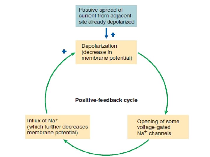
The stronger a triggering event is, the larger the resultant graded potential Graded potentials are usually produced by a specific triggering event that causes gated ion channels to open in a specialize region of the excitable cell membrane. The resultant ion movement produces the graded potential, which most commonly is depolarization resulting from net Na+ entry. The graded potential →→→ is confined to this small, specialized region of the total plasma membrane.
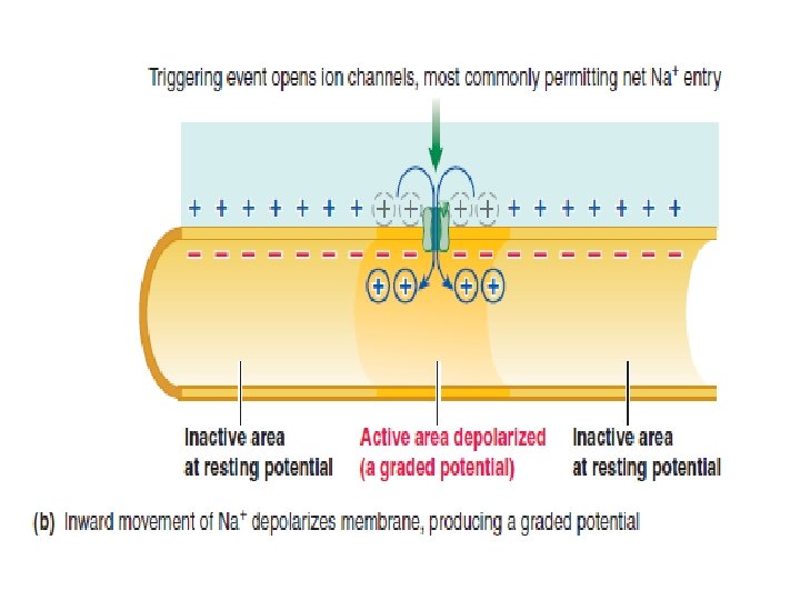

The stronger the triggering event, the more gated channels that open, the greater is the positive charge entering the cell, and the larger is the depolarizing graded potential at the point of origin. Also, the longer the duration of the triggering event, the longer is the duration of the graded potential.

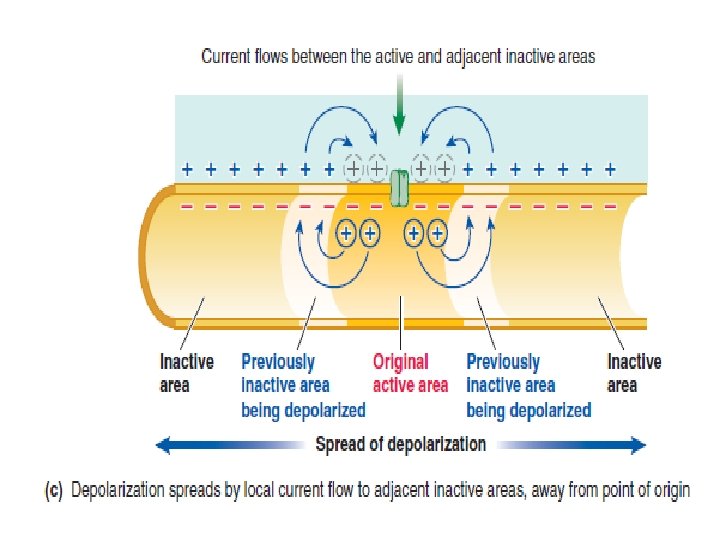
Graded potentials die out over short distances The spread of a graded potential is decremental (gradually decreases). The graded potentials also have limited signaling distance, they are critically important to the body’s function. The following are all graded potentials: postsynaptic potentials, receptor potentials, end-plate potentials, pacemaker potentials, and slow-wave potentials.
 changes in membrane")
Action Potentials Action potentials are brief, rapid, large (100 m. V) changes in membrane potential during which the potential actually reverses, so that the inside of the excitable cell transiently becomes more positive than the outside.

Action potentials are conducted, or propagated, throughout the entire membrane in nondecremental fashion; that is, they do not diminish in strength as they travel from their site of initiation throughout the remainder of the cell membrane.

Depolarization from the resting potential of -70 m. V proceeds slowly at first, until it reaches a critical level known as threshold potential, typically between -50 and- 55 m. V
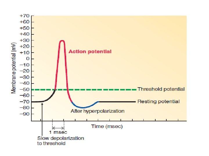

The action potential is the entire rapid change in potential from threshold to peak and then back to resting. Unlike the variable duration of a graded potential, the duration of an action potential is always the same in a given excitable cell. In a neuron, an action potential lasts for only 1 msec (0. 001 sec). It lasts longer in muscle, with the duration depending on the muscle type.

Action potentials take place as a result of the triggered opening a subsequent closing of two specific types of channels: voltage-gated Na+ channels and voltage-gated K+ channels.
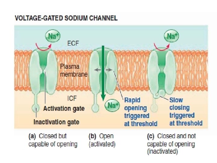
 The rapid opening of the Na+")
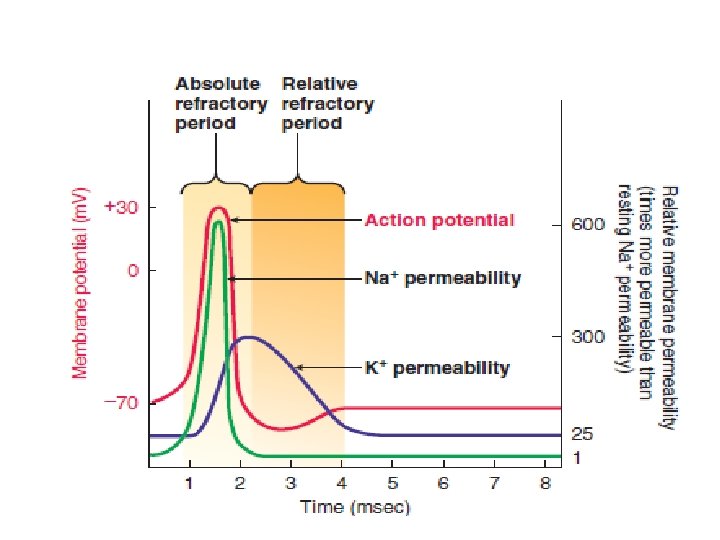
Three action potential-related events occur at threshold: (1) The rapid opening of the Na+ activation gates, which permits Na+ to enter, moving the potential from threshold to its positive peak. (2) The slow closing of the Na+ inactivation gates, which halts further Na+ entry after a brief time delay, thus keeping the potential from rising any further. (3) The slow opening of the K+ gates, which is in large part responsible for the potential plummeting from its peak back to resting.

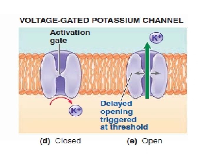
The membrane potential would gradually return to resting after closure of the Na+ channels as K+ continued to leak out but no further Na+ entered. However, the return to resting is hastened by the opening of K+ gates at the peak of the action potential.
 to about")
Opening of the voltage-gated K+ channels greatly increases K+ permeability (designated PK+) to about 300 times the resting PNa+. This marked increase in PK+ causes K+ to rush out of the cell down its electrochemical gradient, carrying positive charges back to the outside.

Note that at the peak of the action potential, the positive potential inside the cell tends to repel the positive K+ ions, so the electrical gradient for K+ is outward, unlike at resting potential. Of course the concentration gradient for K+ is always outward. The outward movement of K+ rapidly restores the negative resting potential.

The Na+ – K+ pump gradually restores the concentration gradients disrupted by action potentials At the completion of an action potential, the membrane potential has been restored to its resting condition, but the ion distribution has been altered slightly. Sodium entered the cell during the rising phase, and a comparable amount of K+ left during the falling phase. The Na+ –K+ pump restores these ions to their original locations in the long run, but not after each action potential.

The active pumping process takes much longer to restore Na+ and K+ to their original locations than it takes for the passive fluxes of these ions during an action potential. However, the membrane does not need to wait until the concentration gradients are slowly restored before it can undergo another action potential.

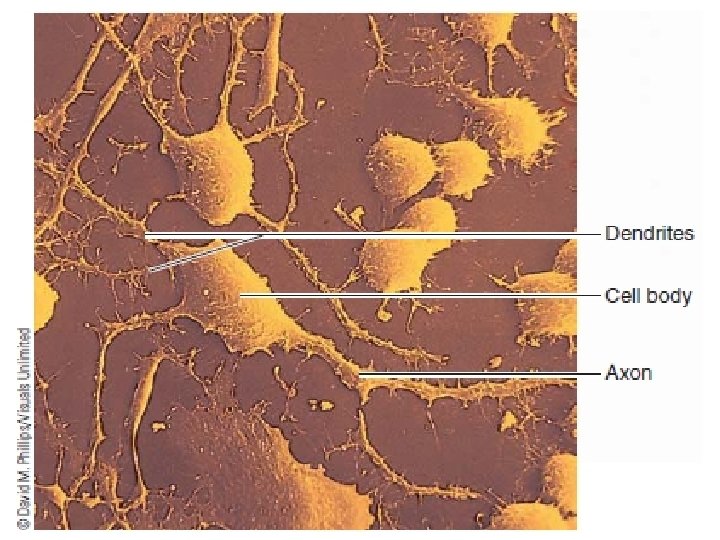
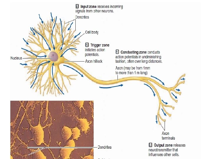
Action potentials are propagated from the axon hillock to the axon terminals A single neuron typically consists of three basic parts: The cell body. The dendrites. The axon.

Once initiated, action potentials are conducted throughout a nerve fiber Once an action potential is initiated at the axon hillock, no further triggering event is necessary to activate the remainder of the nerve fiber. The impulse is automatically conducted throughout the neuron without further stimulation by one of two methods of propagation: contiguous conduction or saltatory conduction.

Contiguous conduction involves the spread of the action potential along every patch of membrane down the length of the axon (contiguous means “touching” or “next to in sequence”).

The refractory period ensures one-way propagation of action potentials and limits their frequency. A time period when a recently activated patch of membrane is completely refractory (meaning unresponsive) to further stimulation is known as the absolute refractory period.

Following the absolute refractory period is a relative refractory period, during which a second action potential can be produced only by a triggering event considerably stronger than usual.

Action potentials occur in all-or-none fashion An excitable membrane either responds to a triggering event with a maximal action potential that spreads nondecrementally throughout the membrane, or it does not respond with an action potential at all. This property is called the all-or-none law.

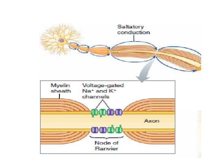
Myelination increases the speed of conduction of action potentials Myelinated fibers are axons covered with myelin, a thick layer composed primarily of lipids, at regular intervals along their length.
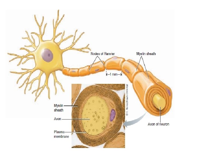

Myelin is not actually a part of the neuron but consists of separate myelin-forming cells that wrap themselves around the axon in jelly-roll fashion. These myelinforming cells are Schwann cells in the peripheral nervous system (PNS).

Between the myelinated regions, at the nodes of Ranvier, the axonal membrane is bare and exposed to the ECF. Current can flow across the membrane only at these bare spaces to produce action potentials.
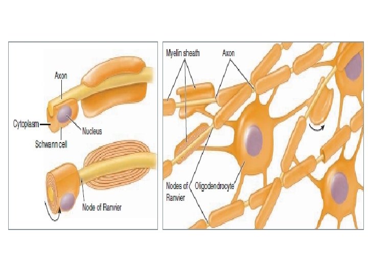

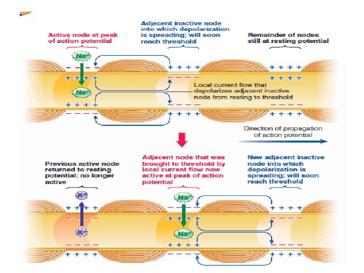
When an action potential occurs at one node, local current flow between this node and the oppositely charged adjacent node reduces the adjacent node’s potential to threshold. So that it undergoes an action potential, and so on. Consequently, in a myelinated fiber, the impulse “jumps” from node to node, skipping over the myelinated sections of the axon This process is called saltatory conduction (saltare means “to jump or leap”)

A neuron may terminate on one of three structures: a muscle, a gland, or another neuron. Therefore, depending on where a neuron terminates, it can cause a muscle cell to contract, a gland cell to secrete, another neuron to convey an electrical message along a nerve pathway, or some other function.

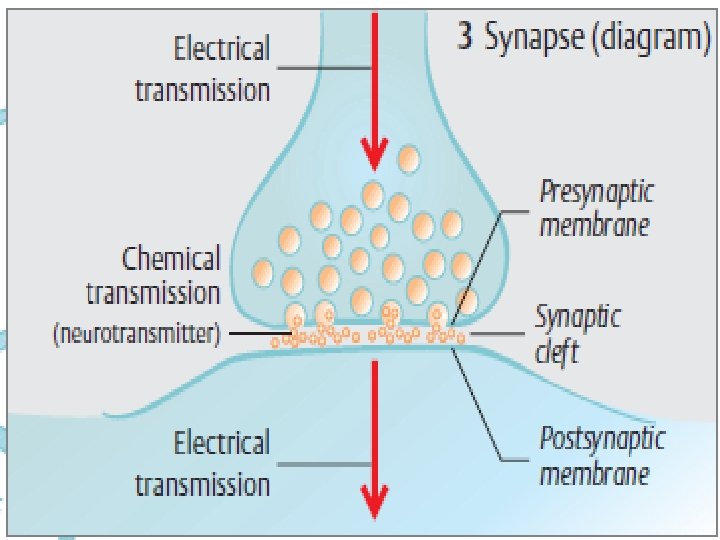
When a neuron terminates on a muscle or a gland, the neuron is said to innervate, or supply, the structure. The junction between two neurons is called a synaps.

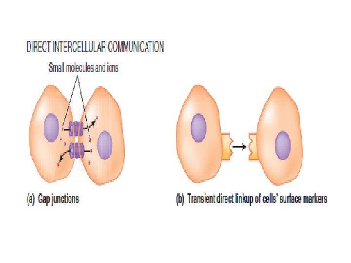
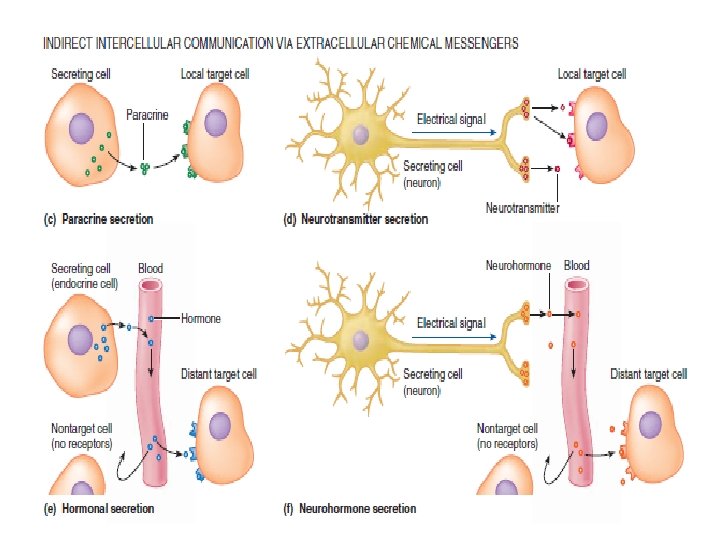
Intercellular Communication and Signal Transduction Intercellular communication can take place either directly or indirectly. Direct intercellular communication involves physical contact between the interacting cells:

The most common means by which cells communicate with one another is indirectly through extracellular chemical messengers or signal molecules, of which there are four types: paracrines, neurotransmitters, hormones, and neurohormones.

Thank you
- Slides: 67