
Melanoma Authors Georgia Beasley MD Level Basic Academic
Melanoma Author(s): Georgia, Beasley, MD Level: Basic Academic Affiliation : Duke University, ( Resident, fellow) Version No: 1. 0 Submitted: April 2012 Editors’ Review:
2 Disclaimer/Liability • The information provided in the VAP is made available in good faith and is derived from sources believed to be reliable and accurate at the time of release. • The materials presented on the VAP may include links to external Internet sites. These external information sources are outside the control of Duke-NUS. The user of the Internet links is responsible for making his or her own decision about the accuracy, reliability and correctness of the information found. • In no event shall Duke-NUS be liable for any indirect, special, incidental, or consequential damages arising out of any use of reliance of any information contained in the VAP. Nor does Duke-NUS assume any responsibility for failure or delay in updating or removing the information contained in the VAP. • Moreover, information provided on the VAP does not constitute medical advice or treatment nor should it be considered as a replacement of the patient/physician relationship or a physician’s professional judgment. Duke-NUS expressly disclaims all liability for treatment, diagnosis, decisions and actions taken or not taken in reliance upon information contained in the VAP. This work is licensed under a Creative Commons Attribution-Non. Commercial-No. Derivs 3. 0 Unported License To view a copy of this license, visit [http: //creativecommons. org/licenses/by-nc-nd/3. 0/]
3 Financial Disclosures (past 3 years) • “No Disclosures”.
4 Learning Objectives • Identify important epidemiologic factors and risk factors for melanoma. • Understand the clinical and pathologic diagnoses of melanoma. • Learn generalized treatment approach to melanoma.
5 Outline • Definition of Melanoma • Epidemiology • Risk Factors • Clinical Diagnoses • Pathologic Diagnoses/Staging • Treatment Options

6 Defintion • Melanoma: malignant tumor of melancoytes. Melanocytes are cells that produce melanin found in skin (most common), bowel, inner eye, bone, meninges, and the eye. • melanocytes comprise of 5 -10% of cells in the stratum basal layer of the epidermis • While cutaneous melanoma is the most common, melanoma can develop wherever melanocytes are present (ie, uveal melanoma, anal melanoma)
7 Epidemiology • American Cancer Society statistics for 2012: 76, 250 new melanomas (about 44, 250 in men and 32, 000 in women). About 9, 180 expected deaths (about 6, 060 men and 3, 120 women). • Incidence rates for melanoma have been rising for at least 30 years. One of fastest increasing incidences of malignancies. • World Health Organization: 48, 000 melanoma related deaths occur worldwide per year.
8 Risk Factors • UV radiation • Fair skin, light hair, light eye color • Dysplastic nevi • Personal or family history of melanoma • Genetic mutations (CDKN 2 A, MC 1 R and many more)
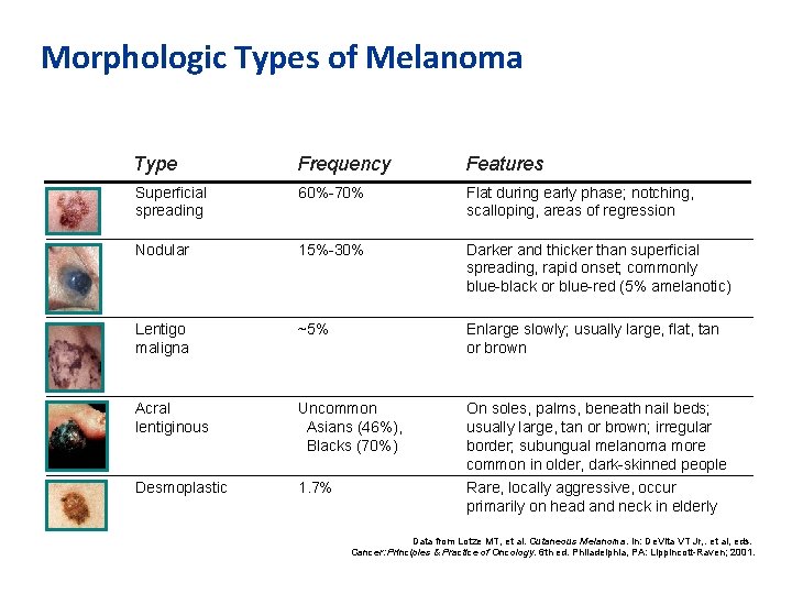
Morphologic Types of Melanoma Type Frequency Features Superficial spreading 60%-70% Flat during early phase; notching, scalloping, areas of regression Nodular 15%-30% Darker and thicker than superficial spreading, rapid onset; commonly blue-black or blue-red (5% amelanotic) Lentigo maligna ~5% Enlarge slowly; usually large, flat, tan or brown Acral lentiginous Uncommon Asians (46%), Blacks (70%) On soles, palms, beneath nail beds; usually large, tan or brown; irregular border; subungual melanoma more common in older, dark-skinned people Desmoplastic 1. 7% Rare, locally aggressive, occur primarily on head and neck in elderly Data from Lotze MT, et al. Cutaneous Melanoma. In: De. Vita VT Jr, . et al, eds. Cancer: Principles & Practice of Oncology. 6 th ed. Philadelphia, PA: Lippincott-Raven; 2001.
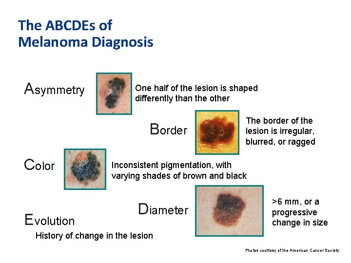
The ABCDEs of Melanoma Diagnosis Asymmetry One half of the lesion is shaped differently than the other Border Color Evolution The border of the lesion is irregular, blurred, or ragged Inconsistent pigmentation, with varying shades of brown and black Diameter >6 mm, or a progressive change in size History of change in the lesion Photos courtesy of the American Cancer Society.
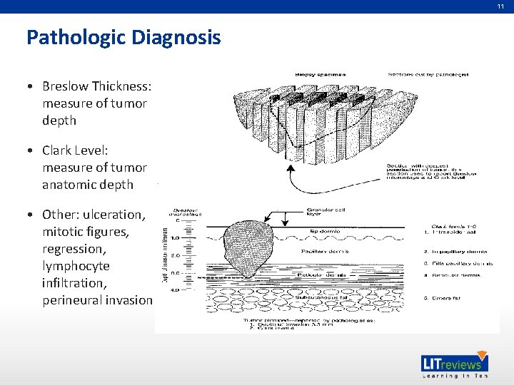
11 Pathologic Diagnosis • Breslow Thickness: measure of tumor depth • Clark Level: measure of tumor anatomic depth • Other: ulceration, mitotic figures, regression, lymphocyte infiltration, perineural invasion
12 Staging of Cutaneous Melanoma • American Joint Committee on Cancer Staging 2009. • Features: T (tumor), N (lymph node), M (metastases) staging • Stage I/II (several subtypes): NO nodal or distant disease • Stage III (subtypes): includes all T stages plus varying degrees of nodal involvement • Stage IV (subtypes): any distant metastatic disease
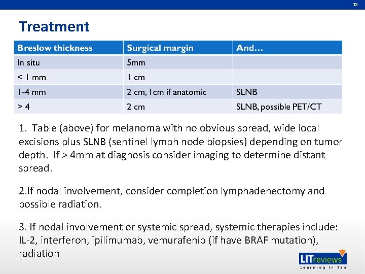
13 Treatment 1. Table (above) for melanoma with no obvious spread, wide local excisions plus SLNB (sentinel lymph node biopsies) depending on tumor depth. If > 4 mm at diagnosis consider imaging to determine distant spread. 2. If nodal involvement, consider completion lymphadenectomy and possible radiation. 3. If nodal involvement or systemic spread, systemic therapies include: IL-2, interferon, ipilimumab, vemurafenib (if have BRAF mutation), radiation
14 Key References • Balch CM, Gershenwald JE, Soong S-J, et al. Final version of 2009 AJCC melanoma staging and classification. J Clin Oncol, 2009; 27(36): 6199 -206.
15 Acknowledgements • Douglas S. Tyler, MD • Hilliard Seigler, MD • Scott K. Pruitt, MD

Partners in Academic Medicine
- Slides: 16