MEIOSIS OR REDUCTION DIVISION Meiosis preceded by duplication

MEIOSIS OR REDUCTION DIVISION � Meiosis preceded by duplication of chromosomes which is followed by meiosis I and meiosis II. These two divisions result in four daughter cells each with half the number of chromosomes that of the parents. � In this division single pair of homologous chromosomes in diploid cell, that both members of the pair are duplicated and the copies sorted into four haploid daughter cells.

HISTORY OF MEIOSIS � � � In 1876 meiosis was first discovered and described in sea urchin eggs by the German biologist Oscar Hertwig. In 1883 it was described at the level of chromosomes, by the Belgian zoologist Edouard Van Beneden. In 1890 the significance of meiosis for reproduction and inheritance, was described by German biologist August Weismann, who noted that two cell divisions were necessary to transform one diploid cell into four haploid cells if the number of chromosomes had to be maintained. In 1911 the American geneticist Thomas Hunt Morgan observed crossover in Drosophila melanogaster meiosis and provided the first genetic evidence that genes are transmitted on chromosomes. In 1905 the term meiosis was introduced by J. B. Farmer and J. E. S. Moore. Meiosis is derived from the Greek word meaning 'lessening'

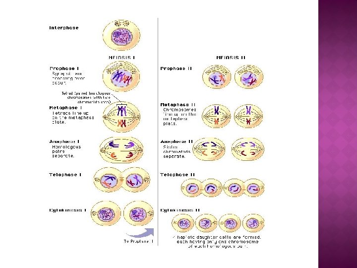
Meiosis is a special type of cell division necessary for sexual reproduction in eukaryotes such as animals, plants and fungi. The number of sets of chromosomes reduced to half the original number, typically from two sets (diploid) to one set (haploid). The cells produced by meiosis are either gametes. In many organisms gametes are called sperm and egg cells. � Meiotic division occurs in two stages, meiosis I and meiosis II. � Meiosis I: The first stage begins with a diploid cell that has two copies of each type of chromosome, one from each the mother and father, called homologous chromosomes. All homologous chromosomes pair up and may exchange genetic material with each other in a process called crossing over. Each pair then separates as two haploid cells are formed, each with one chromosome from every homologous pair. �

� Meiosis II: In the second stage, each chromosome splits into two, with each half, called a sister chromatid, being separated into two new cells, which are still haploid. Therefore from each original cell, four genetically distinct haploid cells are produced. These cells can mature into gametes
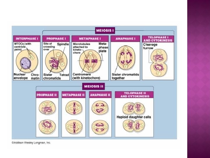

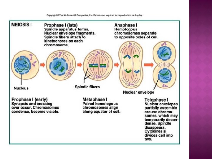
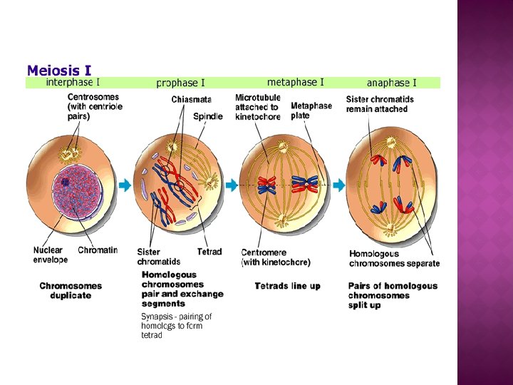
MEIOSIS I � Meiosis I separates homologous chromosomes, producing two haploid cells and thus meiosis I is referred to as a reductional division. � A regular diploid human cell contains 46 (2 n) chromosomes because it contains 23 pairs of homologous chromosomes. However, after meiosis I 46 chromatids, it is only considered as being N, with 23 chromosomes. This is because later, in Anaphase I, the sister chromatids will remain together as the spindle fibers pull the pair toward the pole of the new cell. � In meiosis II, an equational division like mitosis will occur whereby the sister chromatids are finally split, creating a total of 4 haploid cells (23 chromosomes, N) - two from each daughter cell from the first division.

Prophase I It is the longest phase of meiosis. � Chromosomes begin to condense � Homologous chromosomes pair along their length aligned gene by zipper like protein called synaptonemal complex and state is called synapsis. � Crossing over between non sister chromatids leading to exchange of segments of DNA. Chiasmata forms at the site of crossover. � At mid prophase synapsis ends and chromosomes in each pair moved apart. � Centrosomes move, spindle forms and nuclear envelope breakdown. � Microtubules of each pole attach to the kinetochores (protein) at centromeres of two homologs � Homologous pairs move towards the metaphase plate. These events occur in various stages of prophase I. �

STAGES OF MEIOSIS I AND MEIOSIS II

1. Leptotene � It is the first stage of prophase I. � Also known as leptonema, from Greek origin meaning "thin threads”. � In this stage individual chromosomes each consisting of two sister chromatids become condense into visible strands within the nucleus. � However the two sister chromatids are tightly bound that they are indistinguishable from one another. � The synaptonemal complex assemble. � Leptotene is of very short duration and progressive condensation and coiling of chromosome fibers takes place

2. Zygotene � The zygotene or zygonema, from Greek origin meaning "paired threads”. � Homologous chromosomes line up with each other into pairs. � At this stage, the synapsis (pairing/coming together) of homologous chromosomes takes place from the centromere the chromosome ends or portion. � Individuals of a pair are equal in length and in position of the centromere. Thus pairing is highly specific and exact. � The paired chromosomes are called bivalent or tetrad chromosomes.

3. Pachytene � The pachytene or pachynema, from Greek words meaning "thick threads“. � It is the stage when chromosomal crossover(crossing over) occurs. � Nonsister chromatids of homologous chromosomes may exchange segments over regions of homology. � Sex chromosomes, however, are not wholly identical, and only exchange information over a small region of homology. � At the site of exchange chiasmata form. Recombination of information occurs. � The actual act of crossing over is not perceivable through the microscope, and chiasmata are not visible until the next stage.

4. Diplotene � Diplotene or diplonema, from Greek words meaning "two threads", � The synaptonemal complex degrades and homologous chromosomes separate from one another a little. � However, the homologous chromosomes of each bivalent remain tightly bound at chiasmata, the regions where crossing-over occurred. The chiasmata remain on the chromosomes until they are severed in anaphase.

5. Diakinesis � Chromosomes condense further during the diakinesis stage, from Greek words meaning "moving through". � This is the first point in meiosis where the four parts of the tetrads are actually visible. � Chiasmata clearly visible. � The nucleoli disappear. � The nuclear membrane disintegrates into vesicles � The meiotic spindle begins to form.

� Synchronous processes Two centrosomes, containing a pair of centrioles in animal cells, migrate to the two poles of the cell. � These centrosomes, function as microtubule organizing centers. � The microtubules attaches at the kinetochore. The kinetochore functions as a motor, pulling the chromosome along the attached microtubule toward the originating centriole. � Microtubules that attach to the kinetochores are known as kinetochore microtubules. � Microtubules that interact with microtubules from the opposite centriole are called nonkinetochore microtubules or polar microtubules. � A third type of microtubules, the aster microtubules, radiates from the centrosome into the cytoplasm. �

Metaphase I Homologous pairs move together along the metaphase plate � Homologous chromosomes align along an equatorial plane that bisects the spindle. � Anaphase I Kinetochore (bipolar spindles) microtubules shorten, severing the recombination nodules and pulling homologous chromosomes apart. � Whole chromosomes are pulled toward opposing poles, forming two haploid sets. � Each chromosome still contains a pair of sister chromatids. � During this time disjunction occurs, which is one of the processes leading to genetic diversity. � Nonkinetochore microtubules lengthen, pushing the centrioles farther apart. �

Telophase I The first meiotic division ends with the arrival of chromosomes at the poles. � Each daughter cell now has half the number of chromosomes but each chromosome consists of a pair of chromatids. � Microtubules spindle network disappear � New nuclear membrane synthesize � The chromosomes uncoil back into chromatin. � Cytokinesis Pinching of the cell membrane in animal cells or the formation of the cell wall in plant cells, occurs � Creation of two daughter cells � Sister chromatids remain attached during telophase � Cells may enter in interphase II. � No DNA replication occurs during this stage. �

STAGES OF PROPHASE I OF MEIOSIS

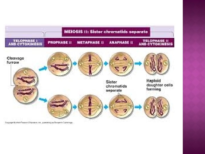
Meiosis II � Meiosis II is the second part of the meiotic process, also known as equational division. � Mechanically, the process is similar to mitosis, though its genetic results are fundamentally different. � The end result is production of four haploid cells (23 chromosomes, N in humans). � The four main steps of Meiosis II are: Prophase II, Metaphase II, Anaphase II, and Telophase II.

Prophase II : � Nucleoli and nuclear envelope disappears. � Shortening and thickening of the chromatids. � Centrioles move to the polar regions and arrange spindle fibers for the second meiotic division. Metaphase II: � The centromeres contain two kinetochores that attach to spindle fibers from the centrosomes (centrioles) at each pole. � The new equatorial metaphase plate is rotated by 90 degrees when compared to meiosis I, perpendicular to the previous plate.

Aanaphase II � The centromeres are cleaved � Microtubules attached to the kinetochores to pull the sister chromatids apart. � The sister chromatids by convention are now called sister chromosomes as they move toward opposing poles Telophase II � Marked by uncoiling and lengthening of the chromosomes. � Disappearance of the spindle. � Nuclear envelopes reform � Cleavage or cell wall formation eventually produces a total of four daughter cells, each with a haploid set of chromosomes

LIGHT MICROSCOPIC IMAGE OF MEIOSIS
- Slides: 26