MEIOSIS FOLDABLE NOTES INTERPHASE G FUNCTION This is

MEIOSIS FOLDABLE NOTES

INTERPHASE – G
")
FUNCTION This is the part of the cell cycle (and first part of interphase) where the cell is increasing in size (by synthesizing cytoplasmic components). It contains one copy of each chromosome in this stage (or 23 pairs of homologous chromosomes).

INTERPHASE – S
")
FUNCTION This is the part of the cell cycle (and second part of Interphase) where the DNA is replicated to create two copies of each chromosome. Identical copies are attached to one another at a midpoint called the centromere to create a duplicated chromosome. For animal cells, the centriole pair is also duplicated at this stage. Plant cells do not contain centrioles.

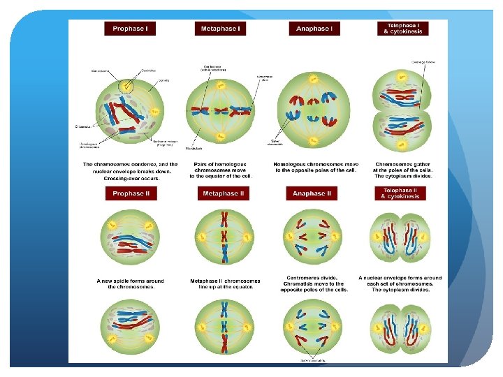
PROPHASE I

FUNCTION This is the beginning of Meiosis I, the first meiotic division. Synapsis occurs when the homologous chromosomes migrate towards one another and pair up. They line up closely next to one another to form a tetrad (tetrad refers to 4 sister chromatids). When the tetrad forms, the paternal and maternal homologues exchange DNA at various loci called chiasmata. This process of forming recombinant DNA is called crossing over.

Since crossing over is random, this process results in random genetic variation in the resulting sex cells (egg/sperm). Other events that occur: the chromosomes begin to thicken and condense, the nuclear envelope begins to dissolve, the nucleolus begins to disappear, centriole pairs begin to migrate to opposite poles and spindle fibers begin to form between them.

METAPHASE I

FUNCTION The tetrads are pulled to the equatorial/metaphase plate by spindle fibers and are lined up along this plate so that each homologue in the tetrad faces an opposing pole. This is where random genetic variation occurs again. The orientation of the paternal and maternal homologues on the equatorial/metaphase plate is random, so they can face either pole. This means that when they are separated in Anaphase I, there is variation in which maternal and paternal chromosomes end up in which pole. This random sorting of homologues is called independent assortment. Other events that occur: the centriole pairs are fully migrated to opposite poles, the spindle apparatus is fully formed and the nuclear membrane is completely dissolved.

ANAPHASE I
 attached to each homologue shorten and pull homologues to")
FUNCTION The spindle fibers (microtubules) attached to each homologue shorten and pull homologues to their opposite poles. (Unlike in Anaphase of mitosis, the sister chromatids are not pulled apart at the centromere in Anaphase I. ) The cell enlarges and elongates in preparation for division.

TELOPHASE I

FUNCTION Telophase I signals the end of the first meiotic division. Two distinct poles of the cell begin to form with one set of (non-identical) DNA on each side. Each side contains a haploid number of duplicated chromosomes. A cleavage furrow forms to divide the cell in half. This marks the beginning of cytokinesis which results in two non-identical daughter cells which will each enter meiosis II.

Other events that occur: the DNA decondenses back into stringy chromatin, the nuclear envelope reforms around each haploid set of DNA, the nucleolus reforms and the spindle fibers dissolve. Between Telophase I and Prophase II, the centriole pairs in both poles are duplicated again. Note: The DNA does not replicate again before entering Meiosis II.

PHROPHASE II

FUNCTION This is the beginning of Meiosis II, the second meiotic division. The stages of Meiosis II are very similar to mitosis except that the resulting daughter cells are non-identical in meiosis (as opposed to identical in mitosis). The nuclear membrane begins to dissolve; the chromosomes condense and thicken, the nucleolus begins to disappear and the centriole pairs migrate to opposite poles as spindle fibers form between them.

METAPHASE II

FUNCTION Spindle fibers move the chromosomes to the equatorial/metaphase plate so that they are lined up along it. This is where random genetic variation occurs again. The orientation of the sister chromatids at the equatorial/metaphase plate is random, so they can face either poles. This means that when they are separated in Anaphase II, there is random variation in which chromatid ends up in which pole. The centrioles are fully migrated to opposite poles. The spindle apparatus is fully formed. The nuclear membrane is completely dissolved.

ANAPHASE II

FUNCTION The spindle fibers attached to the sister chromatids shorten and pull at the sister chromatids which separates them at their centromeres. The cell elongates in preparation for cell division.

TELOPHASE II

FUNCTION Telophase II signals the end of the second meiotic division. The DNA de-condenses back into stringy chromatin. A nuclear envelope reforms around each set of DNA. The nucleolus begins to reappear. The spindle fibers dissolve. A cleavage furrow forms to divide the cell in half. This marks the beginning of cytokinesis which will lead to two non-identical daughter cells.

GAMATES If meiosis occurs as a part of spermatogenesis, then 4 sperm are created at the end of meiosis (as shown in the foldable). If meiosis occurs as a part of oogenesis, then 1 egg and 3 polar bodies are created at the end of meiosis. NOTE: Each gamete produced is different from the other and is a result of the 3 events during meiosis that lead to genetic variation (crossing over and the independent assortment in Metaphase I and Metaphase II. )

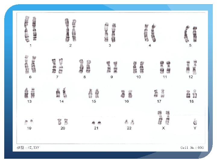
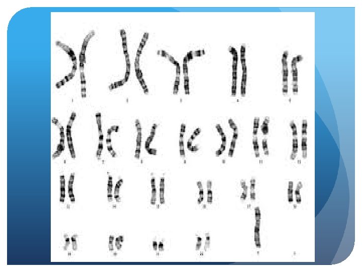
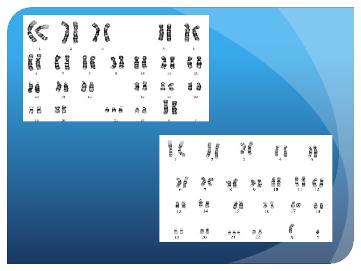
Karyotype Shows all of our chromosomes 1 -22 are out autosomal chromosomes 23 is out sex chromosomes

Male

Female

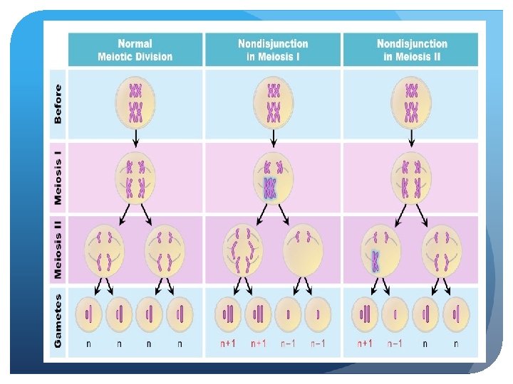
Chromosomal Disorders can be causes by nondisjunction, Non-disjunction is when a chromosomes or chromatid does not separate properly during Meiosis.
 during cell division, so")
Non-Disjunction Nondisjunction: Failure of paired chromosomes to separate (to disjoin) during cell division, so that both chromosomes go to one daughter cell and none go to the other. Nondisjunction causes errors in chromosome number,

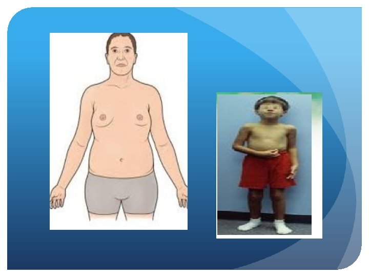
Klinefelter’s Syndrome Klinefelter syndrome occurs as a result of a random error that causes a male to be born with an extra sex chromosome. . Males have an X and a Y sex chromosome (XY). Klinefelter syndrome can be caused by: One extra copy of the X chromosome in each cell (XXY), the most common cause. Often called a trisomy (three chromosomes) disorder. Men with Klinefelter’s syndrome have less body/facial hair, narrow shoulders, thicker breast tissue, infertile due lack of production of sperm

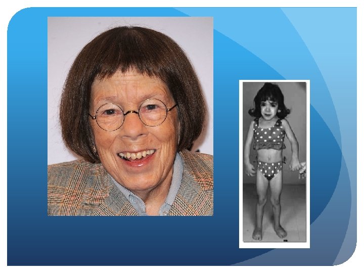
Turner’s Syndrome In girls, one copy of the X chromosome is missing, partially missing or altered. The genetic alterations of Turner syndrome may be one of the following: Monosomy (1 Chromosome). The complete absence of an X chromosome generally occurs because of an error in the father's sperm or in the mother's egg. Women with Turner’s Syndrome are shorter in stature, appearance of shorter neck – due to webbing, broad chest/shoulder, very little breast tissue, poor vision, and infertile.

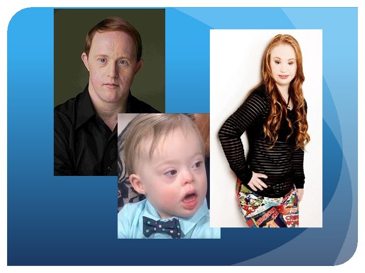
Down Syndrome Can occur in both male or females. The most common form of Down syndrome is known as trisomy 21, a condition where individuals have 47 chromosomes in each cell instead of 46. Trisomy 21 is caused by an error in cell division called nondisjunction. This leaves a sperm or egg cell with an extra copy of chromosome 21 before or at conception

Down syndrome is a genetic disorder that leads to chronic developmental delays and other problems. Down syndrome does range in severity, so these developmental delays may occur on a spectrum from moderate to severe. Behavioral Symptoms: Poor judgment, Impulsiveness, Delayed speech and language development, Short attention span Physical Symptoms: Distinctive, flattened facial features, Short neck, Protruding tongue, Poor muscle tone, Relatively shortened fingers Heart, intestine, ear, breathing problems Cognitive Symptoms: Moderate to severe developmental delays, cognitive impairment, slowed learning, below average intelligence, slowed pace of developmental milestones
- Slides: 41