Megaloblastic Anemia Introduction The megaloblastic anemias are a

Megaloblastic Anemia

Introduction • The megaloblastic anemias are a group of disorders characterized by the presence of distinctive morphologic appearances of the developing red cells in the bone marrow. • The marrow is usually cellular and the anemia is based on ineffective erythropoiesis. • The cause is usually a deficiency of either cobalamin (vitamin B 12) or folate, but megaloblastic anemia may occur because of genetic or acquired abnormalities that affect the metabolism of these vitamins or because of defects in DNA synthesis not related to cobalamin or folate.
 exists in a number of different chemical forms.")
Cobalamin • Cobalamin (vitamin B 12) exists in a number of different chemical forms. All have a cobalt atom at the center of a corrin ring. • In nature, the vitamin is mainly in the 2 -deoxyadenosyl (ado) form, which is located in mitochondria. The other major natural cobalamin is methylcobalamin, the form in human plasma and in cell cytoplasm. It is the cofactor for methionine synthase. • There also minor amounts of hydroxocobalamin to which methyland adocobalamin are converted rapidly by exposure to light. • Dietary Sources and Requirements: • the only source for humans is food of animal origin, e. g. , meat, fish, and dairy products. Cobalamin is stable and resists high-temperature cooking processes • Vegetables, fruits, and other foods of non-animal origin are free from cobalamin. • Adult daily losses (mainly in the urine and feces) are 1– 3µg, and as the body does not have the ability to degrade cobalamin, daily requirements are also about 1– 3 µg. • Body stores are of the order of 2– 3 mg, sufficient for 3– 4 years if supplies are completely cut off.

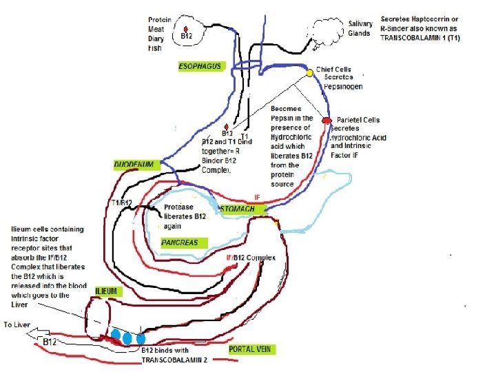
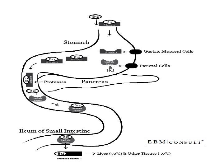
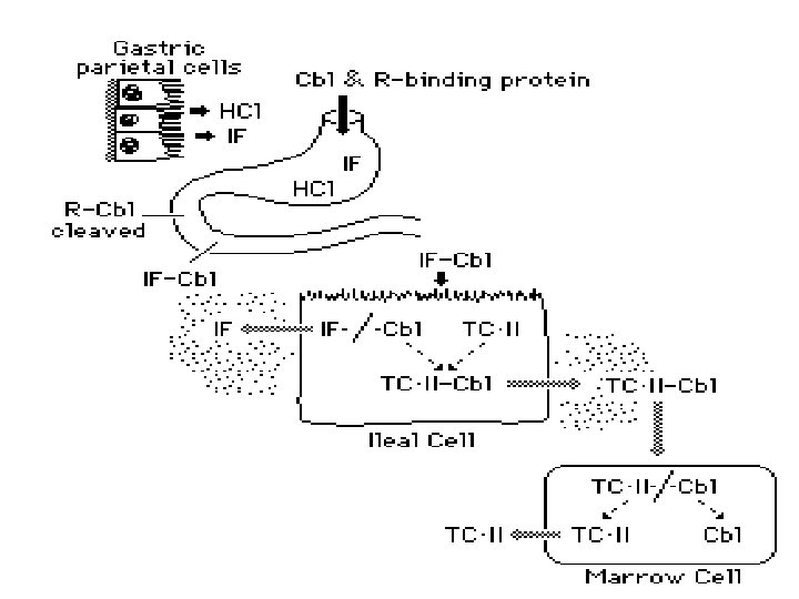
• Absorption: • Two mechanisms exist for cobalamin absorption. • One is passive, occurring equally through buccal, duodenal, and ileal mucosa; it is rapid but extremely inefficient, with <1% of an oral dose being absorbed by this process. • The normal physiologic mechanism is active; it occurs through the ileum and is efficient for small (a few micrograms) oral doses of cobalamin, and it is mediated by gastric intrinsic factor (IF). • Dietary cobalamin is released from protein complexes by enzymes in the stomach, duodenum, and jejunum; it combines rapidly with a salivary glycoprotein that belongs to the family of cobalamin-binding proteins known as haptocorrins (HCs). • In the intestine, the haptocorrin is digested by pancreatic trypsin and the cobalamin is transferred to IF.
 is")
• IF (gene at chromosome 11 q 13 coding for 9 exons) is produced in the gastric parietal cells of the fundus and body of the stomach, and its secretion parallels that of hydrochloric acid. • Normally, there is a vast excess of IF. The IF-cobalamin complex passes to the ileum, where IF attaches to a specific receptor (cubilin) on the microvillus membrane of the enterocytes. • The cobalamin-IF complex enters the ileal cell, where IF is destroyed. • After a delay of about 6 h, the cobalamin appears in portal blood attached to transcobalamin (TC) II

• Because of the appreciable amount of cobalamin undergoing enterohepatic circulation, cobalamin deficiency develops more rapidly in individuals who malabsorb cobalamin than it does in vegans, in whom reabsorption of biliary cobalamin is intact.

• Transport • Two main cobalamin transport proteins exist in human plasma. • TC I is derived primarily from the specific granules in neutrophils. • Normally, it is about two-thirds saturated with cobalamin, which it binds tightly. • TC I does not enhance cobalamin entry into tissues

• The other major cobalamin transport protein in plasma is TC II. • TC II is synthesized by liver and by other tissues, including macrophages, ileum, and vascular endothelium. • It normally carries only 20– 60 ng of cobalamin per liter of plasma and readily gives up cobalamin to marrow, placenta, and other tissues, which it enters by receptor-mediated endocytosis.

 acid Dietary Folate: Most foods contain some folate. The highest concentrations")
Folate Folic (pteroylglutamic) acid Dietary Folate: Most foods contain some folate. The highest concentrations are found in liver, yeast, spinach, other greens, and nuts (>100 µg/100 g). • Folate is easily destroyed by heating, particularly in large volumes of water. • Total-body folate in the adult is 10 mg, with the liver containing the largest store. • Daily adult requirements are 100 µg, and so stores are sufficient for only 3– 4 months in normal adults and severe folate deficiency may develop rapidly. • •

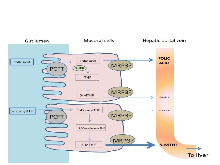
• Absorption: • Folates are absorbed rapidly from the upper small intestine. • All dietary folates (polyglutamate or monoglutamets) are converted to 5 -methyl. THF (5 -MTHF) within the small-intestinal mucosa before entering portal plasma. • About 60– 90 µg of folate enters the bile each day and is excreted into the small intestine. • Pteroylglutamic acid (folic) at doses >400µg is absorbed largely unchanged and converted to natural folates in the liver. Lower doses are converted to 5 MTHF during absorption through the intestine.

• Transport: • Folate is transported in plasma; about 1/3 is loosely bound to albumin, and 2/3 is unbound. • Two types of folate-binding protein are involved in the entry of MTHF into cells • In all body fluids (plasma, cerebrospinal fluid, milk, bile).
 act as coenzymes.")
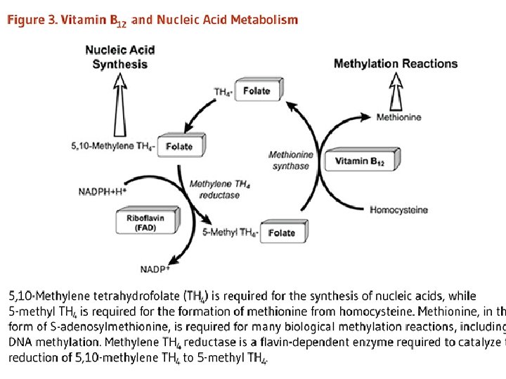
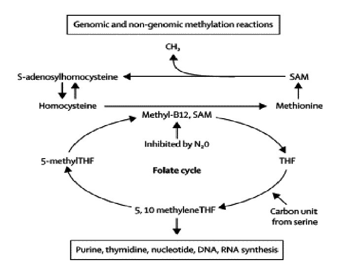
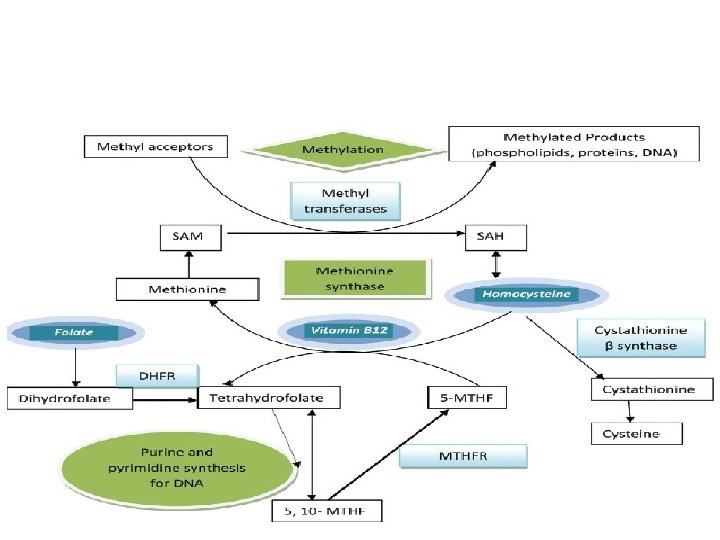
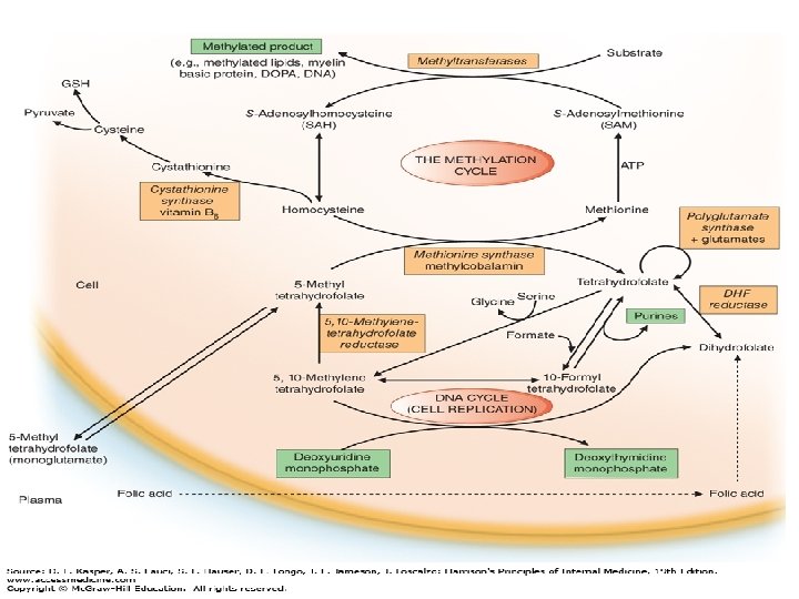
• Biochemical Functions: • Folates (as the intracellular polyglutamate derivatives) act as coenzymes. • Two of these reactions are involved in purine synthesis and one in pyrimidine synthesis necessary for DNA and RNA replication. • Folate is also a coenzyme for methionine synthesis, in which methylcobalamin is also involved and in which THF is regenerated. • Methionine, the other product of the methionine synthase reaction, is the precursor for Sadenosylmethionine (SAM), the universal methyl donor.
. •")
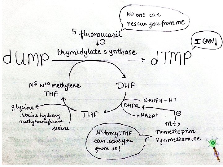
• During thymidylate synthesis, 5, 10 methylene-THF is oxidized to DHF (dihydrofolate). • The enzyme DHF reductase converts this to THF. • The drugs methotrexate, pyrimethamine, and (mainly in bacteria) trimethoprim inhibit DHF reductase and so prevent formation of active THF coenzymes from DHF.

• Biochemical Basis of Megaloblastic Anemia • The common feature of all megaloblastic anemias is a defect in DNA synthesis that affects rapidly dividing cells in the bone marrow. • All conditions that give rise to megaloblastic changes have in common a disparity in the rate of synthesis or availability of the four immediate precursors of DNA • In deficiencies of either folate or cobalamin, there is failure to convert deoxyuridine monophosphate (d. UMP) to deoxythymidine monophosphate (d. TMP), the precursor of d. TTP

Clinical Features • Many symptomless patients are detected through the finding of a raised mean corpuscular volume (MCV) on a routine blood count. • The main clinical features in more severe cases are those of anemia. • Anorexia is usually marked, and there may be weight loss, diarrhea, or constipation. Glossitis, angular cheilosis, a mild fever in more severely anemic patients, jaundice (unconjugated), and reversible melanin skin hyperpigmentation also may occur with a deficiency of either folate or cobalamin. • Thrombocytopenia sometimes leads to bruising, and this may be aggravated by vitamin C deficiency or alcohol in malnourished patients. • The anemia and low leukocyte count may predispose to infections, particularly of the respiratory and urinary tracts. • Cobalamin deficiency has also been associated with impaired bactericidal function of phagocytes.

General Tissue Effects of Cobalamin and Folate Deficiencies • • Epithelial Surfaces After the marrow, the next most frequently affected tissues are the epithelial cell surfaces of the mouth, stomach, and small intestine and the respiratory, urinary, and female genital tracts. The cells show macrocytosis, with increased numbers of multinucleate and dying cells Complications of Pregnancy The gonads are also affected, and infertility is common in both men and women with either deficiency. Maternal folate deficiency has been implicated as a cause of prematurity, and both folate deficiency and cobalamin deficiency have been implicated in recurrent fetal loss and neural tube defects, . Neural Tube Defects Folic acid supplements at the time of conception and in the first 12 weeks of pregnancy reduce by 70% the incidence of neural tube defects (NTDs) (anencephaly, meningomyelocele, encephalocele, and spina bifida) in the fetus. Most of this protective effect can be achieved by taking folic acid, 0. 4 mg daily at the time of conception.

• The incidence of cleft palate and harelip also can be reduced by prophylactic folic acid. • NTDs also can be caused by anti-folate and antiepileptic drugs.
 increase risk vascular")
• Cardiovascular Disease • severe homocystinuria (˃blood levels 100 mol/L) increase risk vascular disease, e. g. , ischemic heart disease, cerebrovascular disease, or pulmonary embolus as teenagers or in young adulthood. • Malignancy • Prophylactic folic acid in pregnancy has been found in some but not all studies to reduce the subsequent incidence of acute lymphoblastic leukemia (ALL) in childhood.

• Neurologic Manifestations • Cobalamin deficiency may cause a bilateral peripheral neuropathy or degeneration (demyelination) of the posterior and pyramidal tracts of the spinal cord and, less frequently, optic atrophy or cerebral symptoms. • The patient, more frequently male, presents with paresthesias, muscle weakness, or difficulty in walking and sometimes dementia, psychotic disturbances, or visual impairment. • Long-term nutritional cobalamin deficiency in infancy leads to poor brain development and impaired intellectual development. • Psychiatric disturbance is common in both folate and cobalamin deficiencies. ( dopamine methylation defect) and neurotransmitters • Associations between lower serum folate or cobalamin levels and higher homocysteine levels and the development of decreased cognitive function and dementia in Alzheimer's disease have been reported.

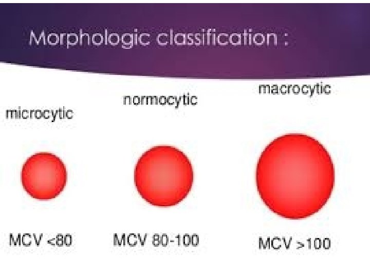
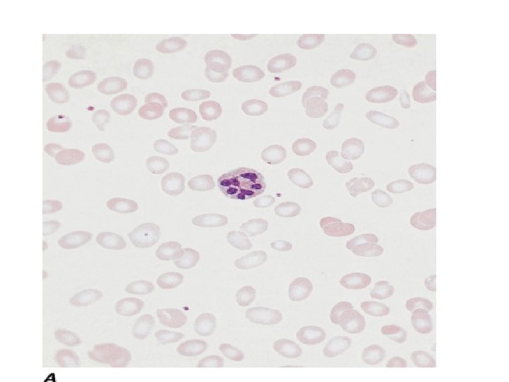
• Hematologic Findings • Peripheral Blood • Oval macrocytes, usually with considerable anisocytosis and poikilocytosis, are the main feature. • The MCV is usually >100 f. L unless a cause of microcytosis (e. g. , iron deficiency or thalassemia trait) is present. • Some of the neutrophils are hypersegmented (more than five nuclear lobes). • There may be leukopenia due to a reduction in granulocytes and lymphocytes, but this is usually >1. 5 x 10⁹/L; the platelet count may be moderately reduced, rarely to <40 x 109/L. • The severity of all these changes parallels the degree of anemia. • In a nonanemic patient, the presence of a few macrocytes and hypersegmented neutrophils in the peripheral blood may be the only indication of the underlying disorder.

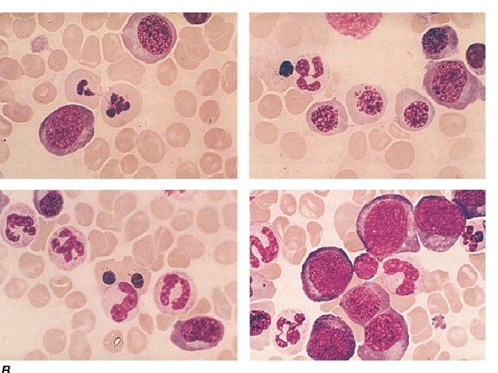
• Bone Marrow • In a severely anemic patient, the marrow is hypercellular with an accumulation of primitive cells due to selective death by apoptosis of more mature forms. • The erythroblast nucleus maintains a primitive appearance despite maturation and hemoglobinization of the cytoplasm. The cells are larger than normoblasts, and an increased number of cells with eccentric lobulated nuclei or nuclear fragments may be present. nuclearcytoplasmic asynchrony (or dissociation). • Giant and abnormally shaped metamyelocytes and enlarged hyperpolyploid megakaryocytes are characteristic. • In less anemic patients, the changes in the marrow may be difficult to recognize. The terms intermediate, mild, and early have been used. The term megaloblastoid does not mean mildly megaloblastic. It is used to describe cells with both immature-appearing nuclei and defective hemoglobinization and is usually seen in myelodysplasia.

• Chromosomes • Bone marrow cells, transformed lymphocytes, and other proliferating cells in the body show a variety of changes, including random breaks, reduced contraction, spreading of the centromere, and exaggeration of secondary chromosomal constrictions and over prominent satellites. • Similar abnormalities may be produced by antimetabolite drugs (e. g. , cytosine arabinoside, hydroxyurea, and methotrexate) that interfere with either DNA replication or folate metabolism and that also cause megaloblastic appearances

• Ineffective Hematopoiesis • There is an accumulation of unconjugated bilirubin in plasma due to the death of nucleated red cells in the marrow (ineffective erythropoiesis). • Other evidence for this includes raised urine urobilinogen, reduced haptoglobins and positive urine hemosiderin, and a raised serum lactate dehydrogenase. • A weakly positive direct antiglobulin test due to complement can lead to a false diagnosis of autoimmune hemolytic anemia.


• Cobalamin deficiency is usually due to malabsorption. • Inadequate Dietary Intake • Adults • Dietary cobalamin deficiency arises in vegans who omit meat, fish, eggs, cheese, and other animal products from their diet. • Dietary cobalamin deficiency may also arise rarely in non-vegetarian individuals who exist on grossly inadequate diets because of poverty or psychiatric disturbance. • Infants • Cobalamin deficiency has been described in infants born to severely cobalamin-deficient mothers. • These infants develop megaloblastic anemia at about 3– 6 months of age, presumably because they are born with low stores of cobalamin and because they are fed breast milk with low cobalamin content. The babies have also shown growth retardation, impaired psychomotor development, and other neurologic sequelae.
")
• Gastric Causes of Cobalamin Malabsortion • Pernicious Anemia • Pernicious anemia (PA) may be defined as a severe lack of IF due to gastric atrophy. • The disease occurs more commonly than by chance in close relatives and in persons with other organ-specific autoimmune diseases, e. g. , thyroid diseases, vitiligo, hypoparathyroidism, and Addison's disease. • It is also associated with hypogammaglobulinemia, with premature graying or blue eyes, and persons of blood group A. • An association with human leukocyte antigen (HLA) 3 has been reported in some but not all series and, in those with endocrine disease, with HLA-B 8, -B 12, and -BW 15. • Life expectancy is normal in women once regular treatment has begun. • Men have a slightly subnormal life expectancy as a result of a higher incidence of carcinoma of the stomach than in control subjects. • Gastric output of hydrochloric acid, pepsin, and IF is severely reduced. The serum gastrin level is raised, and serum pepsinogen I levels are low.

• Gastric Biopsy • This usually shows atrophy of all layers of the body and fundus, with loss of glandular elements, an absence of parietal and chief cells and replacement by mucous cells, a mixed inflammatory cell infiltrate, and perhaps intestinal metaplasia. • The infiltrate of plasma cells and lymphocytes contains an excess of CD 4 cells. • The antral mucosa is usually well preserved. • Helicobacter pylori infection occurs infrequently in PA, but it has been suggested that H. pylori gastritis occurs at an early phase of atrophic gastritis and presents in younger patients as iron-deficiency anemia but in older patients as PA. • H. pylori is suggested to stimulate an autoimmune process directed against parietal cells, with the H. pylori infection then being gradually replaced, in some individuals, by an autoimmune process.

• Serum Antibodies • Two types of IF immunoglobulin G antibody may be found in the sera of patients with PA. • One, the "blocking, " or type I, antibody, prevents the combination of IF and cobalamin, whereas the "binding, " or type II, antibody prevents attachment of IF to ileal mucosa. • Type I occurs in the sera of 55% of patients, and type II in 35%. • IF antibodies cross the placenta and may cause temporary IF deficiency in a newborn infant.

• Parietal cell antibody is present in the sera of almost 90% of adult patients with PA. • The parietal cell antibody is directed against the and subunits of the gastric proton pump (H+, K+-ATPase). • Juvenile Pernicious Anemia • This usually occurs in older children and resembles PA of adults. Gastric atrophy, achlorhydria, and serum IF antibodies are all present, although parietal cell antibodies are usually absent. • About one-half of these patients show an associated endocrinopathy such as autoimmune thyroiditis, Addison's disease, or hypoparathyroidism; in some, mucocutaneous candidiasis occurs.

• Congenital Intrinsic Factor Deficiency or Functional Abnormality • An affected child usually presents with megaloblastic anemia in the first to third year of life; a few have presented as late as the second decade. • The child usually has no demonstrable IF but has a normal gastric mucosa and normal secretion of acid. The inheritance is autosomal recessive. • Parietal cell and IF antibodies are absent. • Variants have been described in which the child is born with IF that can be detected immunologically but is unstable or functionally inactive, unable to bind cobalmin or to facilitate its uptake by ileal receptors.

• Gastrectomy • After total gastrectomy, cobalamin deficiency is inevitable, and prophylactic cobalamin therapy should be commenced immediately after the operation. • After partial gastrectomy, 10– 15% of patients also develop this deficiency. The exact incidence and time of onset are most influenced by the size of the resection and the preexisting size of cobalamin body stores. • Food Cobalamin Malabsorption • Failure of release of cobalamin from binding proteins in food is believed to be responsible for this condition, which is more common in the elderly. • It is associated with low serum cobalamin levels, with or without raised serum levels of MMA and homocysteine. • Typically, these patients have normal cobalamin absorption, as measured with crystalline cobalamin, but show malabsorption when a modified test using food-bound cobalamin is used. The frequency of progression to severe cobalamin deficiency and the reasons for this progression are not clear.

• Intestinal Causes of Cobalamin Malabsorption • Intestinal Stagnant Loop Syndrome • Malabsorption of cobalamin occurs in a variety of intestinal lesions in which there is colonization of the upper small intestine by fecal organisms. • This may occur in patients with jejunal diverticulosis, enteroanastomosis, or an intestinal stricture or fistula or with an anatomic blind loop due to Crohn's disease, tuberculosis, or an operative procedure. • Ileal Resection • Removal of 1. 2 m of terminal ileum causes malabsorption of cobalamin. • In some patients after ileal resection, particularly if the ileocecal valve is incompetent, colonic bacteria may contribute further to the onset of cobalamin deficiency.

• Tropical Sprue • Absorption of cobalamin usually improves after antibiotic therapy and, in the early stages, folic acid therapy. • Fish Tapeworm Infestation • The fish tapeworm (Diphyllobothrium latum) lives in the small intestine of humans and accumulates cobalamin from food, rendering the cobalamin unavailable for absorption. • Individuals acquire the worm by eating raw or partly cooked fish. • Megaloblastic anemia or cobalamin neuropathy occurs only in those with a heavy infestation. • Gluten-Induced Enteropathy • Malabsorption of cobalamin occurs in 30% of untreated patients (presumably those in whom the disease extends to the ileum). • Cobalamin deficiency is not severe in these patients and is corrected with a gluten-free diet.

• • Severe Chronic Pancreatitis In this condition, lack of trypsin is thought to cause dietary cobalamin attached to gastric non-IF (R) binder to be unavailable for absorption. It also has been proposed that in pancreatitis, the concentration of calcium ions in the ileum falls below the level needed to maintain normal cobalamin absorption. • HIV Infection • • Zollinger–Ellison Syndrome Malabsorption of cobalamin has been reported in the Zollinger–Ellison syndrome. It is thought that there is a failure to release cobalamin from R-binding protein due to inactivation of pancreatic trypsin by high acidity, as well as interference with IF binding of cobalamin. Radiotherapy Both total-body irradiation and local radiotherapy to the ileum (e. g. , as a complication of radiotherapy for carcinoma of the cervix) may cause malabsorption of cobalamin. Graft-versus-Host Disease This commonly affects the small intestine. Malabsorption of cobalamin due to abnormal gut flora, as well as damage to ileal mucosa, is common. Drugs The drugs that have been reported to cause malabsorption of cobalamin Megaloblastic anemia due to these drugs is, however, rare • •

Causes of folate deficiency

Causes of Folate Deficiency

• Nutritional • Dietary folate deficiency is common. Indeed, in most patients with folate deficiency a nutritional element is present. • Malabsorption of dietary folate occurs in tropical sprue and in gluteninduced enteropathy. • In the rare congenital syndrome of selective malabsorption of folate due to mutation of the protein-coupled folate transporter (PCFT), there is an associated defect of folate transport into the cerebrospinal fluid, and these patients show megaloblastic anemia, which responds to physiologic doses of folic acid given parenterally but not orally. • They also show mental retardation, convulsions, and other central nervous system abnormalities. • Minor degrees of malabsorption may also occur after jejunal resection or partial gastrectomy, in Crohn's disease, and in systemic infections, but in these conditions, if severe deficiency occurs, it is usually largely due to poor nutrition. • Malabsorption of folate has been described in patients receiving salazopyrine, cholestyramine, and triamterene.

• Excess Utilization or Loss • Pregnancy • Folate requirements are increased by 200– 300 µg to ~400 µg daily in a normal pregnancy, partly because of transfer of the vitamin to the fetus but mainly because of increased folate catabolism due to cleavage of folate coenzymes in rapidly proliferating tissues. • Prematurity • A newborn infant, whether full term or premature, has higher serum and red cell folate concentrations than does an adult. However, a newborn infant's demand for folate has been estimated to be up to 10 times that of adults on a weight basis, and the neonatal folate level falls rapidly to the lowest values at about 6 weeks of age. The falls are steepest and are liable to reach subnormal levels in premature babies, a number of whom develop megaloblastic anemia responsive to folic acid at about 4– 6 weeks of age. • This occurs particularly in the smallest babies (<1500 g birth weight) and those who have feeding difficulties or infections or have undergone multiple exchange transfusions. In these babies, prophylactic folic acid should be given.

• • Hematologic Disorders Folate deficiency frequently occurs in chronic hemolytic anemia, particularly in sickle cell disease, autoimmune hemolytic anemia, and congenital spherocytosis. In these and other conditions of increased cell turnover (e. g. , myelofibrosis, malignancies), folate deficiency arises because it is not completely reutilized after performing coenzyme functions. Inflammatory Conditions Chronic inflammatory diseases such as tuberculosis, rheumatoid arthritis, Crohn's disease, psoriasis, exfoliative dermatitis, bacterial endocarditis, and chronic bacterial infections cause deficiency by reducing the appetite and increasing the demand for folate. Systemic infections also may cause malabsorption of folate. Severe deficiency is virtually confined to the patients with the most active disease and the poorest diet. Long-Term Dialysis As folate is only loosely bound to plasma proteins, it is easily removed from plasma by dialysis. In patients with anorexia, vomiting, infections, and hemolysis, folate stores are particularly likely to become depleted. Routine folate prophylaxis is now given. Congestive Heart Failure, Liver Disease Excess urinary folate losses of >100 g per day may occur in some of these patients. The explanation appears to be release of folate from damaged liver cells.

• Antifolate Drugs • A large number of epileptics who are receiving long-term therapy with phenytoin or primidone, with or without barbiturates, develop low serum and red cell folate levels. The exact mechanism is unclear. Alcohol may also be a folate antagonist, • The drugs that inhibit DHF reductase include methotrexate, pyrimethamine, and trimethoprim. • Methotrexate has the most powerful action against the human enzyme, whereas trimethoprim is most active against the bacterial enzyme and is likely to cause megaloblastic anemia only when used in conjunction with sulphamethoxazole in patients with preexisting folate or cobalamin deficiency. • The activity of pyrimethamine is intermediate. • The antidote to these drugs is folinic acid (5 -formyl-THF). • Congenital Abnormalities of Folate Metabolism • Some infants with congenital defects of folate enzymes (e. g. , cyclohydrolase or methionine synthase) have had megaloblastic anemia

• Diagnosis of Deficiencies Cobalamin and Folate

• The diagnosis of cobalamin or folate deficiency has traditionally depended on the recognition of the relevant abnormalities in the peripheral blood analysis of the blood levels of the vitamins. • Serum Cobalamin: • This is measured by an automated enzyme-linked immunosorbent assay (ELISA). • Normal serum levels range from 118– 148 pmol/L (160 – 200 ng/L) to 738 pmol/L (1000 ng/L). • In patients with megaloblastic anemia due to cobalamin deficiency, the level is usually <74 pmol/L (100 ng/L).

• Serum Methylmalonate and Homocysteine: • In patients with cobalamin deficiency sufficient to cause anemia or neuropathy, the serum MMA level is raised. Sensitive methods for measuring MMA and homocysteine in serum have been introduced and recommended for the early diagnosis of cobalamin deficiency, even in the absence of hematologic abnormalities or subnormal levels of serum cobalamin. • Serum homocysteine is raised in both early cobalamin and folate deficiency but may be raised in other conditions, e. g. , chronic renal disease, alcoholism, smoking, pyridoxine deficiency, hypothyroidism, and therapy with steroids, cyclosporine, and other drugs. • Other Tests • Studies of cobalamin absorption once were widely used, but difficulty in obtaining radioactive cobalamin and ensuring that IF preparations are free of viruses has made these tests obsolete. • Tests to diagnose PA include serum gastrin, which is raised, and serum pepsinogen I, which is low in PA (90– 92%) but also in other conditions. • Tests for IF and parietal cell antibodies are also used as well as tests for individual intestinal diseases.

• Serum Folate • This is also measured by an ELISA technique. In most laboratories, the normal range is from 11 nmol/L (2 g/L) to 82 nmol/L (15 g/L). The serum folate level is low in all folate-deficient patients. • Red Cell Folate • The red cell folate assay is a valuable test of body folate stores. • It is less affected than the serum assay by recent diet and traces of hemolysis. In normal adults, concentrations range from 880– 3520 mol/L (160– 640 g/L) of packed red cells. • Subnormal levels occur in patients with megaloblastic anemia due to folate deficiency but also in nearly two-thirds of patients with severe cobalamin deficiency. • False-normal results may occur if a folate-deficient patient has received a recent blood transfusion or if a patient has a raised reticulocyte count.

• Treatment: Megaloblastic Anemia

• It is usually possible to establish which of the two deficiencies, folate or cobalamin, is the cause of the anemia and to treat only with the appropriate vitamin. • In patients who enter the hospital severely ill, however, it may be necessary to treat with both vitamins in large doses once blood samples have been taken for cobalamin and folate assays and a bone marrow biopsy has been performed (if deemed necessary). • Transfusion is usually unnecessary and inadvisable. If it is essential, packed red cells should be given slowly, one or two units only, with the usual treatment for heart failure if present. • Potassium supplements have been recommended to obviate the danger of the hypokalemia but are not necessary. Occasionally, an excessive rise in platelets occurs after 1– 2 weeks of therapy. • Antiplatelet therapy, e. g. , aspirin, should be considered if the platelet count rises to >800 x 109/L.

• Cobalamin Deficiency • It is usually necessary to treat patients who have developed cobalamin deficiency with lifelong regular cobalamin injections. • In a few instances, the underlying cause of cobalamin deficiency can be permanently corrected, e. g. , fish tapeworm, tropical sprue, or an intestinal stagnant loop that is amenable to surgery. • The indications for starting cobalamin therapy are a well-documented megaloblastic anemia or other hematologic abnormalities and neuropathy due to the deficiency. • Patients with borderline serum cobalamin levels but no hematologic or other abnormality may be followed to make sure that the cobalamin deficiency does not progress. • Cobalamin should be given routinely to all patients who have had a total gastrectomy or ileal resection. • Patients who have undergone gastric reduction for control of obesity or who are receiving long-term treatment with proton pump inhibitors should be screened and, if necessary, given cobalamin replacement.

• Replenishment of body stores should be complete with six 1000 -g IM injections of hydroxocobalamin given at 3 - to 7 day intervals. More frequent doses are usually used in patients with cobalamin neuropathy, • For maintenance therapy, 1000 g hydroxocobalamin IM once every 1: 3 months is satisfactory. • Because a small fraction of cobalamin can be absorbed passively through mucous membranes even when there is complete failure of physiologic IF-dependent absorption, large daily oral doses (1000– 2000 g) of cyanocobalamin have been used in PA for replacement and maintenance of normal cobalamin status in, e. g. , food malabsorption of cobalamin. • Sublingual therapy has also been proposed for those in whom injections are difficult because of a bleeding tendency and who may not tolerate oral therapy. • If oral therapy is used, it is important to monitor compliance, particularly with elderly, forgetful patients.

• Folate Deficiency • Oral doses of 5– 15 mg folic acid daily are satisfactory, as sufficient folate is absorbed from these extremely large doses even in patients with severe malabsorption. • The length of time therapy must be continued depends on the underlying disease. It is customary to continue therapy for about 4 months, when all folate-deficient red cells will have been eliminated and replaced by new folate-replete populations. • Before large doses of folic acid are given, cobalamin deficiency must be excluded and, if present, corrected; otherwise cobalamin neuropathy may develop despite a response of the anemia of cobalamin deficiency to folate therapy • Long-term folic acid therapy is required when the underlying cause of the deficiency cannot be corrected and the deficiency is likely to recur, e. g. , in chronic dialysis or hemolytic anemias. It may also be necessary in gluten-induced enteropathy that does not respond to a gluten-free diet. • Where mild but chronic folate deficiency occurs, it is preferable to encourage improvement in the diet after correcting the deficiency with a short course of folic acid.
 This is a stable form of fully")
• • Folinic Acid (5 -Formyl-THF) This is a stable form of fully reduced folate. It is given orally or parenterally to overcome the toxic effects of methotrexate or other DHF reductase inhibitors. Prophylactic Folic Acid In many countries, food is fortified with folic acid (in grain or flour) to prevent neural tube defects. It is also used in chronic dialysis patients and in parenteral feeds. Prophylactic folic acid has been used to reduce homocysteine levels to prevent cardiovascular disease, but further data are needed to assess the benefit for this and for cognitive function in the elderly. Pregnancy Folic acid, 400µ g daily, should be given as a supplement before and throughout pregnancy. In women who have had a previous fetus with a neural tube defect, 5 mg daily is recommended when pregnancy is contemplated and throughout the subsequent pregnancy. Infancy and Childhood The incidence of folate deficiency is so high in the smallest premature babies during the first 6 weeks of life that folic acid (e. g. , 1 mg daily) should be given routinely to those weighing <1500 g at birth and to larger premature babies who require exchange transfusions or develop feeding difficulties, infections, or vomiting and diarrhea.
- Slides: 63