Medulla Oblongata Cranial Nerve Nuclei 9 10 11


Medulla Oblongata Cranial Nerve Nuclei 9, 10, 11, 12 q The most important part of the brain, It regulates the breathing, heart and blood vessels, digestion and swallowing
; 1 -")
• Functional Arrangement of the Cranial Nerve Nuclei A- Motor (efferent); 1 - General Somatic Efferent (GSE) (3, 4, 6, 12) striated muscles in the head 2 - Special Visceral Efferent (SVE) (5, 7, 9, 10, 11) muscles of pharyngeal arches 3 - General Visceral Efferent (GVE) (10, 9, 7, 3) parasympathetic B- Sensory (Afferent) 1 - General Somatic Afferent (GSA) (5) general sensations from head and face. 2 - Special Somatic Afferent (SSA) (8) special sensation from the head N. B: The optic nerve described by some authors to belong to this group. 3 - General Visceral Afferent (GVA) (9, 10) general sensations from the viscera 4 - Special Visceral Afferent (SVA) (7, 9, 10) taste sensations N. B; the olfactory nerve described by some authors to belong to this group.
 1 - Nucleus Ambiguus")
Motor nuclei (9 th, 10 th, 11 th, 12 th) 1 - Nucleus Ambiguus (S. V. E = pharyngeal arch) - Upper part → g. Iossopharyngeal nerve → to stylopharyngeus muscle. - Middle part → vagus nerve. - Lower part → cranial part of accessory nerve. N. B; The vagus and accessory form pharyngeal plexus supply all muscles of pharynx except stylopharyngeus muscle, all muscles of larynx and palate except tensor palati (Mandibular N). 2 - Hypoglossal Nucleus (G. S. E) → to the hypoglossal nerve → all muscles of the tongue except palatoglossus (supplied by the pharyngeal plexus) - Injury of hypoglossal nerve → paralysis of the muscles of tongue on the same side (The tongue deviated to the side of paralysis).
 1 - Inferior salivary nucleus - It gives parasympathetic")
Parasympathetic nuclei (G. V. E) 1 - Inferior salivary nucleus - It gives parasympathetic → glossopharyngeal nerve → relay in the otic ganglia → to the parotid gland. 2 - Dorsal Nucleus of Vagus a- It is a parasympathetic fibers to the smooth muscles and glands of the digestive, respiratory tracts and cardiac muscle. b- It receives general sensation from the mucous membrane of the digestive, respiratory tract and heart.
: It receives taste sensation from:")
Sensory nuclei 1 - Solitary Nucleus (S. V. A): It receives taste sensation from: a- Anterior 2/3 of the tongue through facial nerve (chorda tympani) and oral surface of the soft palate (greater petrosal nerve). b- Posterior 1/3 of the tongue through glossopharyngeal nerve (lingual nerve). c- Root of the tongue through vagus nerve (internal laryngeal nerve). 2 - Spinal nucleus of trigeminal nerve (general sensation) (G. S. A): It receives pain, temperature and general sensations from the head and face (5 th, 9 th & 10 th). 3 - Inferior Vestibular Nuclei (S. S. A): It receives fibres of the vestibular organs of the inner ear.

NON CRANIAL NERVE NUCLEI OF MEDULLA OBLONGATA

internal arcuate fibers sensory decussation

• • Non-cranial nuclei Gracile and CUneate Nuclei: 2 nd order neuron for the conscious proprioceptive sensation and fine touch (sensation of position & movement, Stereogenosis (recognition of shape with close eye)). Gracile nucleus receives from the lower limb and lower 1/2 of the trunk Cuneate receives from the Upper limb and upper 1/2 of the trunk. Axons of the gracile and cuneate nuclei ( internal arcuate fibers) → Sensory decussation → medial lemniscus → end into the posterolateral ventral nucleus (PLVN) of the thalamus →Sensory area of cerebral cortex • Accessory Cuneate Nucleus: dorsal external arcuate fibers→ cuneocerebellar tract → ICP → cerebellum. - Lesion of gracile and cuneate leading to loss of proprioceptive sensation as in Tabes dorsalis disease (syphilis). - Patient has a characteristic Stamping gait ( During walking the patient raises his foot high up from the ground and then pushes his foot vigorously down to the ground)

Afferent 3 2 4 1 q Olivary Complex Nuclei Efferent
: -")
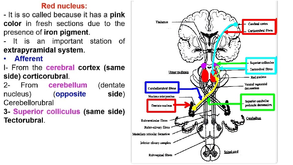
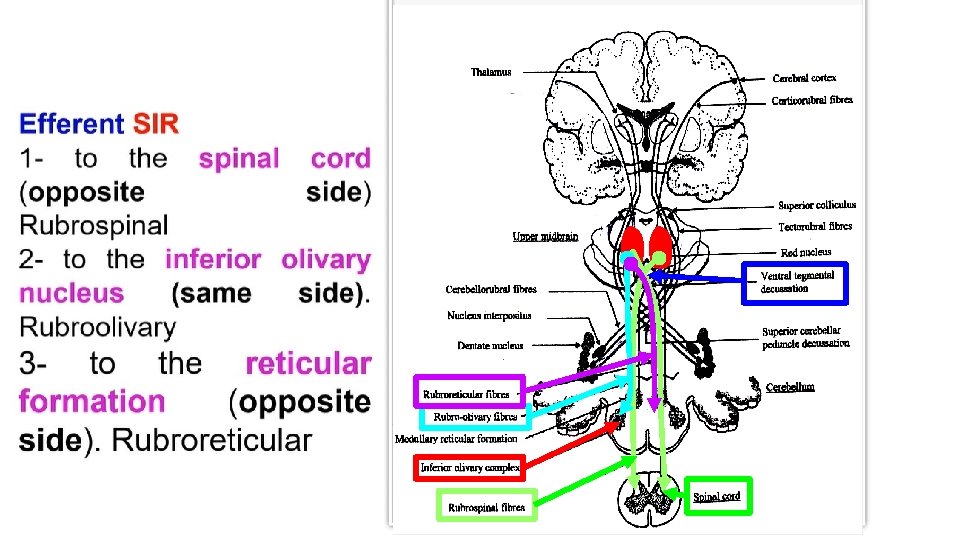
• Olivary complex Nuclei (large corrugated sac with its opening directed medially): - It forms an elevation on the front of the medulla called the olive. - Its function is associated with voluntary muscle movements. ** Afferent from 1 - Spinal cord (Spino-olivary tract). 2 - Red nucleus 3 - Globus pallidus. 4 - Reticular formation ** Efferent; Olivo-cerebellar the fibers cross to the opposite side to the cerebellar cortex through the inferior cerebellar peduncle. q Arcuate nucleus: Ventral external arcuate fibers→ ICP→ cerebellum (capable of chemosensitivity & have a proven role in the respiratory center)

The pyramidal decussation pyramid of Medulla oblongata Motor decussation Uncrossed fibers Lateral corticospinal tract Anterior corticospinal tract (80 -85%) cross the midline (15 -20%) don't cross the midline
 decussation - In the lower part of medulla, most of")
• Motor (pyramidal) decussation - In the lower part of medulla, most of the fibers of corticospinal tract (80 -85%) decussate the midline forming Lateral cortico-spinal tract descends in the lateral column of the white matter to relay in the anterior horn cells (AHC) of the spinal cord • Uncrossed pyramidal tract • 15 -20% does not cross midline forming Ventral (Anterior) cortico-spinal tract, descends in anterior white column of white matter. Its fibers cross midline to relay in anterior horn cells of the opposite side. • Injury of pyramidal tract→ contralateral hemiplegia U. M. N. L (paralysis of the muscles on the opposite ½ of the body). N. B; Some of fibers (1%) remain in the same side without crossing to end around the medial motor nuclei (nuclei that supply the muscles of the trunk and respiration). So, these muscles receive fibers from both sides. This explains absence of respiratory and trunk paralysis in cases of hemiplegia.

Pons Cranial Nerve Nuclei th th th 5 , 6 , 7 , 8 th
; for")
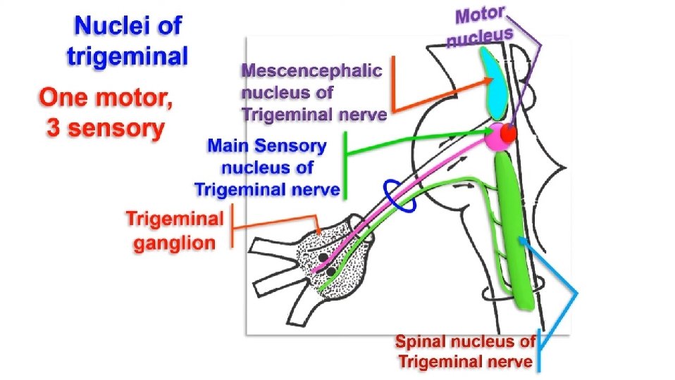
§ Nuclei of the trigeminal nerve 1 - Motor nucleus (S. V. E); for the muscles developed from the 1 st pharyngeal arch. 2 - Main sensory nucleus (G. S. A); - It receives sensory fibers of crude touch from the face and scalp. 3 - Spinal tract (nucleus) of trigeminal (G. S. A); - It lies in the lower part of the pons and descends along the whole length of the medulla oblongata to be continuous with SGR in the spinal cord. - It receives pain and temperature sensations from the scalp and face. N. B: Sensation from lower part of face to the upper part of the nucleus and vice versa. - Sensation from medial part of face to lateral part of the nucleus and vice versa. 4 - Mesencephalic nucleus (G. S. A): extends up to the midbrain -It receives proprioceptive sensations from the scalp, face and muscles of mastication. N. B; the only first order neuron that present inside the central nervous system.
 - It is encircled by the facial")
• Abducent Nucleus (G. S. E) - It is encircled by the facial nerve (looping around it) forming facial colliculus. - It is the motor nerve for the lateral rectus muscle. - LR 6 (SO 4)3 Nerve supply of extraocular muscles.
; muscles developed from")
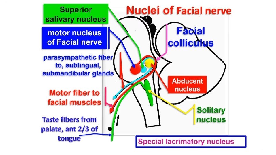
• Facial Nuclei 1 - Motor nucleus (S. V. E); muscles developed from the 2 nd pharyngeal arch. • Upper part of motor nucleus receives fibres from both sides of corticobulbar tracts Lower part receives fibers from opposite side of corticobulbar tract. 2 - Parasympathetic nuclei (G. V. E) a- Superior Salivary Nucleus; (SSS) → facial nerve → chorda tympani → lingual nerve → submandibular ganglion → submandibular & sublingual glands b- Special lacrimatory nucleus (SSL) → facial nerve → greater petrosal nerve → sphenopalatine ganglion → lacrimal, nasal, palatine and pharyngeal glands. 3 - Solitary nucleus (SVA), receives taste sensation.
 superior, medial and lateral")
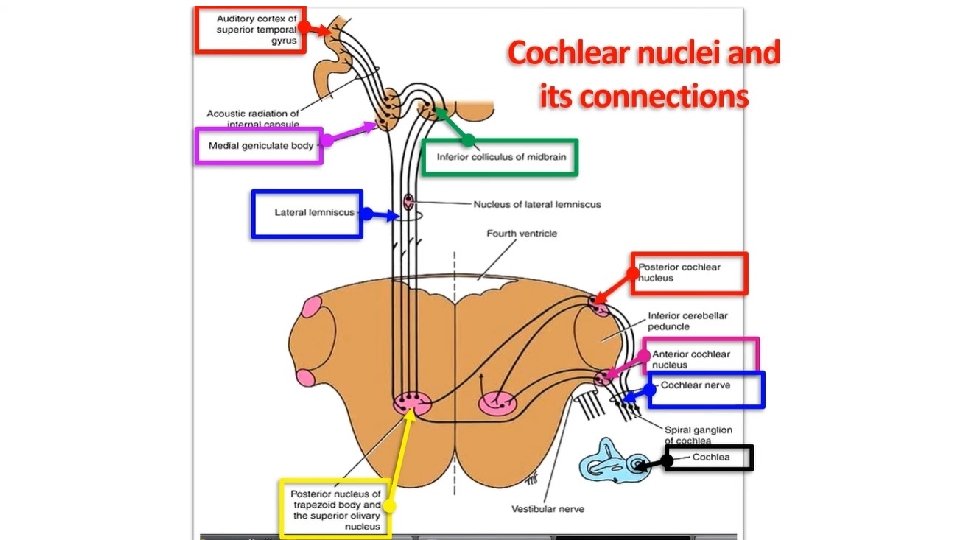
• Vestibulo-cochlear nerve a- Vestibular Nuclei (S. S. A) superior, medial and lateral - They receive equilibrium from inner ear along vestibular nerve - They connected to cerebellum by efferent and afferent fibers. b- Cochlear Nuclei (S. S. A) dorsal and ventral - They receive hearing impulses from the cochlea of the inner ear through the cochlear nerve. - The fibres cross to the opposite side as auditory decussation (trapezoid body) and ascend as lateral leminscus to the medial geniculate body of the thalamus.
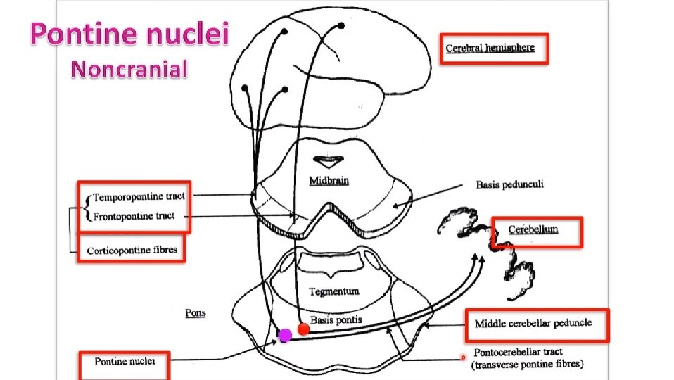

• Non-cranial nerve nuclei 1 - Pontine nuclei: Cortico-ponto-cerebellar tract • Cerebral cortex (same side) ----- Pontine nuclei ----cross to the opposite side ----- middle cerebellar peduncle ------ to the cerebellum 2 - Reticular formation nuclei.

Midbrain Cranial Nerve Nuclei th th th 5 , 6 , 7 , 8 th
 for all")
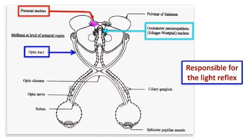
• Nuclei of occulomotor nerve: a- Motor nucleus (G. S. E) for all the extra ocular muscles except lateral rectus and superior oblique. b- Parasympathetic (Edinger-westphal nucleus) (G. V. E) → inferior division of oculomotor nerve → nerve to inferior oblique → ciliary ganglia → to ciliary muscle and constrictor pupillae muscle. § Motor nucleus of the trochlear nerve (G. S. E): - It supplies the superior oblique muscle of the eyeball. - LR 6 (SO 4)3 • Mesencephalic nucleus of trigeminal nerve. - It receives the proprioceptive sensation from the muscle of mastication.
 - It contains melanin")
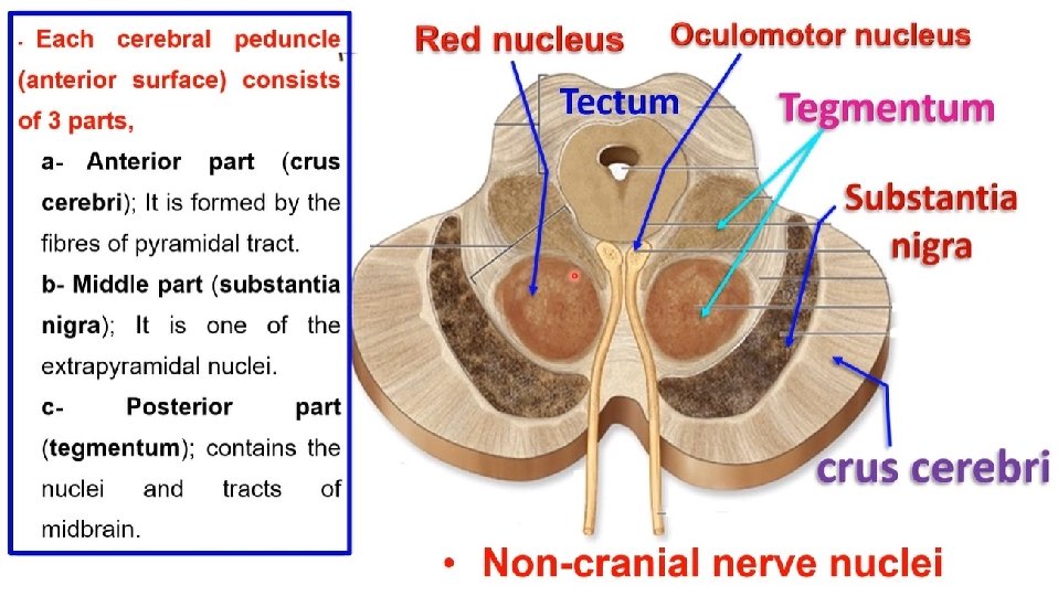
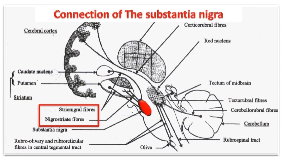
• Non-cranial nerve nuclei - Substantia Nigra (extrapyramidal center) - It contains melanin pigment, hence its name nigra (black). - is concerned with muscle tone ** Afferent = 2 C 1 - From corpus striatum (basal nuclei) 2 - From the cerebral cortex. ** Efferent = To the corpus striatum. - Degeneration of the substantia nigra leading to absence of dopamine secretion causing Parkinson's disease
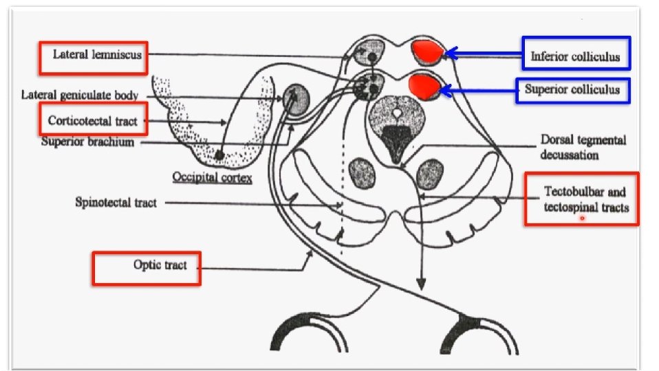
. a- Superior Colliculus")
3 - Tectal nuclei that lie in the tectum (posterior surface). a- Superior Colliculus (Visual) * Afferent : 1 - From the optic tract. 2 - From visual area of the cerebral cortex. b- Inferior Colliculus (Auditory) * Afferent: 1 - From the cochlear nerve. 2 - From auditory area of the cerebral cortex. * Efferent of superior and inferior colliculus to: - Tecto-bulbar: Ocular motor nuclei (3 rd, 4 th, 6 th) responsible for movement of the eyes in relation to the visual and auditory stimuli. - Tecto-spinal: upper segments of the spinal cord responsible for the movement of the head in relation to the visual and auditory stimuli (Spinal part of accessory nerve).

Th ank Qu you est ion s I/Azzam - 2004
- Slides: 34