MEDICINE GRAND ROUNDS Hypertrophic Osteoarthropathy a pretender a


 pain on wrists, toes (+) enlargement &")
 increase in pain severity Advised")
 pallor, anorexia, weight loss HEENT: (-) signs of head")

 Gastric CA – father (+) Colon CA - paternal grandfather")


 digital clubbing on all extremities (+)bilateral tibial tenderness, non erythematous,")


 digital clubbing both upper")


")
")

")
 POSITIVE, TTF 1 INDETERMINATE, �CK")
 for malignancy: defer Thoracotomy & proceed")


 �The pulmonary lesions were generally larger than 5 cm �However,")


- Slides: 104
MEDICINE GRAND ROUNDS Hypertrophic Osteoarthropathy: a pretender, a cheater Margaret Elaine J. Villamayor, RMT, MD Makati Medical Center April 8, 2010
Objective �To present a case of a patient presenting with joint pains as a symptom of a distant pathology
GENERAL DATA EB, 52/male, married, Filipino, Engineer, admitted last January 13, 2010
CHIEF COMPLAINT Joint pains
HISTORY OF PRESENT ILLNESS 2 months Intermittent pain on tibial & ankles, sharp, deep-seated, 5/10, not related to physical activity, no specific timing and lasting more than 30 minutes Self-medicated : Ibuprofen and Meloxicam
HISTORY OF PRESENT ILLNESS 1 month Consult: Normal Blood tests Impression: Gouty Arthritis Unrecalled pain meds Diet modification
HISTORY OF PRESENT ILLNESS 2 weeks (+) pain on wrists, toes (+) enlargement & pain on fingertips (+) difficulty in ambulating and with fine movements (+) sore throat & fever Consult at MMC
HISTORY OF PRESENT ILLNESS 1 week On follow-up: (+) increase in pain severity Advised admission for further workup
REVIEW OF SYSTEMS General: (-) pallor, anorexia, weight loss HEENT: (-) signs of head injury, (-) redness, itchiness, lacrimation of eyes, (-) deafness, tinnitus, and ear discharge, (-) colds, nasal stuffiness, epistaxis, (-) mouth sores, sore throat, gum bleeding, (-) stiffness, lumps, tenderness, adenopathy Respiratory: (-) cough, wheezing, shortness of breath, hemoptysis Cardiovascular: (-) chest pain, palpitation, syncope, cyanosis Gastro-intestinal: (-) nausea, vomiting, hematemesis, dysphagia, diarrhea, constipation Renal: (-) hematuria, no dysuria Neurologic: (-) fainting Psychiatric: (-) history of depression
PAST MEDICAL HISTORY �No serious illness during childhood. �No known co-morbidities �Diagnosed with Cholelithiasis in 2006 �No history of previous surgeries, blood transfusions, trauma, allergies
FAMILY HISTORY (+) Gastric CA – father (+) Colon CA - paternal grandfather
PERSONAL AND SOCIAL HISTORY � 36 pack years smoking history �Occasional alcoholic beverage drinker �Denies illicit drug use & multiple sexual partners �Patient works as an engineer in Saudi Arabia for the past 20 years �Plays golf every week.
PHYSICAL EXAMINATION General Survey: conscious, coherent, oriented to person, place and time, ambulates with assistance BP 120/80, HR 72 bpm, RR 18 cpm, T 36. 9 C, Height: 162 cm Weight: 68. 18 kg BMI: 25. 59 kg/m 2 HEENT: anicteric sclerae, pink conjunctivae, no naso-aural discharge, no cervical lymphadenopathies, no tonsilopharyngeal congestion, no neck vein distention, no carotid bruits Chest and Lungs: No gross lesions, no retractions, no point tenderness, equal chest expansion, equal fremiti, lungs resonant, clear breath sounds
PHYSICAL EXAMINATION �Heart: adynamic precordium, AB at 5 th LICS MCL, regular rate & rhythm, no friction rub, no murmurs �Abdomen: flabby abdomen, non-distended, normoactive bowel sounds, soft, tympanitic, nontender, no hepatosplenomegaly, no Murphy’s sign, no costovertebral angle tenderness, no peritoneal signs
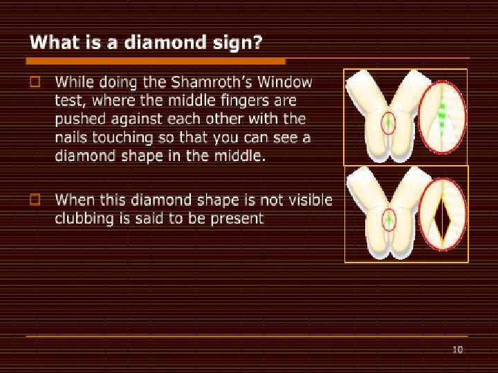
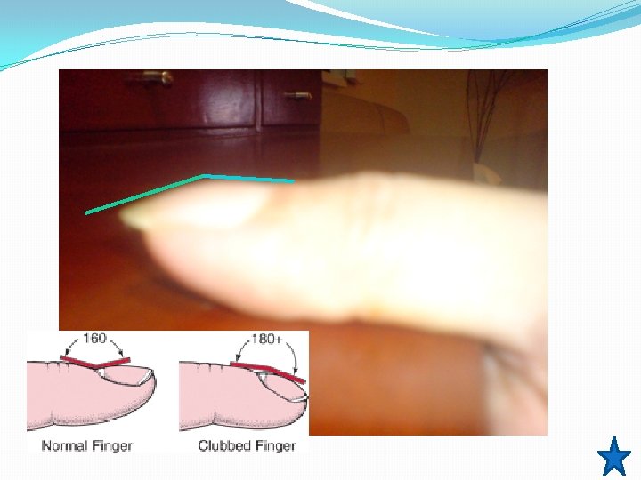
PHYSICAL EXAMINATION �Extremities: (+) digital clubbing on all extremities (+)bilateral tibial tenderness, non erythematous, not warm to touch (+) limitation of movement on wrist & ankle flexion & finger flexion and opposition (-) cyanosis, crepitus Shamroth’s test
Lovibond angle Normal Angle = less than or equal to 160°. Flat/ >180 = clubbing Swartz MH. Textbook of Physical Diagnosis: History and Examination. 2 nd ed. Philadelphia, Pa: WB Saunders; 1994: 76 -8.
NEUROLOGIC EXAMINATION: Mental status: awake, alert, oriented Cranial Nerve Exam: CN I – not assessed CN II, III – pupils 3 mm ERTL, (+) ROR, clear media, no visual field defects CN III, IV, VI – full EOMs CN V – intact V 1 -V 3 CN VII – no facial asymmetry CN VIII – no gross hearing defects CN IX, X – tongue and uvula at midline CN XI – good SCM tone CN XII – tongue at midline
NEUROLOGIC EXAMINATION Cerebellar: no dysdiadochokinesia, no dysmetria Reflexes: +2 on all extremities, no Babinski Meninges: supple neck, no Brudzinski, no Kernig’s Motor : 5/5 on all extremities Sensory: no sensory deficits
SALIENT FEATURES SUBJECTIVE OBJECTIVE 36 pack year smoking history (+) digital clubbing both upper and lower extremities (+) Family History of Cancer (+)bilateral tibial tenderness (+) fever, (-) weight loss, cardiac, gastrointestinal or respiratory symptoms Joint & tibial pain x 2 months - symmetrical, no specific timing, not relieved by rest/ analgesics, >30 mins
Admitting Impression t/c Hypertrophic Osteoarthropathy, etiology to be determined Obese Class I (BMI 25. 59 kg/m 2)
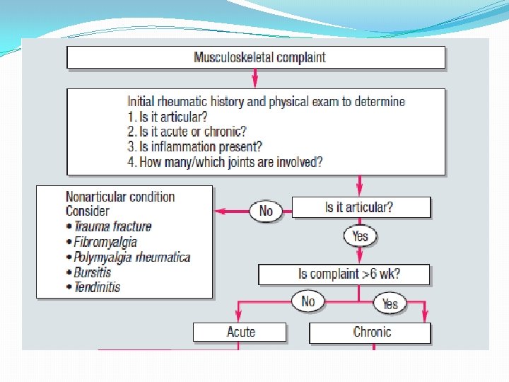
Systemic sxs PE Timing Patient Rheumatic Fever Rheumatoid Arthritis Osteoarthritis HOA Fever, sore throat Weakness, easy fatigability, weight loss, ±fever, None ±fever symmetrical Tibial, wrists, toes digits, wrist, clubbing (-) crepitus Migratory, carditis, symmetrical LS spine, hip, PIP, knee, 1 st MTP MCP/MTP, DIP PIP wrist, elbow, spared are the knee, ankle, wrist, elbow, nodules and ankle (+) crepitus Tibia, wrists, ankles, digits, elbow, clubbing ± Movement, morning , >1 hr ± Movement
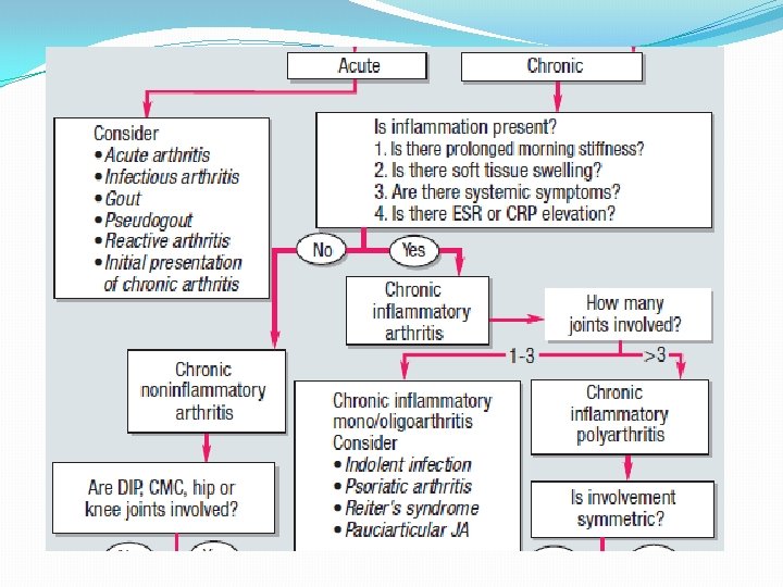
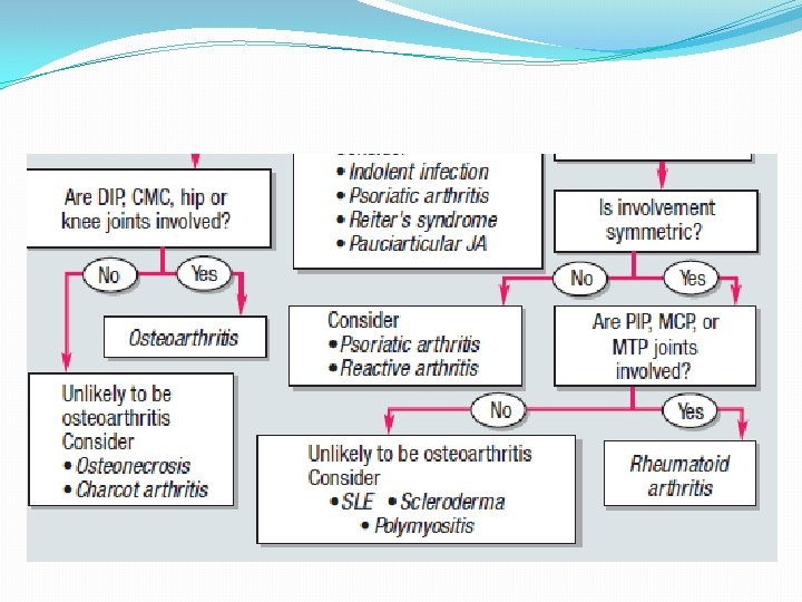
Approach to HOA Pineda C, Fonseca C, Martinez-Lavin M. The spectrum of soft tissue and skeletal abnormalities of hypertrophic osteoarthropathy. J Rheumatol. May 1990; 17(5): 626 -32.
Course in the Wards Jan 13 Hb 12. 7 Hct 37 RBC 4. 42 WBC 15. 25 Seg 76 Lympho 16 Mono 8 Plt 531, 000 ESR CRP Jan 13 93 (+) Sodium Potassium BUN Crea Calcium Total Protein Albumin Globulin A/G ALP AST Total Bili Uric Acid Trigly Chole HDL LDL Jan 13 135 4. 6 10. 12 0. 72 8. 78 6. 9 3. 6 3. 3 1. 09 140 25 7. 8 3. 8 56 185. 57 53. 78 101. 76
Xray of Both Hands �No osseous nor joint abnormalities seen in both hands
Chest Xray
CT-Scan of the Chest: January 13, 2010
CT-Scan of the Chest: January 13, 2010
CT-Scan of the Chest: January 13, 2010
Course in the Wards �TCVS Referral �CT-guided Biopsy �Plan: if NSCLC, mediastinoscopy and possible pneumonectomy of the right lung �Cranial MRI & CT of the Abdomen
Cranial MRI
SMEAR, DIFF-QUIK STAIN, HPO
CELLBLOCK: Right Lung Mass s/p CT-guided Biopsy
CK 7 (+)
CK 20 (-)
TTF – 1 indeterminate
CKHMW (-)
FINAL DIAGNOSIS �POSITIVE FOR MALIGNANT CELLS �CK 7 (+) POSITIVE, TTF 1 INDETERMINATE, �CK 20 - NEGATIVE, CKHMW-FOCALLY POSITIVE �SUPPORTS THE DIAGNOSIS OF NON-SMALL CELL ADENOCARCINOMA
Course in the Wards �Plan: �LN biopsy (+) for malignancy: defer Thoracotomy & proceed with Radiotherapy �LN biopsy (-): Right lung Pneumonectomy and Mediastinal lymph node dissection
PET Scan
PET Scan � Enlarged lymph node is present in the right hilum measuring 1. 3 cm with mild FDG uptake (SUV 2. 0); Mild FDG uptake is also noted in at least 2 unenlarged nodes in the subcarinal area (SUV=2. 0, 2. 1) � An FDG-avid (SUV 11. 0) mass lesion is present in the right hilum measuring 6. 5 x 6. 4 cm.
Quantitative Perfusion Imaging
Perfusion Scan
Course in the wards �February 6, 2010 Mediastinoscopy with Frozen Section and possible right pneumonectomy with mediastinal lymph node dissection.
OR Findings �Bronchoscopic findings: carina & main bronchi free of tumor, slight narrowing of right upper lung bronchus, �Frozen section of the Right tracheobronchial mediastinal lymph node was negative for metastasis.
OR Findings �Exploration revealed a central tumor encompassing all 3 right lung lobes with some enlarged lymph nodes.
Intraoperative Status �Patient was noted to have desaturation 84% on 100% Fi. O 2 �Elevation of mean PA/Swan Ganz �Consistently observed with single lung ventilation �Pneumonectomy was aborted �Section biopsy of the right hilar node done.
CT-Scan of Chest Jan. 14, 2010 Feb. 15. 2010
Course in the Wards �Referral Oncology and Radio-Onco �Induction chemotherapy with radiation therapy
STAGING
STAGING Non-Small Cell Lung Adenocarcinoma Stage IIIA
FINAL DIAGNOSIS �Non-Small Adenosarcinoma Lung Carcinoma Stage IIIA �Hypertrophic Pulmonary Osteoarthropathy secondary to malignancy
DISCUSSION Hypertrophic Osteoarthropathy
Diagnostic Criteria �Clubbing, Joint Pains, Periostosis of the tubular bones �Three other forms of hypertrophic osteoarthropathy are described: (1) clubbing alone (2) periostosis without clubbing in the setting of an illness known to be associated with hypertrophic osteoarthropathy, and (3) pachydermia associated with minor manifestations Martinez-Lavin M, Matucci-Cerinic M, Jajic I, Pineda C. Hypertrophic osteoarthropathy: consensus on its definition, classification, assessment and diagnostic criteria. J Rheumatol. Aug 1993; 20(8): 1386 -7
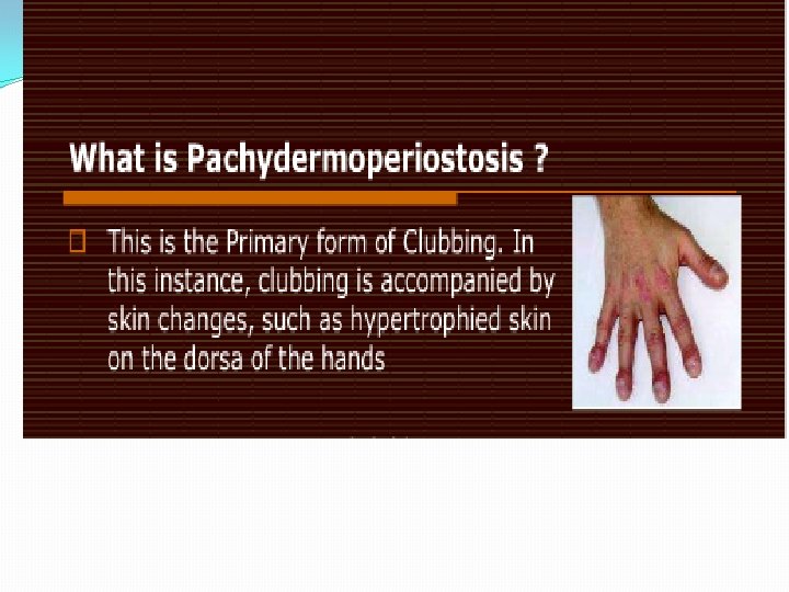
Hypertrophic Osteoarthropathy �Bilateral and symmetric �It may occur as a primary condition, which is familial and affects mainly males � 90% of secondary cases are associated with intra -thoracic pathology Karkuca Murat, Erturk Engin, Capkin Erhan, Akyazi Hikmet, Ozden, Gonca, Tosun Mehmet: Primary hypertrophic osteoarthropathy (pachydermoperiostosis): a case report. Rheumatol Int 2007, 27: 403 -405.
Hypertrophic Osteoarthropathy �HPOA has only been reported in 1– 10% of cases of Lung carcinomas �Among these, HPOA is most commonly found with Non–small cell lung carcinoma Sridhar KS, Lobo CF, Altman RD. Digital clubbing and lung cancer. Chest. Dec 1998; 114(6): 1535 -7.
Clinical Presentation �Mild to severe arthralgias on metacarpal joints, wrists, elbows, knees, ankles. �Range of motion of affected joints may be slightly decreased �When effusions are present, they usually involve the large joints (eg, knees, ankles, wrists). Martinez-Lavin M, Matucci-Cerinic M, Jajic I, Pineda C. Hypertrophic osteoarthropathy: consensus on its definition, classification, assessment and diagnostic criteria. J Rheumatol. Aug 1993; 20(8): 1386 -7
Hypertrophic Pulmonary Osteoarthropathy �The occurrence of HPOA without clubbing of the digits is so rare as to be published only in as few as four case reports in the past 10 years. �It is more common with large central tumors than peripheral tumors, with no predilection for cell type Clarke S, Barnsley L, Peters M, Morgan L, Van der WH. Hypertrophic pulmonary osteoarthropathy without clubbing of the digits. Skeletal Radiol 2001; 30: 652– 5
Hypertrophic Pulmonary Osteoarthropathy �It is a general conception that the malignant neoplasms associated with HPO are primary in the lung and that metastatic tumors to the lungs are rarely associated with HPO. Hammarsten JF, O'Leary J: The features and significance of hypertrophic osteoarthropathy, Arch Intern Med 99: 431 -441, 1956
Hypertrophic pulmonary osteoarthropathy (HPOA) �The pulmonary lesions were generally larger than 5 cm �However, in some cases, the major mass lesion was located in the lower lobes of the lung & was in direct contact with the pleural surfaces, either anteriorly or diaphragmatically
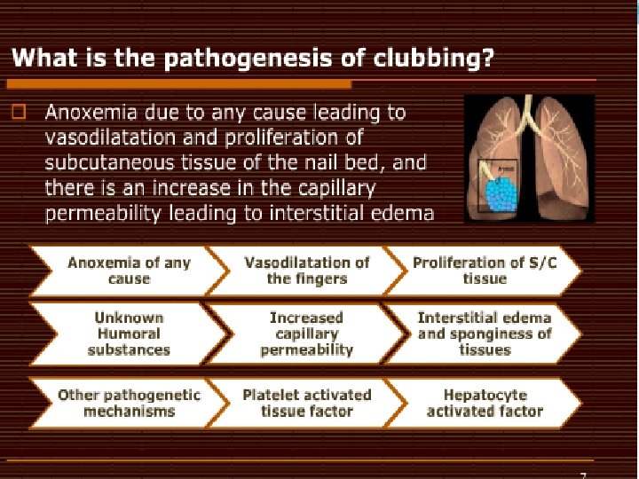
Pathophysiology �Exact mechanism of the syndrome is still unclear. Marinez-Levin M, Pineda C: Hypertrophic osteoarthropathy. Rheumatology 3 rd edition. Edited by: Hochberg MC, Silman AJ, Smolen JS, Weinblatt ME, Weisman MH. Mosby, Edinburgh; 2003: 1763 -1767
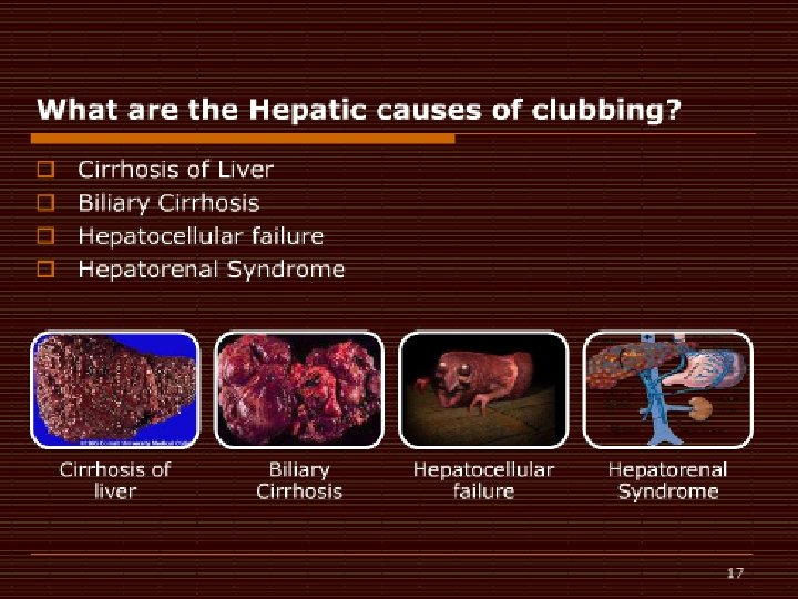
Other secondary causes �Pulmonary Tuberculosis �Congenital Cyanotic Heart Disease �Hepatic and Colorectal Carcinoma �Inflammatory Bowel Disease �Cirrhosis �Pulmonary Fibrosis and Empyema Armstrong David, Mc. Causland Elisabeth, Wright Gary: Hypertrophic pulmonary osteoarthropathy (HPOA) (Pierre Marie-Bamberger syndrome): two cases presenting as acuteinflammatory arthritis. Description and review of the literature. Rheumatol Int 2007, 27
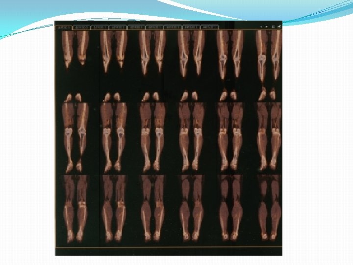
Whole-body bone scintigraphy �Preferred method for diagnosing the syndrome �Increased tracer uptake along the cortical margins of the bilateral lower extremities, which is compatible with the “parallel stripe” sign or “tramline” sign
HPOA vs Metastasis on Bone Scan Sharp et al, Practical Nuclear Medicine 3 rd edition, 2005
HPOA diagnosed by FDG PET-CT: a case report �The CT portion : extensive irregular bilateral periosteal new bone formation in the long bones. �The PET images : diffuse moderately increased FDG uptake in the periostea of the long bones of the legs, with some focal sites of more intense FDG uptake in the thicker portions of the periosteum. Makis. Clin Nucl Med. 2009 Sep; 34(9): 625 -7
Digital Clubbing : Demonstration With PET: A Case Report �The PET scan revealed increased glucose metabolism in all of the patient's fingertips Ward RW, Chin R Jr, Keyes JW Jr, Haponik EF. Digital clubbing. Demonstration with positron emission tomography. Chest. Apr 1995; 107(4): 1172 -3.
Treatment �The only effective treatment for HPOA is treatment of the underlying condition �Atropine, antitumor chemotherapy �In patients with HPOA, tumor resection often results in improvement of symptoms within 2 to 4 weeks and, sometimes, complete resolution by 3 to 6 months Nakayama, Ann Thorac Surg, 2007; 83: 685– 7
Effective Symptomatic Relief of HPOA by VATS Vagotomy �Relief of all joints and she regained full range of movements within 24 hours �Discharged on the third post-op day and remained pain free and fully mobile 3 months after the surgery. Martinez-Lavin M: Hypertrophic osteoarthropathy. Curr Opin Rheumatol 1997; 9: 83– 6.
Current Status: �At present, patient finished 33 sessions of radiotherapy and is on his 5 th cycle of chemotherapy �Patient still experiences intermittent joint pains but lesser in severity
Thank You!
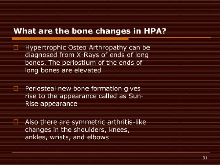
Hypertrophic Osteoarthropathy on Xray �The periostium of the ends of long bones are elevated �Periosteal new bone formation gives rise to the appearance called as “Sun-Rise” appearance
PET vs Bone Scan in Detecting NSCLC Metastasis �Patients and methods: �April 2004 - May 2007, retrospectively reviewed to identify all patients with newly diagnosed NSCLC and who underwent staging with both PET/CT and bone scan prior to the initiation of therapy. �Presence of bone metastases was confirmed by considering all available clinical information. �This search identified 1000 patients, 265 women and 735 men (age range, 18– 89 years; median age, 65 years). Song, et. al, Lung Cancer Journal, Volume 65, Issue 3, Pages 333 -338, September 2009
PET vs Bone Scan in Detecting NSCLC Metastasis �Results �Bone metastases were confirmed in 105 (10. 5%) patients. Accuracy Sensitivity Specificity False (+) False (-) PET/CT 98. 3% 94. 3% 98. 8% 1. 2% 5. 7% Bone Scan 95. 1% 78. 1% 97. 4% 2. 9% 21. 9% p value <0. 001 0. 006 Song, et. al, Lung Cancer Journal, Volume 65, Issue 3, Pages 333 -338, September 2009
Hypertrophic pulmonary osteoarthropathy diagnosed by FDG PET-CT: a case report �The CT portion : extensive irregular bilateral periosteal new bone formation in the long bones. �The PET images : diffuse moderately increased FDG uptake in the periostea of the long bones of the legs, with some focal sites of more intense FDG uptake in the thicker portions of the periosteum. Makis. Clin Nucl Med. 2009 Sep; 34(9): 625 -7
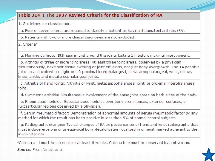
Patient Joints Systemic symptoms Timing Labs Radio findings Rheumatoid Arthritis Tibial area, PIP, MCP/MTP, digits, wrist, elbow, wrist knee, ankle fever Weakness, easy fatigability, anorexia, weight loss, ±fever, nodules ± Movement, morning , >1 hr acute phase reactants (+) RF, elevated acute phase reactants Normal Hand Xray erosions or unequivocal bony decalcification Osteoarthritis Knees, hips None Acute Rheumatic Fever HOA Migratory Tibia, wrists, polyarthritis digits, elbow Fever, sore throat ± No specific timing Normal/
Articular manifestations of hypertrophic pulmonary osteoarthropathy in bronchogenic carcinoma. A clinical and pathologic study �Eight patients with bronchogenic carcinoma presented with painful joint effusions as part of the syndrome of hypertrophic osteoarthropathy. �Elevated sedimentation rates and symptomatic relief with aspirin were common, but synovial fluids all had leukocyte counts less than 500/mm 3. �.
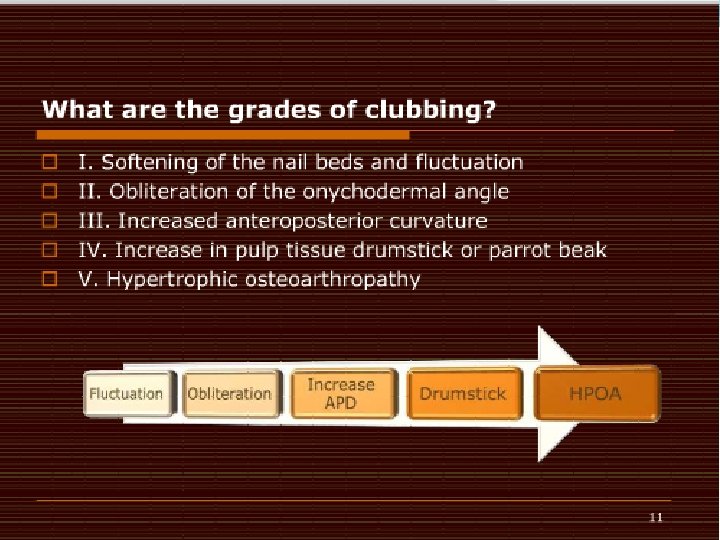
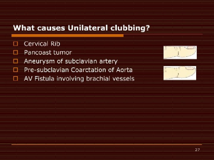
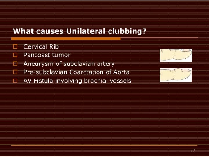
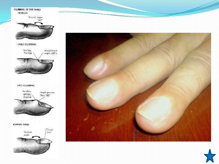
� Clubbing usually progresses through 4 phases. 8 � Fluctuation and softening of the nail bed, with a rocking sensation upon palpation due to increased edema and soft tissue � Loss of the normal 15° angle (Lovibond angle 27 ) between the nail and cuticle � Accentuation of the convexity of the nails and clubbed appearance of the fingertips, with warmth and sweating � Shiny or glossy change in the nail and adjacent skin, with disappearance of the normal creases and appearance of longitudinal striation of the nail � Clubbing can be classified into 3 topographical groups. 28, 29 � Symmetrical: All the fingers and toes are involved, as depicted in the image below. � � � Clubbing associated with hypertrophic osteoarthropathy can be classified into 3 topographical groups (ie, symmetrical, unilateral, unidigital). This is symmetrical clubbing; it involves all the fingers and toes. [ CLOSE WINDOW ] Clubbing associated with hypertrophic osteoarthropathy can be classified into 3 topographical groups (ie, symmetrical, unilateral, unidigital). This is symmetrical clubbing; it involves all the fingers and toes. Unilateral: The fingers or toes of 1 hand or foot are involved. Unidigital: Only 1 finger or toe is involved.
Immunohistochemistry Braunwald, Fauci, Harrison’s Internal Medicine 17 th edition, 2009
Pre-operative Testing ABG Jan 26 p. O 2 96. 2 p. H 7. 50 p. CO 2 34 HCO 3 26. 3 O 2 Sat 97. 9 Base Excess +3. 6 Room air Pre-Rx Best % Pre d 3. 25 106 2. 49 103 77 93 PRE D FVC 3. 05 FEV 1 2. 41 FEV 1/FVC 82 % FEF 253. 03 75% DLCO 14. 3 66 MVV 130 74 57 Best Post-Rx %Pred %Chang e 3. 15 2. 55 81 103 106 98 -3 2 5 55 42 -26
Jones Criteria for Rheumatic Fever MAJOR CRITERIA MINOR CRITERIA Carditis Migratory polyarthiritis Sydenham’s chorea Subcutaneous nodules Erythema marginatum Clinic Fever Arthalgia Laboratory Acute Phase Reactants Prolonged PR interval PLUS Supporting evidence of a recent Group A streptococcal fever 1. GABHS + 2 major and 1 minor criteria, or 1 major and 2 minor criteria 2. carditis or chorea exists with no other cause, 3. previous history of rheumatic fever who have 1 major or 2 minor criteria in association with a recent streptococcal infection.
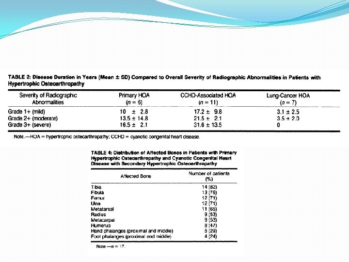
Periostitis in HOA �Primary HOA and congenital heart diseaseassociated HOA had multilayered periostitis vs Lung -cancer HOA, there was only a single layer �Scores indicating severity of periostitis showed that a severe degree was present in a significant percentage of patients with both primary HOA and CCHDassociated HOA compared to HPOA Pineda, Martinez, Periostitis in Hypertrophic Osteoarthropathy: relationship to disease duration
Periostitis in HOA �In our seven patients with lung-cancer HOA, less prominent periosteal changes were noted in a more limited distribution. �Malignancy-associated HOA, a disease process of much shorter duration, the natural evolution of the osseous changes cannot fully be expressed.
Periostitis in HOA
Prediction of Hypoxemia during OLV �Side of Operation �Lung Function Abnormalities �“Paradoxical effect” �Distribution of Perfusion �Central vs Peripheral �Gravity