Medical Virology Third Year Undergraduate Students By Prof

Medical Virology Third Year Undergraduate Students By Prof. Dr. Abdul-Hameed Khalid Khadhair


Medical Virology: - Introduction - History and Technology Development - Theories on origin of viruses - Viruses and virus-like structures

History of Viruses: Events Helped in Virus Discovery: - The first record of a virus infection consists of a hieroglyph from Memphis, the capital of ancient Egypt, drawn in 3700 BC, which depicts a temple priest showing typical clinical signs of paralytic poliomyelitis. Pharaoh Ramses V, who died in 1196 BC and whose extraordinarily well-preserved mummified body is believed to have succumbed to smallpox—a comparison between the pustular lesions on the face of the mummy and those of more recent patients is startling. - Smallpox was endemic in China by 1000 BC and practice of Variolation (similar to Vaccination) was developed. Survivors of smallpox outbreaks were protected from subsequent infection, the Chinese inhaled the dried crusts from smallpox lesions like snuff or, in later modifications, inoculated the pus from a lesion into a scratch on the forearm.
, a Dutch merchant, constructed")
- Invention of microscope by Antony van Leeuwenhoek (1632– 1723), a Dutch merchant, constructed the first simple microscopes and with these identified bacteria as the ‘animalcules’ he saw in his specimens. - However, it was not until Robert Koch and Louis Pasteur in the 1880 s jointly proposed the ‘germ theory’ of disease that the significance of these organisms became apparent. - Koch’s Postulates (proof that an infectious agent is responsible for a specific disease: 1. The agent must be present in every case of the disease 2. The agent must be isolated from the host and grown in vitro 3. The disease must be reproduced when a pure culture of the agent is inoculated into a healthy susceptible host. 4. The same agent must be recovered once again from the experimentally infected host. - Pasteur worked extensively on rabies, which he identified as being caused by a ‘virus’ but despite this he did not discriminate between bacteria and other agents of disease.

First real evidence on virus identity - In 1892, Dimitri Iwanowski, a Russian botanist, showed that extracts from diseased tobacco plants could transmit disease to other plants after passage through ceramic filters fine enough to retain the smallest known bacteria. - A few years later (1898), Martinus Beijerinick confirmed and extended Iwanowski’s results on tobacco mosaic virus (TMV) and was the first to develop the modern idea of the virus, which he referred to as contagium vivum fluidum (‘contagious viral fluids’). - Freidrich Loeffler and Paul Frosch (1898) showed that a similar agent was responsible for foot-and-mouth disease in cattle, but, despite the realization that these new-found agents caused disease in animals as well as plants, people would not accept the idea that they might have anything to do with human diseases. - This resistance was finally dispelled in 1909 by Karl Landsteiner and Erwin Popper, who showed that poliomyelitis was caused by a ‘filterable agent’—the first human disease to be recognized as being caused by a virus.
 and Felix d’Herelle (1917) were the")
New Virus infecting Bacteria: - Frederick Twort (1915) and Felix d’Herelle (1917) were the first to recognize viruses that infect bacteria, which d’Herelle called bacteriophages (‘eaters of bacteria’). - In the 1930 s and subsequent decades, pioneering virologists such as Salvador Luria, Max Delbruck, and many others used these viruses as model systems to investigate many aspects of virology, including virus structure, genetics and replication. - These relatively simple agents have since proven to be very important to our understanding of all types of viruses, including those of humans which are much more difficult to propagate and study

- During the Spanish–American War of the late nineteenth century and the subsequent building of the Panama Canal, the number of American deaths due to yellow fever was great. The disease also appeared to be spreading slowly northward into the continental United States. In 1902, through experimental transmission to mice, Walter Reed demonstrated that yellow fever was caused by a virus spread by mosquitoes. This discovery eventually enabled Max Theiler in 1937 to propagate the virus in chick embryos and to produce an attenuated vaccine—the 17 D strain—which is still in use today. -The success of this approach led many other investigators from the 1930 s to the 1950 s to develop animal systems to identify and propagate pathogenic viruses. - Eukaryotic cells can be grown in vitro (tissue culture) and viruses can be propagated in these cultures, but these techniques are expensive and technically quite demanding.

- Some viruses will replicate in the living tissues of developing embryonated hens eggs, such as influenza virus. Egg-adapted strains of influenza virus replicate well in eggs and very high virus titres can be obtained. Embryonated hens eggs were first used to propagate viruses in the early decades of the twentieth century. This method has proved to be highly effective for the isolation and culture of many viruses, particularly strains of influenza virus and various poxviruses (e. g. , vaccinia virus). Counting the ‘pocks’ on the chorioallantoic membrane of eggs produced by the replication of vaccinia virus was the first quantitative assay for any virus. Animal host systems still have their uses in virology: - To produce viruses that cannot be effectively studied in vitro (e. g. , hepatitis B virus) - To study the pathogenesis of virus infections (e. g. , coxsackie viruses) - To test vaccine safety (e. g. , oral poliovirus vaccine)

Nevertheless, they are increasingly being discarded for the following reasons: 1. Breeding and maintenance of animals infected with pathogenic viruses is expensive. 2. Whole animals are complex systems in which it is sometimes difficult to discern events. 3. Results obtained are not always reproducible due to host variation. 4. Unnecessary or wasteful use of experimental animals is morally not accepted. 5. They are rapidly being overtaken by ‘modern science’—cell culture and molecular biology.

In recent years, an entirely new technology has been employed to study the effects of viruses on host organisms. This involves the creation of transgenic animals and plants by inserting all or part of the virus genome into the DNA of the experimental organism, resulting in expression of virus m. RNA and proteins in somatic cells (and sometimes in the cells of the germ line). Thus, the pathogenic effects of virus proteins, individually and in various combinations, can be studied in living hosts. ‘SCID-hu’ mice have been constructed from immunodeficient lines of animals transplanted with human tissue. These mice form an intriguing model to study the pathogenesis of human immunodeficiency virus (HIV) as there is no real alternative to study the properties of this important virus in vivo. While these techniques often raise the same moral objections as ‘oldfashioned’ experimental infection of animals by viruses, they are powerful new tools for the study of virus pathogenicity. This method will become widely used after solving technical difficulties associated with the construction of transgenic organisms.

CELL CULTURE METHODS They began early in 1907 by Ross Harrison using whole-organ cultures, then progressed to methods involving individual cells, either primary cell cultures (somatic cells from an experimental animal or taken from a human patient which can be maintained for a short period in culture) or immortalized cell lines, which, given appropriate conditions, continue to grow in culture indefinitely. In 1949, John Enders and his colleagues were able to propagate poliovirus in primary human cell cultures. This achievement regarded as the ‘Golden Age of Virology’ and led to the identification and isolation during the 1950 s and 1960 s of many viruses and their association with human diseases.

Plaque Assay Renato Dulbecco in 1952 was the first to quantify accurately animal viruses using a plaque assay. In this technique, dilutions of the virus are used to infect a cultured cell monolayer, which is then covered with soft agar to restrict diffusion of the virus, resulting in localized cell killing and the appearance of plaques after the monolayer is stained. Counting the number of plaques directly determines the number of infectious virus particles applied to the plate.

The same Plaque technique can also be used biologically to clone a virus (i. e. , isolate a pure form a mixture of types). This technique had been in use for some time to quantify the number of infectious virus particles in bacteriophage suspensions applied to confluent ‘lawns’ of bacterial cells on agar plates, but its application to viruses of eukaryotes enabled rapid advances in the study of virus replication to be made. Plaque assays largely replaced earlier endpoint dilution techniques, such as the tissue culture infectious dose (TCID 50) assay, which are statistical means of measuring virus populations in culture; however, endpoint techniques may still be used in certain circumstances—for example, for viruses that do not replicate in culture or are not cytopathic and do not produce plaques, (e. g. , human immunodeficiency virus).

- George Hirst, in 1941, observed haemagglutination of red blood cells by influenza virus (see Chapter 4). This proved to be an important tool in the study of not only influenza but also several other groups of viruses—for example, rubella virus. In addition to measuring the titer (i. e. , relative amount) of virus present in any preparation, this technique can also be used to determine the antigenic type of the virus. Haemagglutination will not occur in the presence of antibodies that bind to and block the virus haemagglutinin. If an antiserum is titrated against a given number of haemagglutinating units, the haemagglutination inhibition titer and specificity of the antiserum can be determined.

In the 1960 s and subsequent years, many improved detection methods for viruses were developed, such as: - Complement fixation tests - Radioimmunoassays mmunofluorescence (direct detection of virus antigens in infected cells or tissue) - Enzyme-linked immunosorbent assays (ELISAs) - Radioimmune precipitation - Western blot assays These techniques are sensitive, quick, and quantitative.
 Technology In 1975, George Kohler and Cesar Milstein isolated the first")
Monoclonal Antibodies (MA) Technology In 1975, George Kohler and Cesar Milstein isolated the first MA from clones of cells selected in vitro to produce an antibody of a single specificity directed against a particular antigenic target. This enabled virologists to look not only at the whole virus, but at specific regions—epitopes—of individual virus antigens as well function of individual virus proteins. MA techniques have more applications in other types of serological assays (e. g. , ELISAs) to increase their reproducibility, sensitivity, and specificity - MA are produced by immunization of an animal with an antigen that usually contains a complex mixture of epitopes. Immature Bcells are later prepared from the spleen of the animal, and these are fused with a myeloma cell line, resulting in the formation of transformed cells continuously secreting antibodies. A small proportion of these will make a single type of monoclonal antibody against the desired epitope.

Ultrastructural & Physical Properties Studies - Physical measurements of virus particles began in the 1930 s with the earliest determinations of their proportions by filtration through colloidal membranes of various pore sizes - The first electron micrograph of a virus (TMV) was published in 1939. Over subsequent years, techniques were developed that allowed the direct examination of viruses at magnifications of over 100, 000 times. The two fundamental types of electron microscope are the transmission electron microscope (TEM) and the scanning electron microscope (SEM) - Studies of the sedimentation properties of viruses in ultracentrifuges in the 1960 s in obtaining purified and highly oncentrated preparations of many different viruses, free of contamination from host cell components that can be subjected to chemical analysis. The relative density of particles, measured in solutions of sucrose or Cs. Cl virus.

Molecular Biology Technology - The term ‘molecular biology’ has taken on the new and different meaning of ‘genetic engineering’ or ‘genetic manipulation. ’ These techniques for manipulating nucleic acids in vitro (that is, outside living cells or organisms) - This powerful new technology has revolutionized virology and, to a large extent, has shifted the focus of attention away from the virus particle onto the virus genome.

Initially, any investigation of a virus genome will usually include questions about the following: Composition—DNA or RNA, singler double-stranded, linear or circular? ? ? - Virus Size and number of segments? ? ? - Virus Terminal structures? ? ? - Virus Nucleotide sequence? ? ? ing capacity—open reading frames? ? ? gulatory signals—transcription enhancers, promoters, and terminators? ? ?

Theories on Origin of viruses: 1. Regressive evolution: This theory states that viruses are degenerate life forms that have lost many functions that other organisms possess and have only retained the genetic information essential to their parasitic way of life. 2. Cellular origins: In this theory, viruses are thought to be subcellular, functional assemblies of macromolecules that have escaped their origins inside cells. 3. Independent entities: This theory suggests that viruses evolved on a parallel course to cellular organisms from the self-replicating molecules believed to have existed in the primitive, prebiotic RNA world. This is similar to what spontaneous generation stated that viruses created from primitive cellular molecules have replication properties such as plasmids.

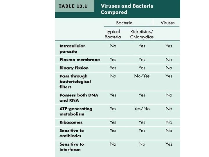
Viruses are: - submicroscopic, obligate intracellular parasites. - particles produced from the assembly of preformed components - particles (virions) themselves do not grow or undergo division. - lacking the genetic information that encodes apparatus necessary for the generation of metabolic energy or for protein synthesis (ribosomes). They are therefore absolutely dependent on the host cell for this function - One view said that inside the host cell viruses are alive, whereas outside it they are merely complex assemblages of metabolically inert chemicals. - Viruses are smallest entities in most cases. While this is true, the largest known virus (Mimivirus, for ‘mimicking microbe’) is 400 nm in diameter, while the smallest mycoplasma (e. g. , Mycoplasma, Ralstonia pickettii) is only 200 -300 nm long

Exceptional Entities: A number of pathogenic entities are more similar to viruses like - Viroids: Viroids are very small (200– 400 nucleotides), circular RNA molecules with a rod-like secondary structure. They have no capsid or envelope and are associated with certain plant diseases. Their replication strategy is like that of viruses—they are obligate intracellular parasites. - Virusoids: Virusoids are satellite, viroid-like molecules, almost larger than viroids (e. g. , approximately 1000 nucleotides); they depend on the presence of virus replication for multiplication (e. g. ‘satellite’); they are packaged into virus capsids as passengers. - Prions: are infectious agents believed to consist of a single type of protein molecule with no nucleic acid component. Confusion arises that the prion protein and the gene that encodes it are also found in normal ‘uninfected’ cells. These agents are associated with ‘slow’ virus diseases such as Creutzfeldt–Jakob disease in humans, scrapie in sheep, and cattles called bovine spongiform encephalopathy (BSE) or mad cow disease.
 = 1/100000,")
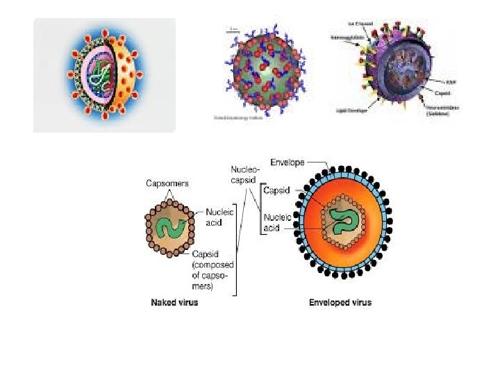
Chemistry of Viruses: Virus Particle: - Submicroscopic entity measured in nanometer (nm) = 1/100000, ranged from 20 -250 nm - Has nucleic acid (RNA or DNA covered with a protective protein coat or lipoprotein. - The protein coat called capsid consists from smaller subunit called capsomers. - Able to organize its own multiplication within the host cell by Replication & Protein synthesis It is dependent on the host’s proteinynthesizing system - They have receptor binding-protein for attaching to cells

Chemical structure and shape of helical Virus particle
 and Deoxyribonucleic (DNA) acids are important genetic materials in")
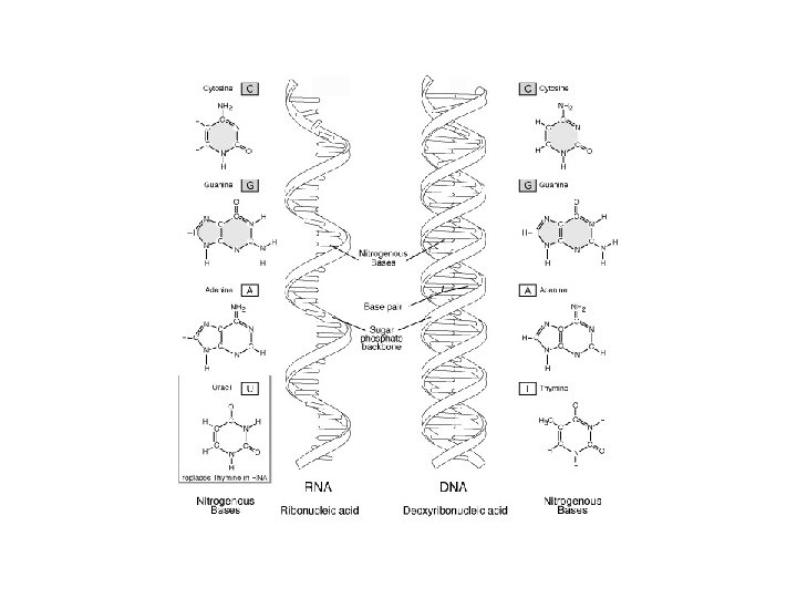
Nucleic Acids: - Ribonucleic (RNA) and Deoxyribonucleic (DNA) acids are important genetic materials in both virus and host. - RNA may take enzyme role during amino acids synthesis to form polypeptides in protein production ost plant viruses are RNA and in human and animals some are DNA viruses but RNA viruses are dominant. - Both are unbranched macromolecules polymers that differ primarily in the structure of their monomers (repeating unit).

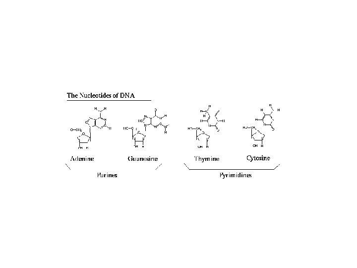
- Each monomer of N. A. is a nucleotide consists of 3 major parts: 1. Phosphates (PO 4 -3) 2. Pentose Sugar (Deoxyribose or Ribose) 3. Nitrogenous Bases: Adenine (A), Guanine (G), Cytosine (C), Thymine (T) in RNA and Uracil (U) in DNA. - Bases A + G= purines (double ringed molecule) nd C + T or U= pyrmidines (single ringed molecule). Nucleotide lacks phosphate root (PO 4 -3) called Nucleoside.

Rellationship between RNA role and amino acids synthesis: - Triplet Code or 3 letter code first suggested by the Physicist Gamow in 1954. It is called now Codon which specify each amino acid: Example: AGA Synthesis of 20 amino acids are determined 4 bases (4 X 4 x 4) of purine or pyramidine which makes distinct triplets (codon) with assistance of transfer RNA.

Proteins: e macromolecules consists of amino acids connected by polypeptides to form the protein coat - The protein coat of virus particle which called capsid consists of capsomers which may be covered by a lipid layer. - Sometimes, there are protein spikes originate from the capsid. - Protein represent the major part of the virus particle about 95% of the size.

- Amino acids are the repeating units in the protein synthesis using the genetic code or codon (triplet letters) as described earlier. The process includes two steps: nscription: ds. DNA orders to make a complimentary copy of RNA GTG CAT CTG ACT CCT GAG AAG GUG CAU CUG ACU CCU GAG AAG Valine Histidin Lucin Trypto Phenyl Gluta Lys 2. Translation: amino acids formation and protein synthesis

Type of Nucleic Acid Strands

CAPSID SYMMETRY AND VIRUS ARCHITECTURE Viral coat protein is the outer shell of which (the capsid) consists of a single, hollow protein molecule, which, as it folds to assume its mature conformation, traps the virus genome inside. In practice, this arrangement cannot occur, for the following reason. The triplet nature of the genetic code (Codon) means that three nucleotides (or base pairs, in the case of viruses with double-stranded genomes) are necessary to encode one amino acid.

Terminologies on viral components
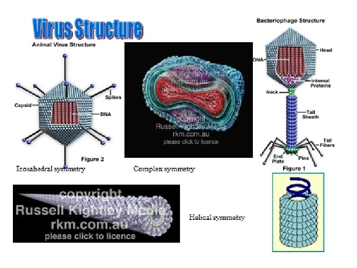

- The forces that drive the assembly of virus particles include hydrophobic and electrostatic interactions—only rarely are covalent bonds involved in holding together the multiple subunits. - There are three main classes of virus structures: helical, icosahedral and complex symmetries - TMV is a typical example on helical symmetry: - The simplest way to arrange multiple, identical protein subunits is to use rotational symmetry and to arrange the irregularly shaped proteins around the circumference of a circle to form a disk. Multiple disks can then be stacked on top of one another to form a cylinder. The virus genome coated by the protein shell or contained in the hollow centre of the cylinder.

RNA Protein subunit capsid Protein shell TMV particles capsomer

- Closer examination of the TMV particle by x-ray crystallography reveals that the structure of the capsid actually consists of a helix rather than a pile of stacked disks. A helix can be defined mathematically by two parameters: its amplitude (diameter) and pitch (the distance covered by each complete turn of the helix) -Helices are simple structures formed by stacking individual protein subunits in a constant form (amplitude and pitch) to repeated one another. - These helices are expressed by the number of subunits per turn of the helix, µ, and the axial rise per subunit, p; therefore, the pitch of the helix, P, is equal to: P = µ X p

- For TMV, µ = 16. 3; there are 16. 3 coat protein molecules per helix turn, and p = 0. 14 nm. Therefore, the pitch of the TMV helix is 16. 3 X 0. 14 = 2. 28 nm. - That helical symmetry is a useful way of arranging a single protein subunit to form a particle is confirmed by the large number of different types of virus that have evolved with this capsid arrangement. Among the simplest helical capsids are those of the well-known bacteriophages of the family Inoviridae, such as M 13 and fd. These phages are about 900 nm long and 9 nm in diameter, and the particles contain five proteins
 Capsids This symmetry generally for 3 -dimensional geometry with 20 equal triangular")
Icosahedral (Isometric) Capsids This symmetry generally for 3 -dimensional geometry with 20 equal triangular sides. Best examples are polio and hepatitis C viruses - The criteria for arranging subunits on the surface of a solid are a little more complex. - In theory, a number of solid shapes can be constructed from repeated subunit. For example, a tetrahedron (4 triangular faces), a cube (6 square faces), an octahedron (8 triangular faces), a dodecahedron (12 pentagonal faces), and an icosahedron, a solid shape consisting of 20 triangular faces arranged around the surface of a sphere
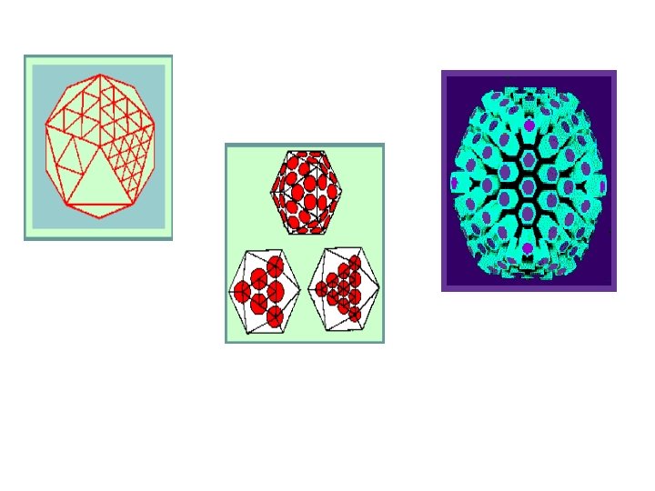

- viruses with 2 -3 -5 symmetry required a multiple of 60 subunit to cover the surface completely. Each unit would be related identically and asymmetrically with its neighbours and none of the units would coincide with an axis of symmetry. Complex Symmetry: This is restricted to viruses with large genomes making a complicated architecture. It has lipids in both envelope and outer membrane of virus particles. These viruses neither helical nor icsahedral shapes which called complex symmetry such as poxviruses

- Although in theory it is possible to construct virus capsids based on simpler symmetrical arrangements. - It is unlikely that a simple tetrahedron consisting of four identical protein molecules would be large enough to contain even the smallest virus genome. If it were, it is probable that the gaps between the subunits would be so large that the particle would be leaky and fail to carry out its primary function of protecting the virus genome. - For an icosahedron, the rules are based on the rotational symmetry of the solid, known as 2– 3– 5 symmetry as in the following forms:
 giving a")
-Therefore the basic with 12 groups of 5 structural subunits (termed pentomers) giving a total 60 structural subunits can be subtriangulated. - Because protein molecules have irregular shape and are not regular equilateral triangles, the simplest icosahedral capsids are built up by using three identical subunits to form each triangular face. This means that 60 identical subunits are required to form a complete capsid (20 X 3).

- In most cases, analysis reveals that icosahedral virus capsids contain more than 60 subunits, for the reasons of genetic economy given above. This presents a difficulty. - A regular icosahedron composed of 60 identical subunits is a very stable structure because all the subunits are equivalently bonded (i. e. , they show the same spacing relative to one another and each occupies the minimum free energy state). - In spherical viruses, there is a problem with limit of 60 protein subunit. A very basic infectious genome would code for a capsid protein (1200 Nucleotides) requires a minimum internal hole of

radius about 9 nm. - A 60 subunit icosahedron made of coat protein of about 20 -30 k. Da (the usual size of a viral coat protein) would have an internal hole of about 6 nm which is not sufficiently large enough to encapsidate most viral genomes. - Most small icosahedral viruses are in the range of 20 -30 k. Da The satellite viruses are the smallest known plant viruses with a particle diameter of bout 17 nm and capsid made up of 17 -21 k. Da. Satellite tobacco necrosis virus made of 60 otein subunit of 21. 3 k. Da

Virus Disease Development Lecture 5

Viral Pathogencity and Disease Development: The disease is any abnormal function in the metabolism of living cell due to infectious or noninfectious agent and virus is the agent here. The capability of infectious virus particle to initiate a disease in a susceptible host is called pathogencity. The stages of initiation infection until symptoms formation is called disease development. Incubation period: time required after virus penetration to symptoms development in the infected host.

Viral Infection: the establishment of a virion in a susceptible host and causing a disease. Latent infection: a delayed infection in the susceptible host due to environmental and/or host-related factors such as age and genetic. Symptoms are external and/or internal expressions of pathological activity in a living host.

Virus Disease Cycle: Disease cycle is the process of disease development in the infected living host. This involves six phases carried out by all types of viruses: 1. The attachment 2. Penetration 3. Uncoating 4. Biosynthesis process (Replication and protein synthesis or gene expression) 5. Assembly and maturation. 6. Release of mature virions from the infected cell

Release Maturation Protein synthesis Replication Penetration Attachment Biosynthesis uncoating Diagrammatic representation of the six phases common to all virus multiplication cycles
 to")
- Virus attachment consists of specific binding of a virus-attachment protein (or ‘antireceptor’) to a cellular receptor molecule (except plant viruses). - Target receptor molecules on cell surfaces may be proteins (usually glycoproteins) or the carbohydrate residues present on glycoproteins or glycolipids. The former are usually specific receptors in that a virus may use a particular protein as a receptor. Carbohydrate groups are usually less specific (not common) receptors because the same configuration of side-chains may occur on many different glycosylated membrane bound molecules.

- Plant viruses face special problems initiating an infection. The outer surfaces of plants have protective layers of waxes and pectin, and each cell is surrounded by a thick wall of cellulose overlying the cytoplasmic membrane. - To date, no plant virus is known to use a specific cellular receptor of the type that animal and bacterial viruses use to attach to cells; instead, plant viruses rely on a mechanical damage integrity of a cell wall to introduce a virus particle directly into a cell. This is achieved either by the vector transmitting the virus or simply by mechanical damage to cells. These entries can be called infectable sites.

Plant Leaf Layers Protective layer: wax-pectin Thick Cellulose wall Nucleus

- Penetration: normally occurs a very short time after attachment of the virus to its receptor in the cell membrane. Unlike attachment, cell penetration is generally an energy-dependent process; that is, the cell must be metabolically active for this to occur. Three main mechanisms are involved: 1. Translocation of the entire virus particle across the cytoplasmic membrane of the cell 2. Endocytosis of the virus into intracellular vacuoles. This is the most common mechanism of a virus entry into cells. It does not require any specific virus proteins (compared to attachment).

- It relies on the normal formation and internalization of coated pits at the cell membrane. Receptor-mediated endocytosis is an efficient process for taking up and concentrating extracellular macromolecules. 3. Fusion: of the virus envelope (only applicable to enveloped viruses) with the cell membrane, either directly at the cell surface or following endocytosis in a cytoplasmic vesicle, which requires the presence of a specific fusion protein in the virus envelope. These proteins promote the joining of the cellular and virus membranes which results in the nucleocapsid deposited directly in the cytoplasm.

- The process of endocytosis is almost universal in animal and human cells. - The formation of coated pits results in the engulfment of a membrane-bounded vesicle by the cytoplasm of the cell. The lifetime of these initial coated vesicles is very short, within seconds, most fuse with endosomes, releasing their contents into these larger vesicles. The release of virus particles from endosomes and their passage into the cytoplasm is intimately connected with (and often impossible to separate from) the process of uncoating.

- Uncoating is a general term for the events that occur after penetration, in which the virus capsid is completely or partially removed and the virus genome exposed, usually in the form of a nucleoprotein complex. The removal of a virus envelope that occurs during membrane fusion is part of the uncoating process. The initial events in uncoating may occur inside endosomes, being triggered by the change in p. H as the endosome is acidified or directly in the cytoplasm.
 and uncoating phases are combined in a single")
For some viruses the entry (penetration) and uncoating phases are combined in a single process. Typically attachment and these two phases do not require the expenditure of energy in the form of ATP hydrolysis. Having made the virus genome available it is now used in the biosynthesis phase when genome replication, transcription of m. RNA, and translation of the m. RNA into protein occur. The process of translation uses ribosomes provided by the host cell and it is this requirement for the translation machinery, as well as the need for molecules for biosynthesis, that makes viruses obligate intracellular parasites.

After uncoating or protein coat removal, two biosynthesis process may occur: 1. Replication 2. Gene Expression: The most critical interaction between a virus and its host cell is the requirement by the virus for the cellular apparatus of nucleic acid and protein synthesis. The course of virus replication is determined by tight control of gene expression. - Therefore, gene expression is a multistep process by which the protein product of a gene is synthesized.

The replication process of any virus depends on the nature of its genetic material. For viruses with RNA genomes in particular, genome replication and the expression of genetic information or gene expression are completely linked; - The control of gene expression determines the overall course of a virus infection: - Latent or masked - Acute or sharp, - Chronic or persistent,

Virus Replication - The objective of a virus after entering a host is to replicate its genetic information. - Replication occurs in eukaryotic cells in nuclear and/or cytoplasmic compartments using genetic information and biochemical capacity between the virus genome and that of the host cell. - Viruses with an RNA lifestyle (i. e. , an RNA genome plus messenger RNAs) have no apparent need to enter the nucleus, although during the course of replication some do. - DNA viruses, mostly replicate in the nucleus, where host-cell DNA is replicated and where the biochemical apparatus necessary for this process

During Gene Expression, viruses make use of the biochemical apparatus of their host cells to express their genetic information as proteins and, consequently, utilize the appropriate biochemical language recognized by the cell. Therefore, viruses of prokaryotes produce polycistronic m. RNAs (a m. RNA that encodes more than one protein which means several different polypeptide chains), while viruses with eukaryotic hosts produce mainly monocistronic m. RNAs (a m. RNA that encodes only one kind of protein which means one polypeptide chain).
")
Gene Expression (protein synthesis)

The process of translation uses ribosomes provided by the host cell and it is this requirement for the translation machinery, as well as the need for molecules for biosynthesis, that makes viruses obligate intracellular parasites. The newly synthesized genomes may then be used as templates for further rounds of replication and as templates for transcription of more virus m. RNA in an amplification process which increases the yield of virus from the infected cells. When the new genomes are produced they come together with the newly synthesized virus proteins to form progeny virus particles in a process called assembly. Therefore, it is obvious that the

Assembly process involves the collection of all the components necessary for the formation of the mature virion at a particular site in the cell. During assembly, the basic structure of the virus particle is formed. The site of assembly depends on the site of replication within the cell and on the mechanism by which the virus is eventually released from the cell and varies for different viruses. For example, - In picornaviruses, poxviruses, and reoviruses, assembly occurs in the cytoplasm; - In adenoviruses, polyomaviruses, and parvoviruses, it occurs in the nucleus.

Maturation is the stage of the life-cycle at which the virus becomes infectious. Maturation usually involves structural changes in the virus particle that may result from specific cleavages of capsid proteins to form the mature products or conformational changes in proteins during assembly. Such events frequently lead to substantial structural changes in the capsid that may be detectable by such criteria as differences in the antigenicity of incomplete and mature virus particles, which in some cases (e. g. , picornaviruses) alters radically. Alternatively, internal structural alterations—for example, the condensation of nucleoproteins with the virus genome—often result in such changes. Virus-encoded proteases are frequently involved in maturation.

Virus Release: viruses escape the cell by one of two mechanisms: - For lytic viruses (such as most non-enveloped viruses), release is a simple process—the infected cell breaks open and releases the virus. - For Enveloped viruses: acquire their lipid membrane as the virus buds out of the cell through the cell membrane or into an intracellular vesicle prior to subsequent release. Virion envelope proteins are picked up during this process as the virus particle is extruded. This process is known as budding.

- Plant viruses face particular difficulties imposed by the structure of plant cell walls when it comes to leaving cells and infecting others. In response, they have evolved particular strategies to overcome this problem using mechanical and vector mechanisms - There is no known plant virus employs a specific cellular receptor of the types that animal and bacterial viruses use to attach to host cells. - Transmission of plant viruses by vectors is the main and most common biological transmission method in the fields to infect and cause disease in healthy plants.

Main transmission methods of human viruses: 1. Blood transfusion like hepatitis virus C 2. Direct shaking contact like herpes viruses 3. Sexual relationship like HIV Using contaminated medical or dental tools and equipments like HIV, herpes 5. By air: Coughing and sneezing like flu viruses 6. Contaminated foods like stomach viral flue sucking blood like mosquitoes transmitting yellow fever virus. Using same utensils of sick people like virus flu or viral stomach flu Using clothes and shoes of infected persons like feet warts.
: Any disease or infection that is naturally transmissible from")
10. Transmission from animals (ZOONOSES): Any disease or infection that is naturally transmissible from vertebrate animals to humans and vice-versa is classified as a zoonosis. Many emergent virus diseases are zoonoses (i. e. , transmitted from animals to humans). Typical example is severe acute respiratory syndrome (SARS) is a type of viral pneumonia, with symptoms including fever, a dry cough, shortness of breath, and headaches. Death may result from progressive respiratory failure due to lung damage. The first SARS outbreak originated in the Guangdong province of China in 2003, where 300 people became ill and at least five died. The cause was found to be a novel corona virus, SARS-Co. V. The SARS virus is believed to be spread by droplets produced by coughing and sneezing, and faecal contamination. Where did the SARS virus come from? Coronaviruses with 99% sequence similarity to the surface spike protein of human SARS isolates have been isolated in Guangdong, China, from healthy masked palm civets (a cat-like mammal closely related to the mongoose).

• Other examples of zoonoses is rabies virus transmitted by dogs as the primary host. Rabies can be passed on infected saliva by biting. • Yellow fever virus transmitted by female mosquito as vectors from monkeys to human. • The other example is swine flue virus which was isolated first from pigs. In all above examples, the virus multiplies in both the vertebrate (animals) and the arthropods (insects).

Mechanisms of Viruses Transmission The process of transfer between hosts is referred to as transmission and this is an important step in the life cycle of viruses. All transmission strategies adopted by viruses have in common the ability to penetrate the outer layer of the skin which is impermeable to viruses, and to bring virus into contact with the naked cells. Person-toperson infections are said to take place by horizontal transmission, while those from mother to baby are put in a separate category and described as vertical transmission such as rubella virus

Some of horizontal transmission routes are: • Respiratory route: occurs commonly, e. g. rhinoviruses, influenza viruses • Conjunctival route: occurrence rates are not known, e. g. respiratory viruses • Fecal route: occurs commonly, e. g. poliovirus • Sexual route: used by specific viruses, e. g. HIV-1, HBV, papillomaviruses • Via urine: used by specific viruses, e. g. Lassa fever virus, cytomegalovirus • Mechanical route: common with tropical arthropods that feed on humans, e. g. arboviruses, and with high risk behaviors, e. g. HIV-1, HBV
- Slides: 77