Medical Virology Lower Respiratory Tract Infections Dr Sameer


- Slides: 19

Medical Virology Lower Respiratory Tract Infections Dr. Sameer Naji, MB, BCh, Ph. D (UK) Dean Assistant Head of Basic Medical Sciences Dept. Faculty of Medicine The Hashemite University

Viral Lower Respiratory Tract Infections Additions are in Italic Green

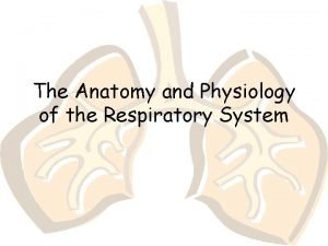
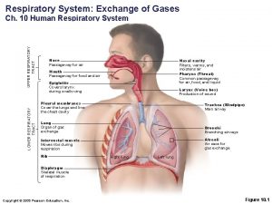
�Structures of the Lower Respiratory System �Components of the lower respiratory system � Larynx � Trachea � Bronchi � Alveoli � Diaphragm � Various protective components �Ciliated mucous membrane, alveolar macrophages, and secretory antibodies (Ig. A)

Structures of the respiratory system-overview

Important Viral Causes of Lower Respiratory Infections �Influenza (by dr. ashraf) �Para-influenza �Respiratory syncytial virus �SARS �MERS �Human metapneumovirus (2001) common � in comprised children and elderly persons �No etiologic agent found (33%)

Para influenza Viruses � It causes Croup (Acute Laryngotracheobronchitis/ Laryngotrachiaites (the lung is involved)) and pneumonia in children � Common cold – like disease in adults and children. � 5 subtypes: 1, 2, 3 (most important) 4 a and 4 b � Surface spikes consist of H(hemagglutinin) , N(neuroaminidase) and fusion proteins. H and N on the same spike while fusion protein is on a different spike.

Epidemiology � Transmission: respiratory droplets, winter months. (viruses have seasonal variations) � Croup is the commonest clinical manifestation of parainfluenza virus infection, caused by subtypes 1 and 2. � It occurs in children (below 3 years). � Parainfluenza 3 is prone to produce bronchiolitis and pneumonia (so it causes infection in more lower areas of the respiratory tract). � The majority of infections with parainfluenza viruses are subclinical.

Clinical Findings � Croup � Harsh cough (larynx is edematous) � Inspiratory stridor (hardly breathing) � Hoarse voice (because of edema ) � SO CROUP IS AN EMERGENCY CASE – send your patient to hospital � Patients are usually afebrile. � About 80% of patients exhibit runny nose (called coriza) 1 to 3 days before the onset of the cough (it can be messed with common cold). Usually, respiratory symptoms subside within 1 or 2 days.

� In addition to croup, parainfluenza viruses cause � common cold, � pharyngitis, � otitis media, � bronchitis � pneumonia. � Other viruses can induce croup, such as influenza viruses, RSV, measles and chickenpox. � Parainfluenza virus infections in adults are relatively uncommon, and symptoms are usually less severe in adults than children.

Laboratory Diagnosis � Croup is a well-defined, easily recognized clinical entity. (if you face a patient with croup outside your clinic, let him breathe a moistened air) � Cell culture isolation � Immunoflurescence (antigen-antibody reaction – needs UV microscope) � Antibody rising titre using HAI or ELISA (there should be 4 x increase in titre)

Treatment � Hospital admission � Nursing in plastic tents supplied with cool, moistened oxygen (first step in treatment) � Severe respiratory obstruction may require endotracheal intubation followed by a tracheotomy.

Respiratory Syncytial Virus Infection �RSV causes Pneumonia and bronchiolitis in infants �(infant<1 year; neonate< 28 days) � (baby<7 months can have pneumonia associated with chlamydia trachomatis) �Fusion protein causes cells to fuse, forming multinucleated giant cells (syncytia) �RSV causes outbreaks of respiratory infections every winter. �RSV is a major nosocomial pathogen in pediatric wards (because it’s a droplet infection). �The pathogen may be introduced by infected infants who are admitted from the outside and adults, especially members of staff with mild infections.

Transmission � Respiratory droplets and direct contact of contaminated hands with the nose or eye � The incubation period is usually 3 - 6 days (in parainfluenza 1 to 2 or 3 days) � The virus spreads along the epithelium of the respiratory tract, mostly by cell-to-cell transfer Pathogenesis � Virus causes syncytia to form in the lungs � Immune response to RSV further damages the lungs

CLINICAL FINDINGS � RSV is the most common cause of severe lower respiratory disease in young infants. It is responsible for 50 - 90% of cases of bronchiolitis, 5 - 40% of pneumonias and bronchitis and less than 10% of croups in young children. � Young children � otitis media � Older children & adults � common cold like – disease � Infants � � Febrile URTI Lower respiratory tract involvement Worsening cough Tachypnoea (fast breathing) and dyspnoea � In bronchiolitis, the respiratory rate may be elevated, with wheezing and hyperinflation. Cyanosis may be present in severe cases (because of tachypnoea ).

Risk groups for fatal RSV infection � Infants with congenital heart disease � Infants with underlying pulmonary disease � especially bronchopulmonary dysplasia � Immunocompromized infants � children who are immunosuppressed or have a congenital immunodeficiency disease. � Nephrotic syndrome (kidney leaking protiens) and cystic fibrosis (a genetic disease – prone to bacterial infection with the mucoid type pseudomonas aeruginosa)

Complications � Apnea (������ ) � occurs in approximately 20% of cases (premature infants). The apnea is non-obstructive and develops at the onset or within the first few days of illness. � The most common complication is prolonged alterations in pulmonary function, which may lead to chronic lung disease in later life.

LABORATORY DIAGNOSIS � Immunoflurescence on smears of respiratory secretions � ELISA for detection of RSV antigens � Isolation in cell culture (multinucleated giant cells or syncytia) � Rise of antibody titer. (4 x)

A syncytium forms when RSV triggers infected cells to fuse with uninfected cells

Treatment � All infants with RSV lower respiratory tract disease are hypoxemic and oxygen should be given to hospitalized infants � Aerosolized ribavirin in severely ill infants � Respi. Gam contains a high concentration of protective antibodies against RSV. It is given for the prevention in children under 24 months with bronchopulmonary dysplasia or a history of premature birth.
 Upper respiratory system
 Lower respiratory tract
 Upper and lower respiratory system
 Introduction to medical virology
 Fields virology
 Duhare
 Virology
 Fields virology
 Extrapyramidal vs pyramidal
 Anterior spinothalamic tract
 Upper respiratory tract
 Air passageway
 Respiratory system nasal cavity
 Anatomy of the upper respiratory tract
 Conclusion of respiratory tract infection
 Upper respiratory tract definition
 Normal flora of respiratory tract
 Classification of upper respiratory tract infection
 Upper respiratory tract
 Mahadev konar
