Medical School Histology Basics Introduction to Microscopy VIBS

Medical School Histology Basics Introduction to Microscopy VIBS 243 lab Larry Johnson Texas A&M University

Objectives • Learn the difference in magnification and resolution • Learn about different types of staining for LM and observe details of cells by EM • Learn how cell/organelles look different at different magnifications • Learn about different types of EM

Use your atlas! pancreas Use your atlas! blood stomach testis Ref code #5

MAGNIFICATION VS. RESOLUTION 1. MAGNIFICATION - INCREASE IN IMAGE SIZE 2. RESOLUTION - SMALLEST DISTANCE BETWEEN TWO POINTS THAT CAN BE SEEN (DISTINGUISHED) RESOLUTION is CALCULATED BY 0. 61 (WAVELENGTH)/NUMERICAL APERTURE 0. 25 um FOR LIGHT MICROSCOPE 0. 1 nm FOR ELECTRON MICROSCOPE

Blood Ref code #5

Blood Ref code #5

Ref code # 12
")
113 Peripheral blood smear (May-Grunwald-Giemsa)
 110")
Peripheral blood smear (Leishman-Giemsa) 110
 basophil, and neutrophils")
110 Peripheral blood smear (Leishman-Giemsa) basophil, and neutrophils

Slide 113 human blood Neutrophils

Slide Histo 021 human blood Platelets Nucleus Neutrophils Red blood cells Neutrophil cytoplasm will merely have a Neutrophils granular appearance

EM 8 f: Peripheral blood cells; 9, 000 x 1. 2. 3. Monocyte Lymphocyte Neutrophil Granules Neutrophils

Ref code #5
 and")
158 Pancreas In H&E staining, the acid dye is eosin (stains proteins red) and the basic dye is a completed form of hematoxylin (stains ribosomes and nuclei blue). Hence, color provides distinguishing characteristics. Islets of Langerhans = light-staining endocrine portion produces insulin Acinar cells = exocrine produces pancreatic enzymes

158 Pancreas Islets of Langerhans Secretory granules are red as they are protein rich with enzymes Base cytoplasm is blue with ribosomes as in RER
 The entire pancreatic acinar cell is blue with varying")
156 Pancreas, monkey (toluidine blue) The entire pancreatic acinar cell is blue with varying intensities depending on the density of structures. Shape, size, and darkness are used to identify structures. Secretory granules

Secretory granules 158 Secretory granules are red as they are protein rich Base cytoplasm is blue with ribosomes as in RER 156 Smooth cytoplasm region = high density of ribosomes in this case

Ref code #5 Mucosa of stomach

145 Fundic stomach Mucosa Connective tissue of submucosa

145 Fundic stomach: mucosa Chief cells Parietal cells Chief cells
 Chief cells Parietal cells")
244 Fundic stomach, rabbit (toluidine blue) Chief cells Parietal cells

244 Secretory granules in chief cells 145 Nuclei Dark spots visible with toluidine blue staining are mitochondria in parietal cells. Mitochondria are not distinguishable with H&E staining

Ref code #5

19680 Toluidine blue staining Human testis - blood and lymph vessels

19709 Transparency of unstained tissue

165 UT 165 human testis H & E staining and right insert toluidine blue staining – note differences in details of cytoplasm Leydig cells 19680

EM 8 f EM 12 a EM 4 c Compare sizes of membranes ribosomes mitochondria as transmission electron microscopy (TEM) provides more cellular detail than light microscopy EM 6 a EM 2 b EM 7

EM 8 f: Peripheral blood cells; 9, 000 x 1. 2. 3. Monocyte Lymphocyte Neutrophil

EM 12 a: Bone marrow; 13, 200 x. Note the reticular cell and developing red blood cells. 1. 2. Reticular cell Developing red blood cell

EM 4 c: Intestinal absorption cell; 60, 000 x 1. 2. 3. 4. 5. 6. 7. Budding RER Coated vesicle Golgi Mitochondria Nucleus Plasma membrane Primary lysosome

EM 2 b: Liver; 60, 000 x; cytoskeletal elements. Microtubes, microfilaments, and intermediate filaments can be compared in this cell, which has a high concentration of cortical microfilaments. 1. 2. 3. Microtube Microfilaments Intermediate filaments

EM 7: Ascites fluid; 80, 000 x. Clear examples of Golgi apparatuses with their cisternae and vesicles are present in this cell 1. 2. 3. 4. Golgi apparatus Ribosomes Lipofuscin Mitochondrion

EM 6 a: Centriole-microtubules; 200, 000 x. Centriolar region of a cell showing both the stable, triplet microtubule arrays within the centriole, and the labile, individual microtubules originating from pericentriolar material. 1. 2. 3. Centriole Stable microtubule Labile microtubule

EM 4 c 60, 000 x EM 8 f 9, 000 x Compare sizes of membranes ribosomes mitochondria EM 12 a 13, 200 x

EM 6 a 200, 000 x EM 2 b 60, 000 x Compare sizes of membranes ribosomes mitochondria EM 7 80, 000 x

Conventional TEM, SEM carbon replica TEM EM 2 b EM 4 a EM 18 b EM 6 EM 7 EM 8
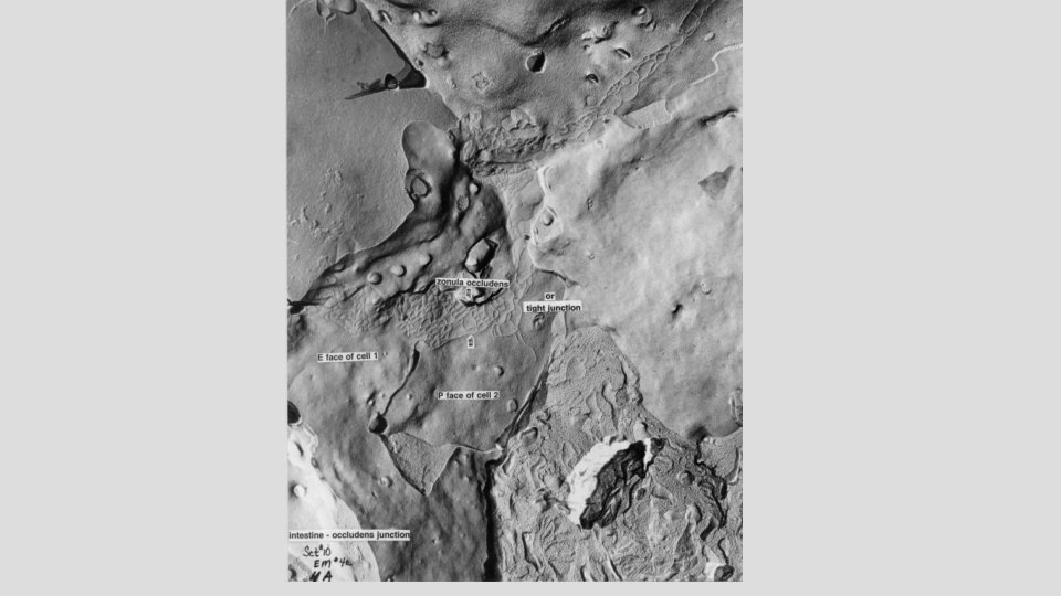
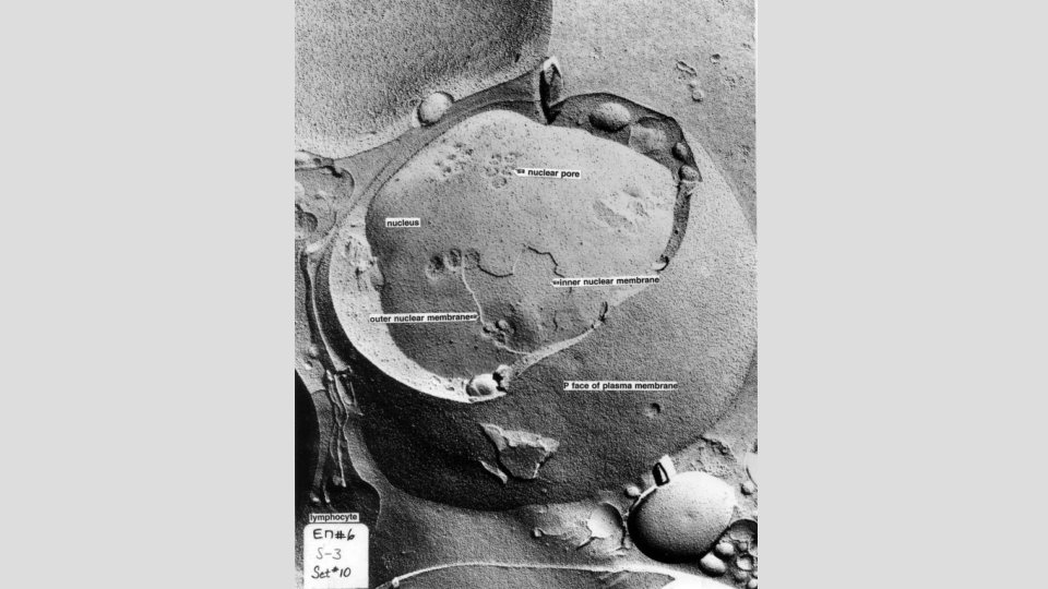
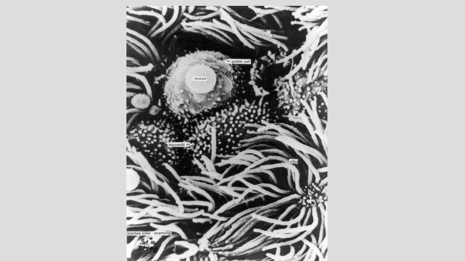
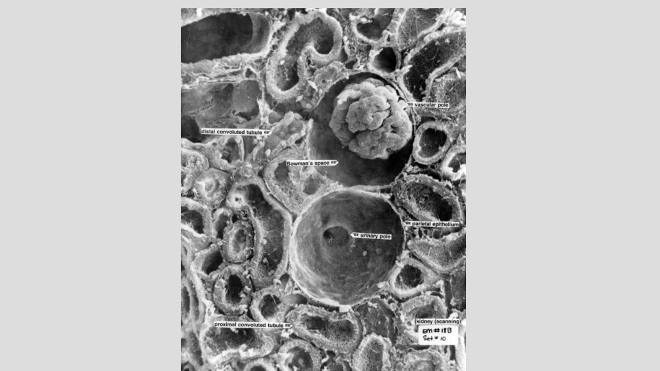

Conventional TEM, SEM carbon replica TEM EM 2 b EM 4 a EM 18 b EM 6 EM 7 EM 8

Conventional TEM, SEM carbon replica TEM EM 2 b EM 4 a EM 18 b EM 6 EM 7 EM 8

Conventional TEM, SEM carbon replica TEM EM 2 b EM 4 a EM 18 b EM 6 EM 7 EM 8

In summary Use your atlas! pancreas blood stomach testis Ref code #5

Many illustrations in these VIBS Histology You. Tube videos were modified from the following books and sources: Many thanks to original sources! 1. Alberts, et al. , 1989. Molecular Biology of the Cell. 2 nd Edition. Garland Publishing, Inc. New York. ISBN 0 -8240 -3695 -6. 2. Alberts, et al. , 1994. Molecular Biology of the Cell. 3 nd Edition. Garland Publishing, Inc. New York. ISBN 0 -8153 -1619 -4. 3. Bloom, W. and Fawcett, D. W. , 1968. A Textbook of Histology. 9 th Edition. W. B. Saunders Company. Philadelphia. Library of Congress #67 -17445. 4. Elias, H. et al. , 1978. Histology and Human Microanatomy. A Wiley Medical Publication. John Wiley & Sons, New York. ISBN 0 -47104929 -8. 5. Eroschenko, V. 2000. Atlas of Histology with Functional Correlations. 9 th Edition. Lippincott Williams & Wilkins. Philadelphia. ISBN 07817 -2676 -X. 6. Fawcett, D. W. , 1986. Bloom and Fawcett. A Textbook of Histology. 11 th Edition. W. B. Saunders Company. Philadelphia. ISBN 0 -72161729 -8. 7. Fawcett, D. W. , 1986. Bloom and Fawcett. A Textbook of Histology. 12 th Edition. Chapman and Hall. New York. ISBN 0 -412 -04691 -1. 8. Guyton, A. C. 1971. Textbook of Medical Physiology. 4 th Edition. W. B. Saunders Company. Philadelphia. Library of Congress # 74118589. 9. Ham, A. W. 1974. Histology. 7 th Edition. J. B. Lippincott Company. Philadelphia. ISBN 0 -397 -52062 -X. 10. Ham, A. W. and Cormack, D. H. 1979. Histology. 8 th Edition. J. B. Lippincott Co. Philadelphia. ISBN 0 -397 -52089 -1. 11. Junqueria, et al. , 1995. Basic Histology. 8 th Edition. Appleton and Lange. Norwalk, Connecticut. ISBN 08385 -0567 -8. 12. Junqueira, et al. , 1998. Basic Histology. 9 th Edition. Appleton and Lange. Stamford, Connecticut. ISBN 0 -8385 -0590 -2. 13. Knobil, E. et al. 1988. The Physiology of Reproduction. Volume 1. Raven Press. New York. ISBN 0 -88167 -281 -5. 14. Langley, et al. , 1974. Dynamic Anatomy and Physiology. 4 th Edition. Mc. Graw-Hill Book Company. New York. ISBN 0 -07 -036274 -2. 15. Mescher, A. L. , 2010. Junqueira’s Basic Histology Text and Atlas. 12 th Edition. Mc. Graw Hill Medical. New York. ISBN 978 -0 -07 -1604314. 16. Tuttle, W. W. and Schottelius, B. A. 1969. Textbook of Physiology. 16 th Edition. The C. V. Mosby Company. Saint Louis. Library of Congress # 7589848. 17. Varner, D. et al. 1991. Diseases and Management of Breeding Stallions. American Veterinary Publications. Goleta, California. ISBN 0939674 -33 -5. 18. Von Hagens, Gunther and A. Whalley, 2007. Body Worlds – The Anatomical Exhibition of Real Human Bodies. ISBN 978 -3 -937256 -04 -7 19. Weiss, L. 1983. Histology: Cell and Tissue Biology. 5 th Edition. Elsevier Biomedical. New York. ISBN 0 -444 -00716 -4. 20. Weiss, L. and Greep, R. 1977. Histology. 4 th Edition. Mc. Graw-Hill Book Company. New York. ISBN 0 -07 -069091 -X.

Questions Which microscope type/staining is/are better for observing cellular details: a. Light microscopy/ H&E b. Light microscopy/ toluidine blue c. Transmission electron microscopy (TEM)/ typical EM staining d. a and b e. a, b, and c

Mexico USA Santa Elena Canyon Big Bend National Park, TX

END OF
- Slides: 49