
Medical Image Registration A Survey Aiming Lu Outline



- Slides: 19
Medical Image Registration: A Survey Aiming Lu
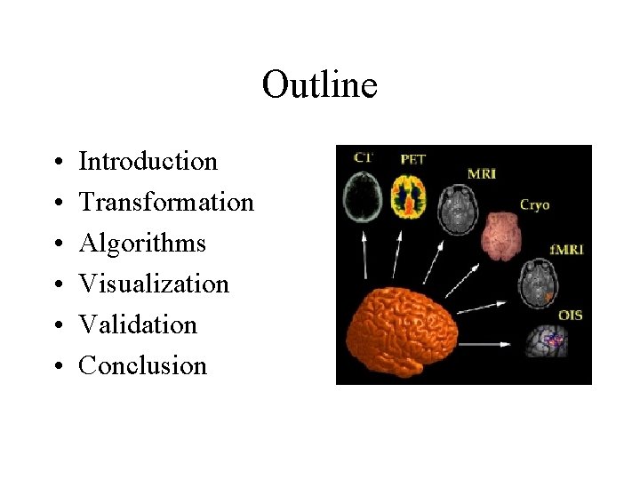
Outline • • • Introduction Transformation Algorithms Visualization Validation Conclusion
Introduction • Image registration – matching two images so that corresponding coordinate points in the two images correspond to the same physical region of the scene being imaged – also referred to as image fusion, superimposition, matching or merge MR SPECT registered
Applications • Diagnosis – Combining information from multiple imaging modalities • Studying disease progression – Monitoring changes in size, shape, position or image intensity over time • Image guided surgery or radiotherapy – Relating pre-operative images and surgical plans to the physical reality of the patient • Patient comparison or atlas construction – Relating one individual’s anatomy to a standardized atlas
Classification • Dimensionality – 2 D-2 D, 3 D-3 D, 2 D-3 D • Nature of registration basis – Image based • Extrinsic, Intrinsic – Non-image based • Nature of the transformation – Rigid, Affine, Projective, Curved • Interaction – Interactive, Semi-automatic, Automatic • Modalities involved – Monomodal, Multimodal, Modality to model • Subject: – Intrasubject – Intersubject – Atlas • Domain of transformation • Local, global • Optimization procedure • Object
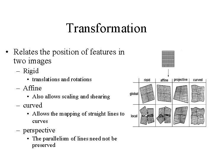
Transformation • Relates the position of features in two images – Rigid • translations and rotations – Affine • Also allows scaling and shearing – curved • Allows the mapping of straight lines to curves – perspective • The parallelism of lines need not be preserved
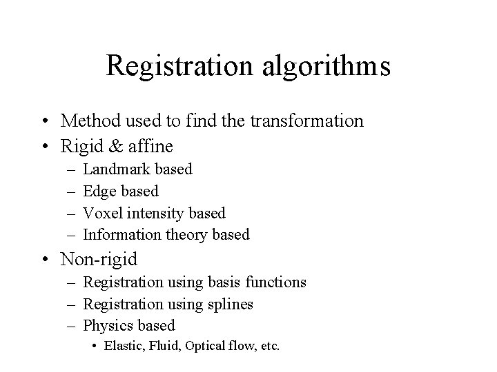
Registration algorithms • Method used to find the transformation • Rigid & affine – – Landmark based Edge based Voxel intensity based Information theory based • Non-rigid – Registration using basis functions – Registration using splines – Physics based • Elastic, Fluid, Optical flow, etc.
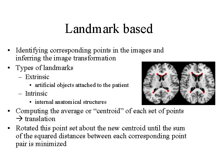
Landmark based • Identifying corresponding points in the images and inferring the image transformation • Types of landmarks – Extrinsic • artificial objects attached to the patient – Intrinsic • internal anatomical structures • Computing the average or “centroid” of each set of points translation • Rotated this point set about the new centroid until the sum of the squared distances between each corresponding point pair is minimized
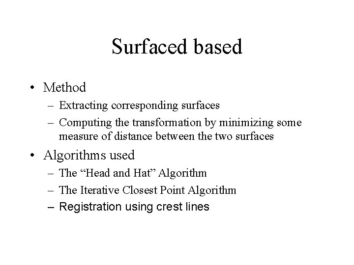
Surfaced based • Method – Extracting corresponding surfaces – Computing the transformation by minimizing some measure of distance between the two surfaces • Algorithms used – The “Head and Hat” Algorithm – The Iterative Closest Point Algorithm – Registration using crest lines
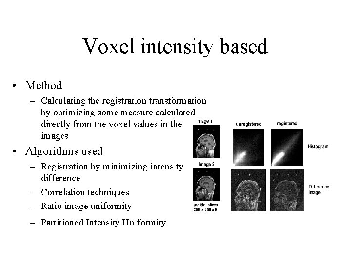
Voxel intensity based • Method – Calculating the registration transformation by optimizing some measure calculated directly from the voxel values in the images • Algorithms used – Registration by minimizing intensity difference – Correlation techniques – Ratio image uniformity – Partitioned Intensity Uniformity
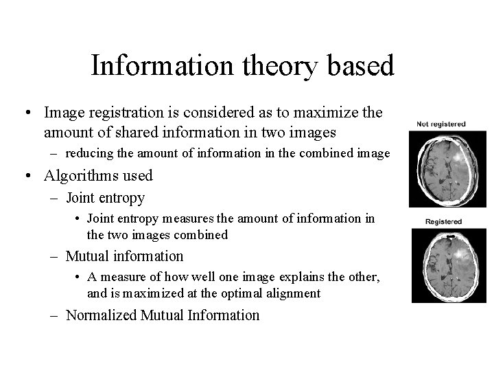
Information theory based • Image registration is considered as to maximize the amount of shared information in two images – reducing the amount of information in the combined image • Algorithms used – Joint entropy • Joint entropy measures the amount of information in the two images combined – Mutual information • A measure of how well one image explains the other, and is maximized at the optimal alignment – Normalized Mutual Information
Registration using basis functions • Represent the deformation field using a set of basis functions – Fourier (trigonometric) basis functions or wavelet basis functions. – Implement smoothness constraint by linear combination of basis functions – The trigonometric basis functions corresponds to a spectral representation of the deformation field where each basis function describes a particular frequency of the deformation.
Registration using splines • Assumption – a set of corresponding points or landmarks (control points) can be identified • At control points, interpolate or approximate the displacements to map the location of the control points in both images • Between control points, they provide a smoothly varying displacement field
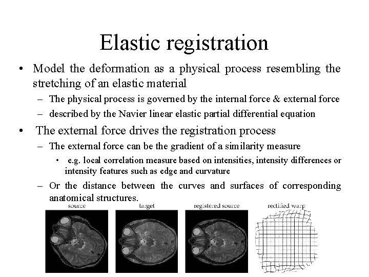
Elastic registration • Model the deformation as a physical process resembling the stretching of an elastic material – The physical process is governed by the internal force & external force – described by the Navier linear elastic partial differential equation • The external force drives the registration process – The external force can be the gradient of a similarity measure • e. g. local correlation measure based on intensities, intensity differences or intensity features such as edge and curvature – Or the distance between the curves and surfaces of corresponding anatomical structures.
Other physics based registration • Fluid registration – The image was modeled as a highly viscous fluid • Registration using mechanical models – using a three-component model to simulate the properties of rigid, elastic and fluid structures. • Registration using optical flow
Optimization • Many registration algorithms require an iterative approach – an initial estimate of the transformation is gradually refined – In each iteration, the current estimate of the transformation is used to calculate a similarity measure – makes another estimate of the transformation, evaluates the similarity measure again, and continues until the algorithm converges • no transformation can be found that results in a better value of the similarity measure, to within a preset tolerance.
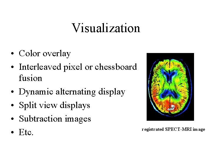
Visualization • Color overlay • Interleaved pixel or chessboard fusion • Dynamic alternating display • Split view displays • Subtraction images • Etc. registrated SPECT-MRI image
Validation • Measurements using computer generated models, images of physical phantoms of accurately known construction and dimensions and images of patients or volunteers. – robustness – Accuracy • Assessment of accuracy – estimate of some geometrical measure of alignment error – compare the system to be validated against a gold standard – Visual assessment • Benefit to the patient and cost
Conclusion • Rigid body registration is now used routinely – image guided surgery systems for neurosurgery and orthopaedic surgery • Challenges: – validation methodologies for non-rigid registration algorithms – More robust similarity measures – distinguish between rigid and deformable structures – Novel display techniques