Medical Helminthology 2020 2021 Lab 7 Cestode Echinococcus
")
Medical Helminthology 2020 -2021 Lab 7 Cestode (Echinococcus granulosus)

Geographical distribution Cosmopolitant, the disease is common in east Africa Disease which causes: Echinococcosis or hydatidosis(hydatid disease)

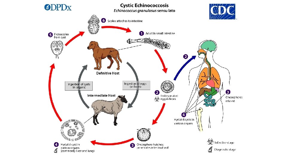
Habitat The adult of Echinococcus granulosus live in the small intestine of carnivores (specially canids) and the larval stage hydatid cyst are found in various organ of mammalian intermediate host.
 1 - Adult worm is a small tape worm and measures 2")
Morphology (adult) 1 - Adult worm is a small tape worm and measures 2 -9 mm in length. 2 - The scolex is pyriform provided with 4 suckers and a rostellum with double crown of large and small hooklets. 3 - It has an attenuated neck; usually one immature proglottid, one mature proglottid, and one gravid proglottid. The morphology of mature proglottid is similar to that of Taenia. 5 - Larva is known as hydatid cyst
")
Echinococcus granulosus (adult)
 1. When egg swallowed by a suitable intermediate host, it will")
Hydatid cyst (larvae) 1. When egg swallowed by a suitable intermediate host, it will hatch in the duodenum, the oncosphere migrate through the intestinal wall, enters the mesenteric venules bed in various organs tissues. They begin to develop a cystic cavity and it becomes young hydatid cyst. 2. After 5 months the hydatid cyst has reached a diameter of 1 cm and has differentiate to (outer layer and inner layer) 3. From the inner layer there are budded out toward the cystic cavity masses of cells which become vacuolated and alter stalked these are the brood capsules which may remain attached or may set free into the fluid of cystic cavity 4. From the inner wall of these capsuled the scolex the free brood capsules and the free scolex collectively referred to hydatid sand

Hydatid cyst
")
Definitive host Adult worm intermediate hosts Hydatid cyst (larvae)

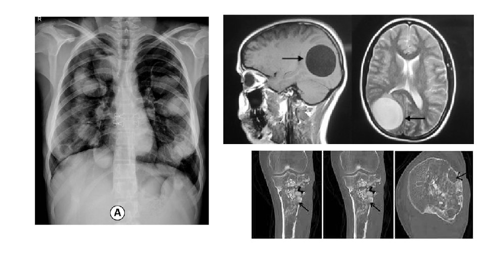
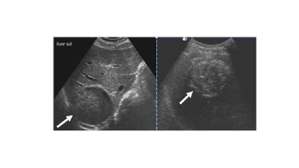
Sign and symptoms The symptoms of hydatid disease depend on which organs are affected. The most commonly affected organ is the liver. The kidneys, brain and lungs are sometimes affected. In rare cases, hydatid cysts may form in the thyroid gland or heart or within bone. Symptoms include: 1. Liver: Hepatomegaly, obstructive jaundice, cholangitis. 2. Lung: dyspnoea, chest pain, Haemoptysis. 3. CNS: space-occupying signs. 4. Bone: an interesting osteolytic cause of knee pain.

Diagnosis of hydatid disease 1. 2. 3. 4. 5. X-ray examination ultrasound CT scan MRI scan examination of blood, urine, sputum, faeces or other bodily fluids if a burst hydatid cyst is suspected 6. blood tests for antibodies to the cysts.

Prevention 1. Stary dogs should be destroyed. 2. Regular treatment of infected dog to reduce worm. 3. Prevention of dogs from eating infected offal of domestic animals(sheep, etc…) in the endemic area. 4. Avoidance of unnecessary contact with infected dogs. 5. Health education and personal hygiene.
- Slides: 14