
Medical Genetics LECTURE 1 R Human Chromosomes GenotypePhenotype
- Slides: 27
Medical Genetics LECTURE 1 R Human Chromosomes: Genotype/Phenotype Muhammad Faiyaz-Ul-Haque, Ph. D, FRCPath
Lecture Objectives: By the end of this lecture, the students should be able to: Ø Describe the number, structure, and classification of human chromosomes. Ø Explain what a Karyotype is and how it is obtained. Ø Describe chromosomal banding and explain its use. Ø Describe the process of in situ hybridization and the information it provides.
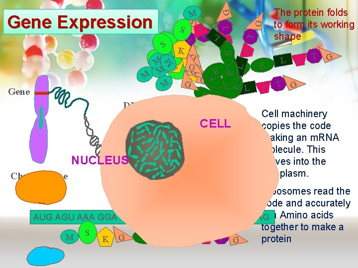
G G T FG S T FL T Gene Expression E F T LL M Gene F E E E G L M M S SS K KK S G S K K M G E E M S GK M G EE G M L E E E DNA G T NUCLEUS Chromosome The protein folds to form its working shape CELL A C T A The order of bases in DNA is a code for making proteins. The code is read in groups of three E F F L T T G G Cell machinery copies the code making an m. RNA molecule. This moves into the cytoplasm. Ribosomes read the code and accurately join Amino acids AUG AGU AAA GGA GAA CUU UUC ACU GGA UAG together to make a S L F T M E E K G protein G
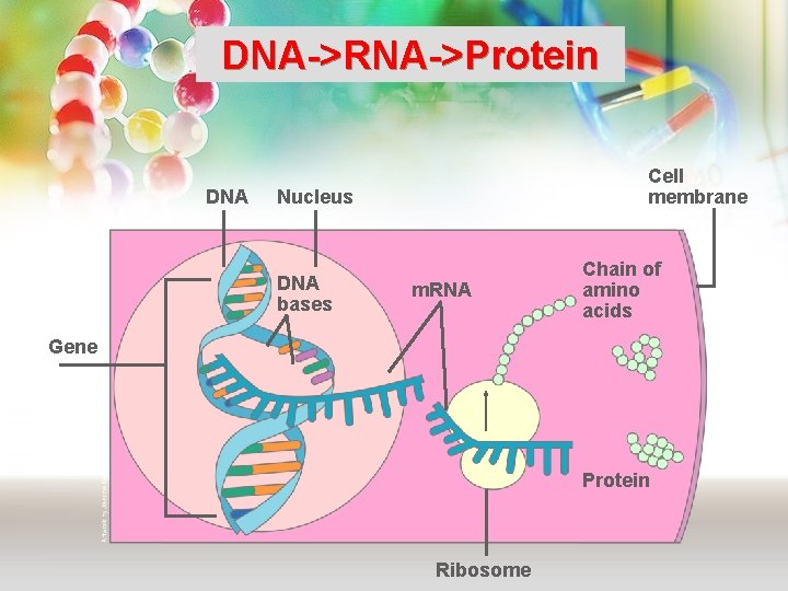
DNA->RNA->Protein DNA Cell membrane Nucleus DNA bases m. RNA Chain of amino acids Gene Protein Ribosome
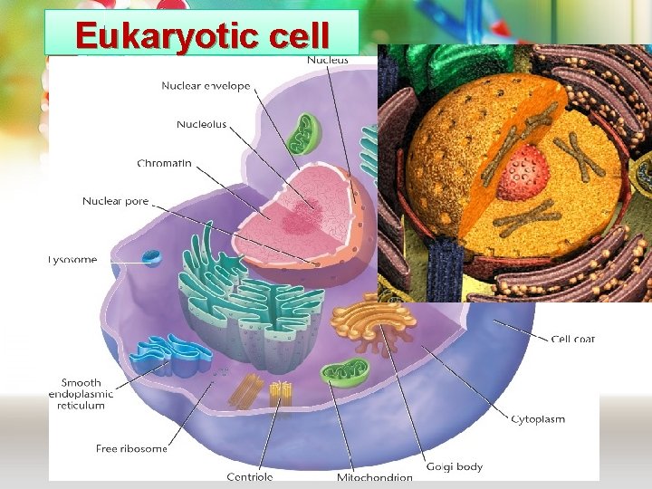
Eukaryotic cell
GENETICS : ■ Cytogenetics: The study of the structure and function of chromosomes and chromosome behaviour during somatic and germline division ■ Molecular genetics: The study of the structure and function of genes at a molecular level and how the genes are transferred from generation to generation.
Cytogenetics: Human Cytogenetics involves the study of human chromosomes in health and disease. Chromosome studies are an important laboratory diagnostic procedure in 1) prenatal diagnosis 2) certain patients with mental retardation and multiple birth defects 3) patients with abnormal sexual development 4) some cases of infertility or multiple miscarriages 5) in the study and treatment of patients with malignancies & hematologic disorders. New techniques allow for increased resolution.
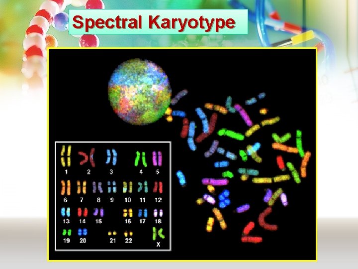
Spectral Karyotype
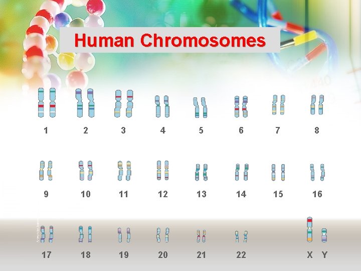
Human Chromosomes 1 2 3 4 5 6 7 8 9 10 11 12 13 14 15 16 17 18 19 20 21 22 X Y
Cytogenetics: ■ Non-Banded Karyotype ■ High resolution Karyotype Molecular cytogenetics: ■ Fluorescent in situ hybridization (FISH).
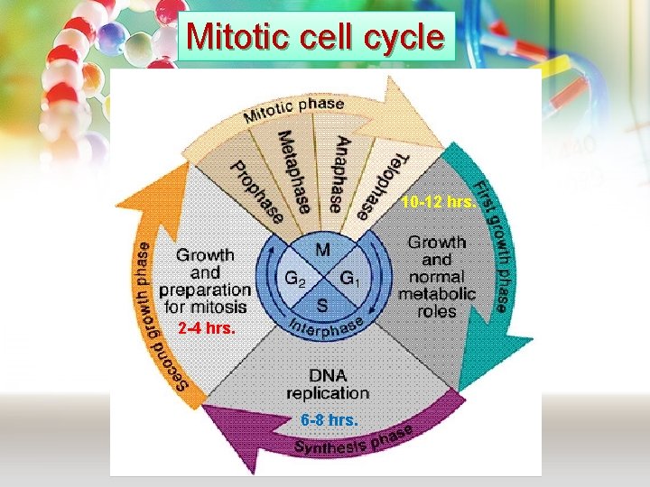
Mitotic cell cycle 10 -12 hrs. 2 -4 hrs. 6 -8 hrs.
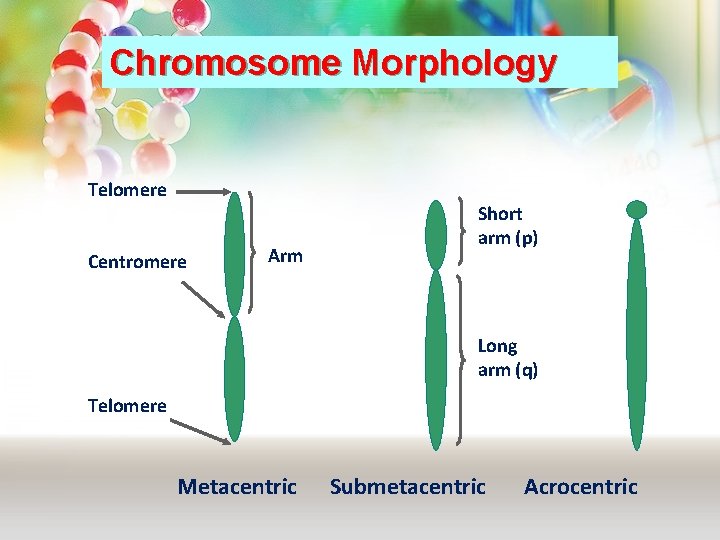
Chromosome Morphology Telomere Centromere Arm Short arm (p) Long arm (q) Telomere Metacentric Submetacentric Acrocentric
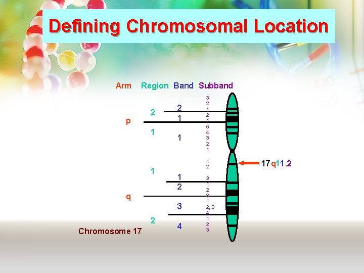
Defining Chromosomal Location Arm Region Band Subband p 2 1 1 1 2 q 3 2 Chromosome 17 3 2 1 5 4 3 2 1 4 3 1 2, 3 4 1 2 3 17 q 11. 2
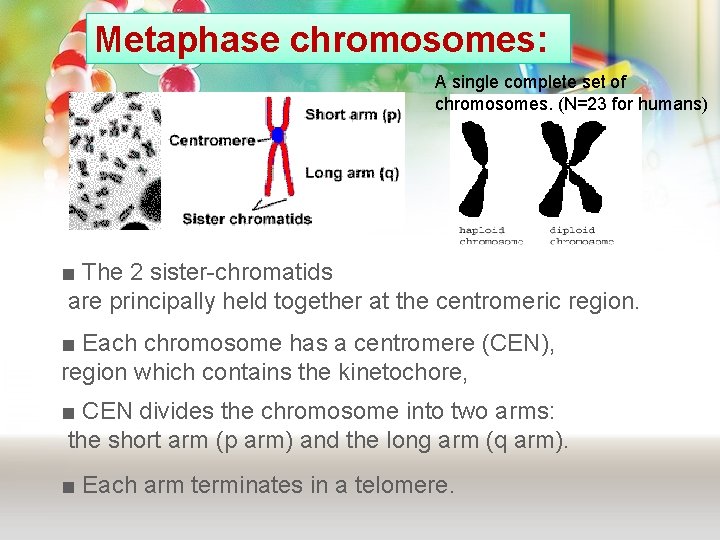
Metaphase chromosomes: A single complete set of chromosomes. (N=23 for humans) ■ The 2 sister-chromatids are principally held together at the centromeric region. ■ Each chromosome has a centromere (CEN), region which contains the kinetochore, ■ CEN divides the chromosome into two arms: the short arm (p arm) and the long arm (q arm). ■ Each arm terminates in a telomere.
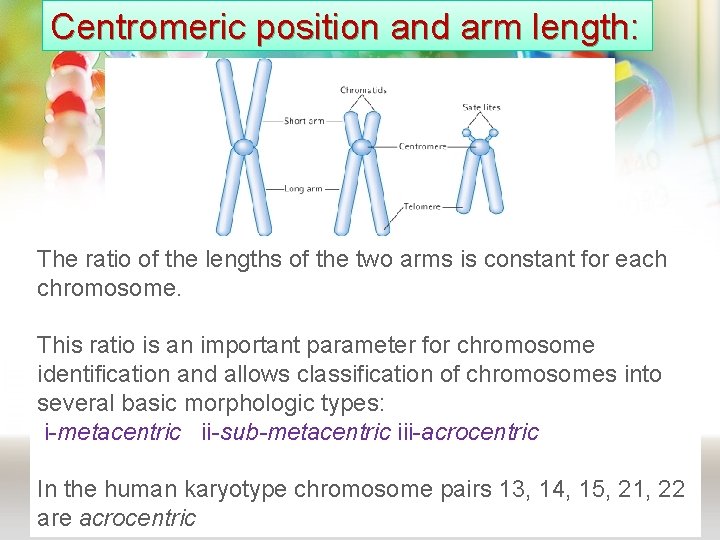
Centromeric position and arm length: The ratio of the lengths of the two arms is constant for each chromosome. This ratio is an important parameter for chromosome identification and allows classification of chromosomes into several basic morphologic types: i-metacentric ii-sub-metacentric iii-acrocentric In the human karyotype chromosome pairs 13, 14, 15, 21, 22 are acrocentric
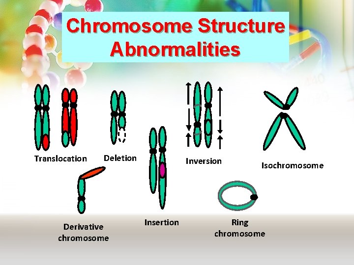
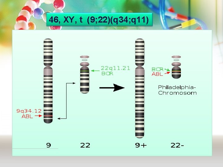
Chromosome Structure Abnormalities Translocation Deletion Derivative chromosome Inversion Insertion Isochromosome Ring chromosome
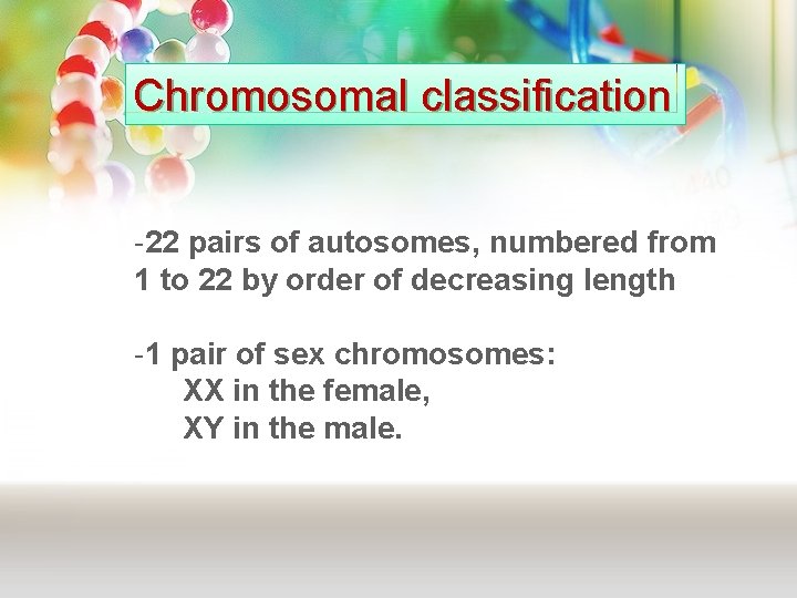
Chromosomal classification -22 pairs of autosomes, numbered from 1 to 22 by order of decreasing length -1 pair of sex chromosomes: XX in the female, XY in the male.
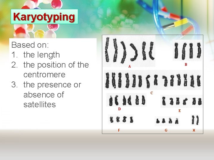
Karyotyping Based on: 1. the length 2. the position of the centromere 3. the presence or absence of satellites B A C D F E G X
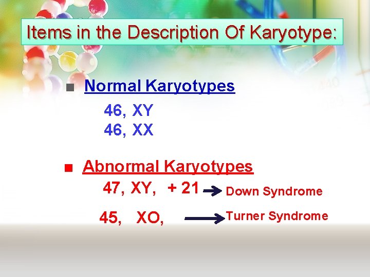
Items in the Description Of Karyotype: ■ Normal Karyotypes 46, XY 46, XX ■ Abnormal Karyotypes 47, XY, + 21 Down Syndrome 45, XO, Turner Syndrome
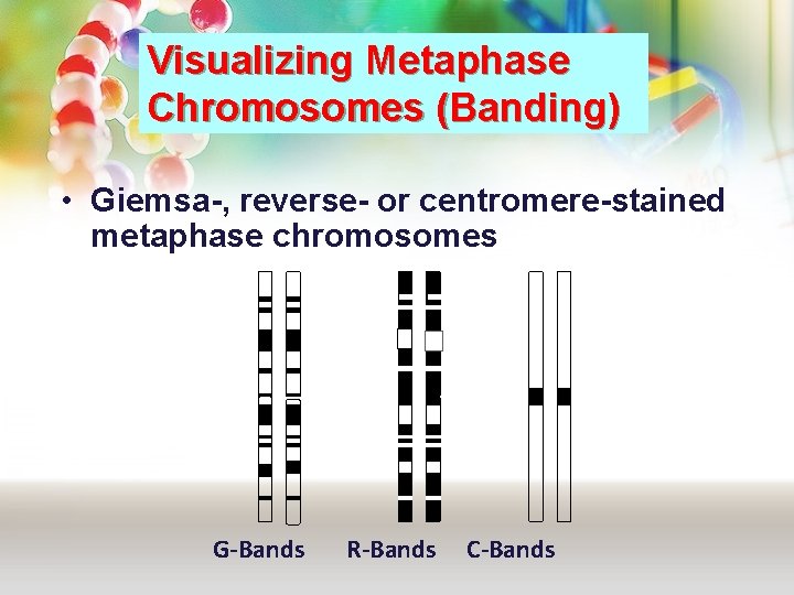
Visualizing Metaphase Chromosomes (Banding) • Giemsa-, reverse- or centromere-stained metaphase chromosomes G-Bands R-Bands C-Bands
46, XY, t (9; 22)(q 34; q 11)
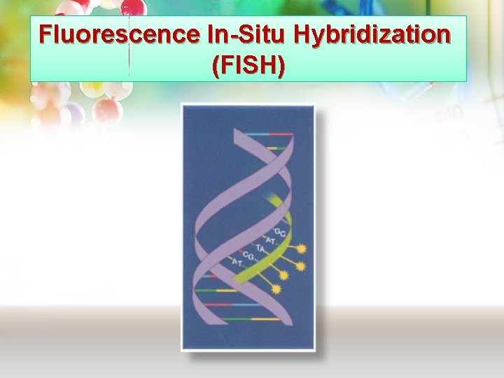
Fluorescence In-Situ Hybridization (FISH)
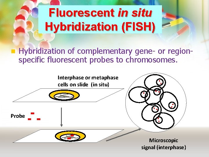
Fluorescent in situ Hybridization (FISH) n Hybridization of complementary gene- or regionspecific fluorescent probes to chromosomes. Interphase or metaphase cells on slide (in situ) Probe Microscopic signal (interphase)
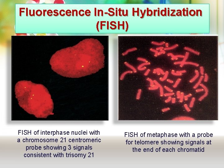
Fluorescence In-Situ Hybridization (FISH) FISH of interphase nuclei with a chromosome 21 centromeric probe showing 3 signals consistent with trisomy 21 FISH of metaphase with a probe for telomere showing signals at the end of each chromatid
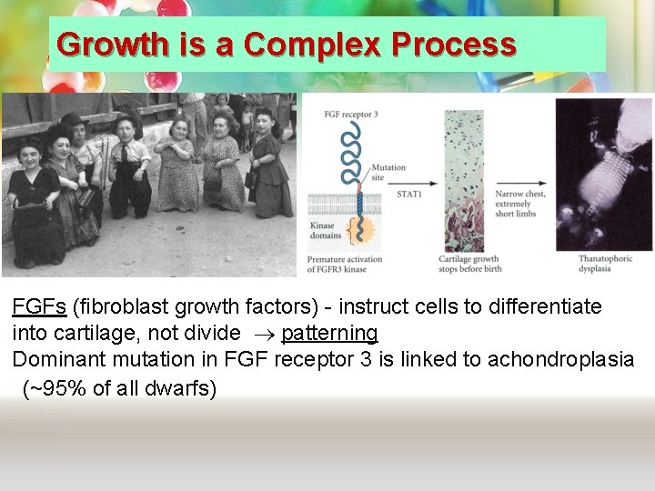
Growth is a Complex Process FGFs (fibroblast growth factors) - instruct cells to differentiate into cartilage, not divide patterning Dominant mutation in FGF receptor 3 is linked to achondroplasia (~95% of all dwarfs)
Take Home Message Ø The packaging of DNA into chromosomes involves several orders of DNA coiling and folding. Ø The normal human karyotype is made up of 46 chromosomes consisting of 22 pairs of autosomes and a pair of sex chromosomes, XX in the female, and XY in the male. Ø Each chromosome consists of a short (p) and a long (q) arm joined at the centromere. Ø Molecular cytogenetic techniques (e. g. FISH) are based on the ability of DNA probes to attach with its complementary target sequence. They can be used to study chromosmes in metaphase or interphase.
THANK YOU