Mediastinum and mediastinal mass Done by Razan Tarawneh

Mediastinum and mediastinal mass Done by : Razan Tarawneh Rawan zedaneean Mallak Aljafari

Mediastinum It’s the central compartment of thoracic cavity , covered on each side by mediastinal pleura , contain all thoracic viscera and structure except the lungs. Division of the mediastinum into four compartments: - superior mediastinum - Inferior mediastinum: anterior mediastinum (prevascular zone), middle mediastinum (peritracheoesophageal zone), posterior mediastinum(paravertebral zone).

Normal Lateral view of Mediastinum on chest X-ray

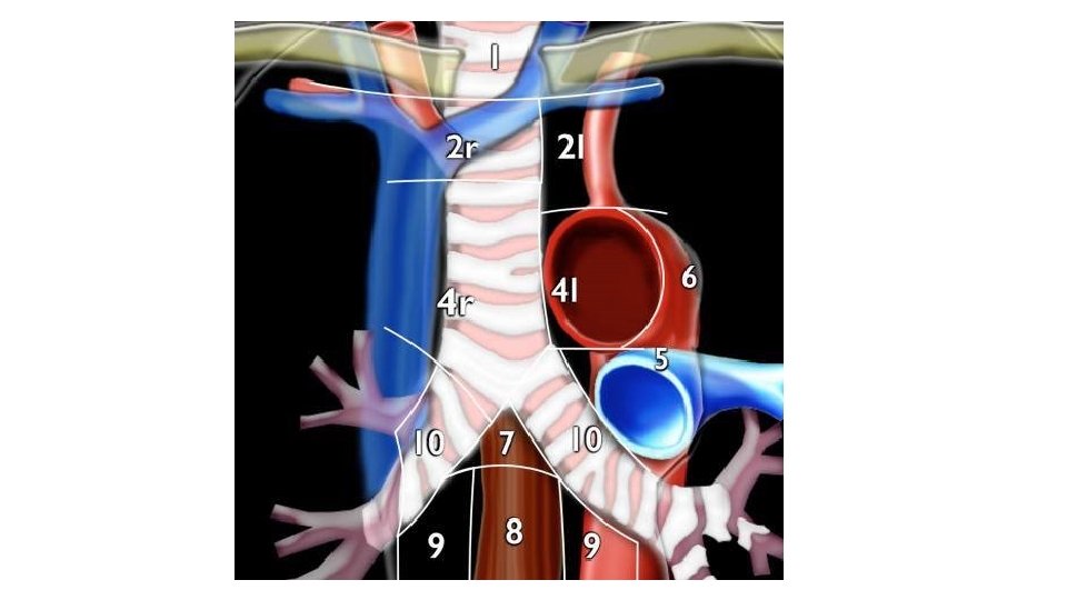
q superior mediastinum q Arteries Aortic arch Brachiocephalic artery Thoracic portions of the left common carotid and the left subclavian. q Veins Brachiocephalic veins Upper half of the superior vena cava Left highest intercostal vein q Nerves Vagus nerve Cardiac nerve Superficial and deep cardiac plexuses Phrenic nerve Left recurrent laryngeal nerve q Trachea with paratracheal and tracheobronchial lymph nodes q Esophagus q Thoracic duct q Remains of the thymus q Some lymph glands q Anterior longitudinal ligament

Anterior Mediastinum contains major contents of this compartment include thymus, lymph nodes, mediastinal fat, and left brachiocephalic vein. 2, 13, 15

Mediastinal masses your goal is to determine the following: • Is it a mediastinal mass? • Is it in the anterior, middle or posterior mediastinum? • Are you able to characterize the lesion by determining whether it has any fatty, fluid or vascular components?

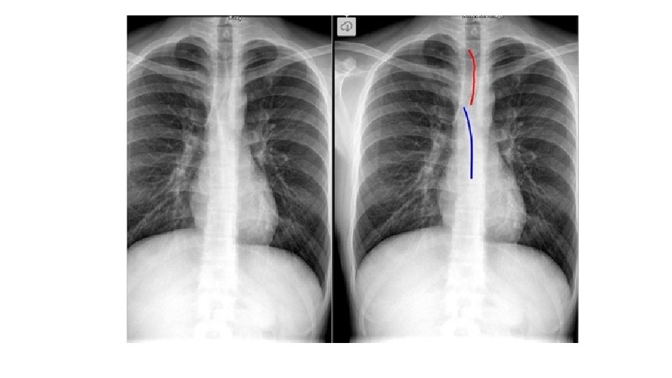
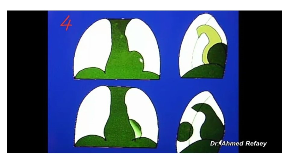
The following characteristics indicate that a lesion originates within the mediastinum: • Unlike lung lesions, a mediastinal mass will not contain air bronchograms. • The margins with the lung will be obtuse. • Mediastinal lines (azygoesophageal recess, anterior and posterior junction lines) will be disrupted. • There can be associated spinal, costal or sternal abnormalities. • LEFT: A lung mass abutts the mediastinal surface and creates acute angles with the lung. RIGHT: A mediastinal mass will sit under the surface of the mediastinum, creating obtuse angles with the lung
. §Thyroid (Retrosternal goiter). §Teratoma. §Thymic")
Anterior mediastinal masses § 5 T’s §Lymphoma (Terrible lymphoma). §Thyroid (Retrosternal goiter). §Teratoma. §Thymic tumor. §Pericardial cyst. §Diaphragmatic hernia (morgagni hernia). The diagnosis of anterior mediastinal mass may be an incidental finding in the asymptomatic patient. However, 60% of patients are symptomatic at presentation • The most common localizing symptom is cough, which is present 60% of the time • Other common obstructive symptoms include chest pain, dyspnea, voice hoarseness, hemoptysis, and dysphagia. • On conventional radiographs look for the signs : • Obliterated retrosternal clear space • Hilum Overlay Sign • Obliterated cardiophrenic angle

Normal chest x-rays

Hilum Overlay Sign in lymphoma in a HIVpositive patient. Obliterated retrosternal clear space in Lymphoma

Mediastinal Teratoma There is a large anterior mediastinal mass measuring 9 cm in size with calcification within it. shift of the mediastinum towards right side Substernal Thyroid Goiter. Frontal chest radiograph shows a large superior/anterior mediastinal mass (white arrows) displacing the trachea (black arrow) to the right of midline

MIDDLE MEDIASTINAL MASSES BOUNDERIES

• : Contents • - pericardium • - heart • - great vessels joining the heart • ascending aorta • pulmonary trunk • right & left pulmonary arteries • the lower half of the superior vena cava • - tracheal bifurcation and both main bronchi • - phrenic nerves • - cardiac plexus • - tracheobronchial lymph nodes

• Causes: • • + Lymph node enlargment : • Lymphoma • Primary TB • Sarcoidosis • + Bronchogenic cyst. • + Aneurysm of aortic arch

lymphoma + common + responsible for 15% of all primary mediastinal masses + the majority are Hodgkin lymphomas + arise from either the thymus or lymph nodes + majority of patients have anterior mediastinal and paratracheal involvement. Isolated hilar nodal involvement is uncommon

+ bilateral widening /unilateral + well-defined margin + lobulated + smooth contour

demonstrates a soft tissue attenuating mass, with smooth or lobulated margins which conform to surrounding structures

Clinical presentation patients are often asymptomatic from the mediastinal component but present with systemic manifestations of lymphoma, most commonly constitutional symptoms

TB Could be unilateral /bilateral

Sarcoidosis + bilateral +symmetrical +hilar enlargement

Clinical presentation is variable. Approximately 50% of patients are asymptomatic. The remainder present with either respiratory symptoms (e. g. cough and dyspnea) or skin changes (e. g. erythema nodosum, scars) diagnosis of pulmonary sarcoidosis is proven by pathology after tissue biopsy

Aortic arch aneurysm

Bronchogenic cyst • +Rare condition Middle mediastinum • +Most common site • +Filled with fluid (water) • +soft-tissue density rounded structures on the x ray • +Rarely multiple

Bronchogenic cyst

Complications fistula formation with the bronchial tree ulceration of the cyst wall secondary bronchial atresia superimposed infection hemorrhage malignant transformation is very rare Clinical presentation asymptomatic. When large may result in bronchial obstruction leading to air trapping and respiratory distress. An alternative presentation may occur when the cyst becomes infected.

Posterior mediastinum

Posterior mediastinal borders • Sup : thoracic plane • Inf : diaphragm • Ant : pericardium • Post : ant border of spine

Posterior mediastinum component q Thoracic part of descending aorta q Azygos vein, hemiazygos and accessory hemiazygos veins q Autonomic nerves (Vagus nerve, splanchnic nerves, sympathetic chain) q Esophagus q Thoracic duct q Fat and lymph nodes
. Aneurysm of descending aorta. Hiatus")
Posterior mediastinum mass Neurogenic tumors (neurofibroma, schwannoma , neuroblastoma). Aneurysm of descending aorta. Hiatus hernia. Paravertebral mass /abscess Meningocele Extramedullary hematopoiesis

• Ant. Med x-ray post. Med x-ray

• Frontal lateral

• Ant. M. masses post. M. masses • Silhouetting cardiac border • Not extend above the clavicle ** Cervicothoracic sign not extend above the clavicle : The anterior mediastinum stops at level of the superior clavicle. Therefore , when a mass extends above the superior clavicle , its located either in the neck or in the posterior mediastinum. When lung tissue comes between the mass and the neck , the mass is probably in the posterior mediastinum.
 ANTERIOR MEDIASTINAL MASS (")
POSITIVE Cervicothoracic sign GOITER ) ANTERIOR MEDIASTINAL MASS (
")
sign NEGATIVE Cervicothoracic POSTERIOR mediastinal mass (schwannoma)

Case study • A 14 years old female patient presented with cough and has sickle cell anemia , her chest x -ray reveal : • Diagnosis: Extramedullary hematopoiesis.

- Slides: 39