Median Arcuate ligament syndrome Imaging Diagnosis and Management


. \"Median arcuate")


 • Increase of at least 30 bpm when going")
 • Theory– Underlying Dysautonomia • Hyper adrenergic POTS –")


- Slides: 53
Median Arcuate ligament syndrome Imaging, Diagnosis and Management Jonathan B. Bard, MD Medical Director, Envision Imaging Mc. Kinney
Jonathan B. Bard, MD • Radiology Residency UCLA • Neuroradiology Fellowship Stanford • Diagnostic Radiology Associates of Dallas – Partner • Solo Practice 2003 - Present with Envision Radiology • Married, 2 boys, 15 and 11 • Hobbies include piano lessons, Texas Hold’em, Bass, Music (Blues) • Disclosure Statement – No financial interest to disclose
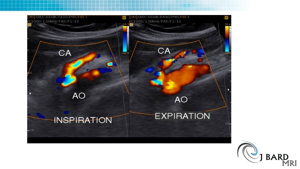
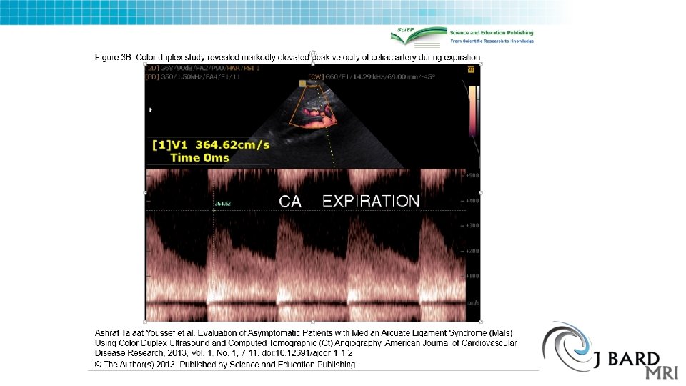
Median Arcuate Ligament syndrome MALS • Celiac artery compression syndrome • Dunbar syndrome, Horjola. Marable syndrome • Compression of the celiac trunk by the median arcuate ligament • Worse with expiration Median arcuate ligament
MALS symptoms Common symptoms Preoperative workup • Post prandial pain • Emesis, bloating, weight loss • Diagnosis of exclusion and with Doppler ultrasound, CTA • Colonoscopy • Upper endoscopy • Testing for Celiac Disease • Testing for H Pylori • Upper GI barium swallow • Ultrasound and HIDA scan • Psychological and Pain evaluation
MALS anatomy Theories • Abnormally inferior and anterior location of MALS • Compresses the celiac axis, causing ischemia • Possible compression of the celiac ganglia • However, this occurs in up to 25% of normal individuals Normal on left, abnormal on right
Celiac axis Anatomy Celiac trunk Left Gastric Splenic artery Common Hepatic artery Gastroduodenal artery
Celiac axis Anatomy Celiac trunk Left Gastric Splenic artery Common Hepatic artery Gastroduodenal artery
Celiac axis Anatomy Celiac trunk Left Gastric Splenic artery Common Hepatic artery Gastroduodenal artery
MALS Management Theory • Compression of the Celiac axis leads to symptoms • Vascular - Bowel ischemia • Neurogenic - Celiac Ganglia compression cause pain • Laparoscopic treatment of MALS can help resolve symptoms by decompression of the artery, but also releasing or destroying the adjacent ganglia • Chronic Functional abdominal pain Syndrome overlaps with MALS • CFAP is similar to IBS, but no bowel changes (diarrhea, constipation)
Web and other Resources • Horton KM, Talamini MA, Fishman EK (2005). "Median arcuate ligament syndrome: evaluation with CT angiography". Radiographics 25 (5): 1177– 82. doi: 10. 1148/rg. 255055001. PMID 16160104. • You. Tube “SMA Syndrome and Median Arcuate Ligament Syndrome: True Syndromes or Fantasy” ctisus • https: //www. youtube. com/watch? v=L 9 ftfucw. LWI • University of Chicago MALS Program • http: //www. ucmals. com/mals. html
http: //www. slideshare. net/pryce 27/rsna-final-2? qid=1560 f 210 -f 9 be-489 a-b 48 f 1231 cf 91008 d&v=qf 1&b=&from_search=6 http: //www. theparisreview. org/blog/2015/09/11/the-most-misread-poem-in-america
The Road Not Taken- Case 1
What has happened? A. Partial resolution of PV thrombosis B. Tumor invasion of PV C. Cavernous transformation of PV
Ultrasound of MALS • Mesenteric/celiac duplex ultrasound interpretation criteria revisited. • J Vasc Surg. 2012 Feb; 55(2): 428 -436. e 6; discussion 435 -6. doi: 10. 1016/j. jvs. 2011. 08. 052. Epub 2011 Dec 21. • he PSV threshold that provided the highest OA for ≥ 50% stenosis was ≥ 240 cm/s and for ≥ 70% stenosis was ≥ 320 cm/s
CTA of MALS • Narrowed Celiac Axis • Post stenotic dilation • Hooked proximal Celiac artery after stenosis • Vessel collaterals • Protocol varies, need 1 mm recons, but arterial phase done in expiration, venous or delayed in normal respiration • Need thin MIPS for diagnosis and stenosis, thick MIPS for collaterals
Motion artifact full expiration
Same Patient in earlier slides – thin MIPS High Grade stenosis of the Celiac trunk
Same Patient – thin MIPS High Grade stenosis of the Celiac trunk
Same Patient – thin MIPS High Grade stenosis of the Celiac trunk
MALS - Collaterals • SMA to Gastroduodenal Collaterals • Same patient
MALS - Collaterals • SMA to Gastroduodenal Collaterals • Same patient
Atherosclerotic Disease
Second Year Case
Which of the following is false? A. B. C. D. E. Red line is Left Gastric Artery Yellow is Left Renal Vein Blue is Duodenum This patient has SMA Syndrome This patient does not have MALS
SMA Angle
Nutcracker phenomenon • Congestion of left renal vein from SMA and Aorta • Some have urinary symptoms • Pelvic congestion syndrome cause? • Renin angiotensin system activation • Treatment
Is this MALS?
Challenges to MALS diagnosis and management • Defining Significant stenosis on CTA • Excluding other diagnoses (Nutcracker, SMA syndrome) • Recurrent symptoms after laparoscopic surgery • Recurrent stenosis • Repeat CTAs are done only on patients with recurrent symptoms • Here are some failures
Initial study March 2015 pre-surgery
Post Surgery
Preoperative MALS
Recurrent post op stenosis
th 4 Year case - Pain
Differential? A. B. C. D. E. F. Sclerosing Mesenteritis Lymphoma Carcinoid Mesothelioma Malignancy All of the above
What would aid in narrowing the differential? A. B. C. D. E. Desmoplastic Reaction Calcification Ascites Hypervascular Liver tumors All of the above
First Year – What Is the most common cause of this finding? A. B. C. D. Medullary sponge kidney Distal Renal tubular acidosis Hyperparathyroidism Hypercalcemia
With What is this finding NOT associated? A. B. C. D. MEN type 2 B Polyhydramnios Immobilization Ehlers-Danlos Syndrome
Ehlers Danlos syndrome Q 79. 6 • Inherited Connective tissue disorder • 6 major types (consolidated from 10) • Multiple genes, can be x-linked or Dominant or Recessive • 1 in 2, 500 -5, 000 people. • Type 3 Hypermobility type most common Joint instability, MSK Symptoms, early arthritis Also Mitral valve prolapse, Ascending Aortic Rupture
EDS Type 3 • 1 in 10 -15, 000, most common subtype • Scoliosis • Fragile skin • Nerve compression disorders • Hiatal Hernia • Ascending Aorta Rupture • Raynaud’s Phenomenon • Postural orthostatic Tachycardia Syndrome
POTS (Postural Orthostatic Tachycardia Syndrome) • Increase of at least 30 bpm when going from supine to upright. • No orthostatic hypotension • Blood pressure does not drop • Chronic fatigue syndrome – up to 50 % may have POTS • EDS type 3 patients have POTS up to %40 comorbidity • Theory – Underlying Dysautonomia
POTS (Postural Orthostatic Tachycardia Syndrome) • Theory– Underlying Dysautonomia • Hyper adrenergic POTS – hypovolemia or autonomic neuropathy • Neuropathic POTS – Denervation of sympathetic nerves, impaired constriction of vessels • Causes • • Genetics Post viral illness or parasitic infection, Lyme, MS, Lupus Many others Syrinx – Chiari type 1
Symptomatic but no stenosis
Recurrent Stenosis Post operative
Summary • MALS Diagnosis is not as straightforward as we thought • Treatment failures cause reevaluation of imaging studies • Is there something on the images that can guide management and alter the outcome? I suspect so • Data are still being collected – 60 patients so far, 65 studies total
THANK YOU
Fellow case - 57 year old with soccer injury
Regarding the rectus femoris muscle tear: A. B. C. D. The bipennate portion is torn The unipennate portion is torn The indirect tendon is torn The bipennate portion surrounds the unipennate portion
3 rd year case - Which is false? A. There is an Os trigonum B. There is a Shephard Fracture C. Tibiotalar arthrosis D. Stieda’s process is fractured E. There is posterior impingement
Potpourri - Which of the following is false? A. Bard-Pic Syndrome is Progressive obstructive jaundice, Large palpable gallbladder (Courvoisier’s sign) and rapid cachexia due to Pancreatic Head Carcinoma B. Bard Syndrome is military pulmonary metastases from Gastric neoplasm C. A Bard was Behind the plate when Barry Bonds hit his 756 th career home run, breaking Hank Aaron’s record D. BARD 1 is a gene on Chromosome 2 that is associated with BRCA 1 mutations and increase risk of cancer
True
The Real Bard syndrome