MECHANISMS OF CARDIAC CONTRACTION AND RELAXATION BY DR

MECHANISMS OF CARDIAC CONTRACTION AND RELAXATION BY DR SHILPI LAHOTY CARDIOLOGY RESIDENT

MICROANATOMY OF CONTRACTILE CELLS AND PROTEINS • Major functions of cardiomyocytes is to execute excitationcontraction-relaxation that depends on the electrical calcium transport and contractile properties • Atrial and ventricular myocytes have cross striations and are often branched. • Each myocyte is bounded by sarcolemma and is filled with myofibrils containing the contractile elements. • The sarcolemma invaginates to form an extensive transverse tubular network that extends the extravascular space into the interior of the cell(T-tubules).
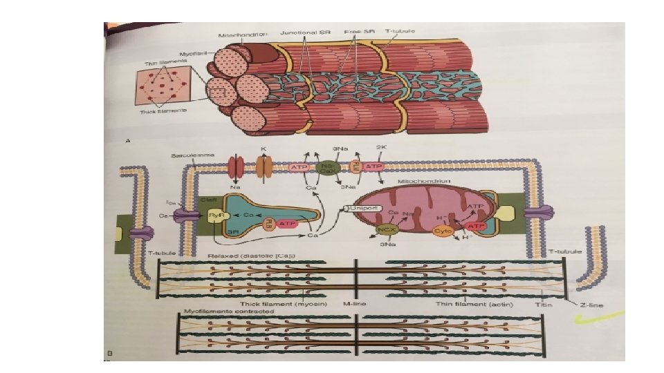

• Sarcoplasmic reticulum , which is a lipid membrane bounded, fine interconnected network spreading throughout the myocytes , is a specialized form of ER that is critical for calcium cycling, which is the on-off switch for contraction. • The calcium release channels(Ry. Rs) are concentrated at the part of the SR that is in very close apposition to the t-tubules Ca 2+ channel called the junctional SR. The rest of the SR consists of ramifying tubules that surrounds the myofilaments and take up Ca 2+ back----so drive relaxation via SERCA This Ca 2+ is stored at high concentration in the SR, in part bound to Ca 2+ buffering proteins including calsequestrin. • SARCOPLASM refers to intracellular fluid and proteins there in but excludes the contents of organelles such as mitochondria, SR and nucleus. • MITOCHONDRIA : each ventricular myocyte has approx. 8000 mitochondria which has an outer and inner mitochondrial membrane (OMM and IMM resp). As per the functions mitochondria can either make ATP or extrude calcium.

• The IMM contains the cytochrome complexes that make up the respiratory chain, including F 0 -F 1 ATP synthase. The space within the IMM, the mitochondrial matrix, contains enzymes of the tricarboxylic acid (TCA) cycle and other key metabolic components. • These components provide reducing equivalent protons that are pumped out of the matrix by the cytochromes, and it is this proton pumping that creates the very negative matrix potential with respect to cytosol (Ψm = − 180 m. V). Such a negative Ψm creates a strong electrochemical gradient for protons, which as they flow down this energy gradient on F 0 -F 1 ATP synthase, are responsible for making ATP. The ATP still needs to get out of the mitochondria, and an adenine nucleotide transporter exchanges mitochondrial ATP for cytosolic adenosine diphosphate (ADP).
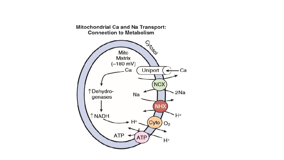

• So calcium overload in the cell SHORT TERM: calcium is taken up by the mitochondria to protect the cell CHRONICALLY: a) this diminishes voltage and occurs at the expense of ATP production b) increased calcium in the mitochondria facilitates opening of mitochondrial permeability transition pore which wipes off the negative voltage and allows matrix contents to be released to the cytosol leading to death of mitochondria as well as the cell. • ATP produced in the mitochondria is transported to the microfilaments and this process is facilitated by the location of creatine kinase, an enzyme that converts creatine phosphate to ATP.

CONTRACTILE PROTEINS • Functional contractile unit is sarcomere and energy for contraction is via ATP • The two chief contractile proteins are the motor protein myosin on the thick filament and actin on the thin filament. • The sarcomere is limited on either side by a Z-line, which with the thin filaments creates a sort of cage around the thick myosin filament that extends from the center of the sarcomere outward toward, but not reaching the Z-line. • During contraction, the myosin heads grab onto actin and pull the actin filaments toward the center of the sarcomere. The thin and thick filaments can thus slide over each other to shorten the sarcomere and cell length (without the individual actin or myosin molecules actually shortening). The interaction of the myosin heads with actin filaments when sufficient Ca 2+ arrives from the SR is called cross -bridge cycling. As the actin filaments move inward toward the center of the sarcomere, they draw the Z-lines closer together so that the sarcomere shortens. The energy for this shortening is provided by the breakdown of ATP, made chiefly in the mitochondria.
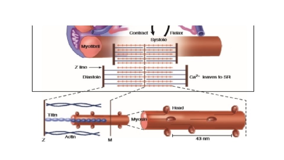

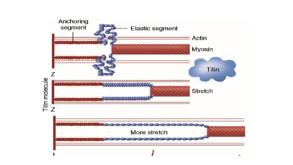
Titin and length sensing • It is the largest protein yet described and is extra ordinarily long, elastic and slender and connects the thick filament to the Z line. • The titin molecule can stretch between 0. 6 and 1. 2 micron metre in length and has multiple functions: • It stabilizes the sarcomeric structure and its elasticity contributes to the stress-strain relationship of cardiac muscle. • The stretched molecular spring limits the overstretching of sarcomeres and end diastolic volume and returns some potential energy during systole as the sarcomeres shorten during cardiac ejection. • Titin may transduce mechanical stretch into growth signals. With sustained diastolic stretch, as in volume overload, the elastic segment of titin is under constant strain and transmits this mechanical signal to the muscle LIM protein (MLP) attached to the terminal part of titin that forms part of the Z -disc complex. MLP is proposed to be a stretch sensor that transmits the signals that result in the myocyte growth pattern characteristic of volume overload. This signal system may be defective in a subset of human dilated cardiomyopathy.

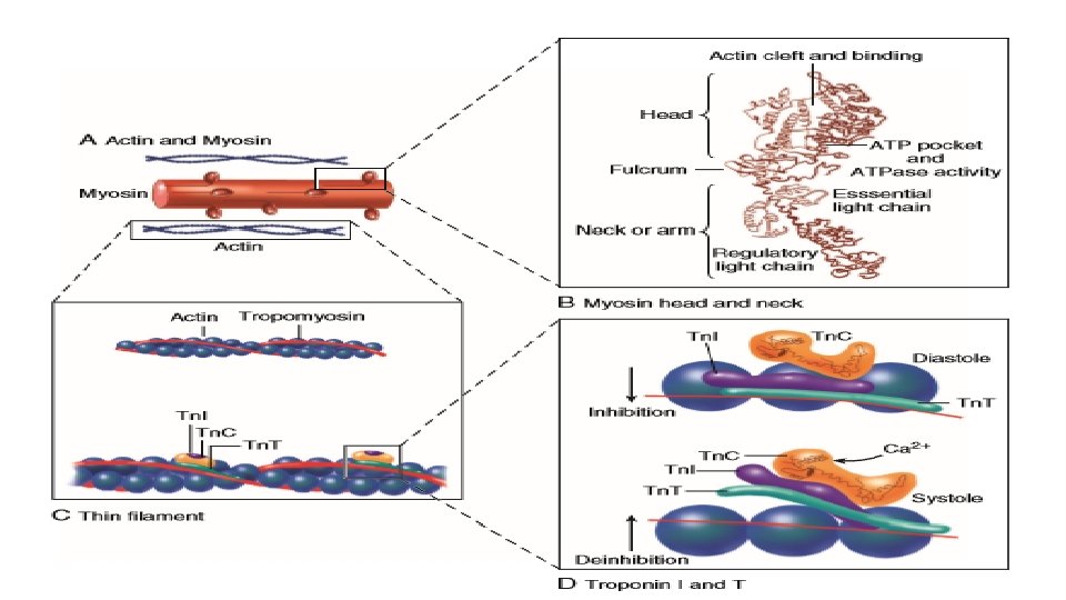
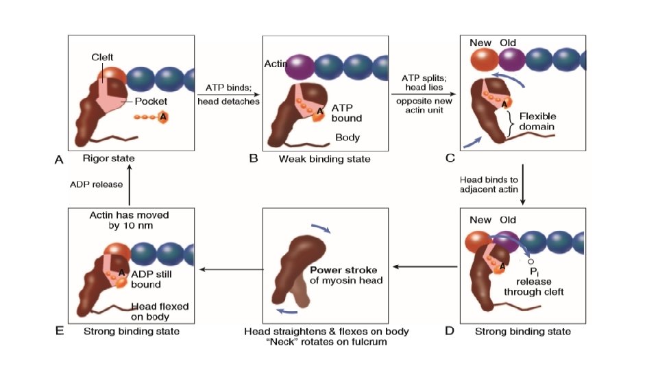
MOLECULAR BASIS OF MUSCULAR CONTRACTION • Calcium enters the cell via T-tubules and via ryanodine receptors in SR Binds to troponin C Troponin C binds more tightly to troponin I and allows tropomyosin to roll deeper into the thin filament groove and this shifts the position of troponin-tropomyosin complex on the actin filament Poised Myosin head forms strong binding cross bridges with actin molecules and use the energy stored in myosin-ADP-Pi

This rotates the myosin head while bound to actin in the power stroke Until ATP binds again to myosin, this will remain in the rigor state ATP binds again to myosin and shifts back to weak binding state allowing cross-bridge detachment and ATP hydrolysis and the cycle continues

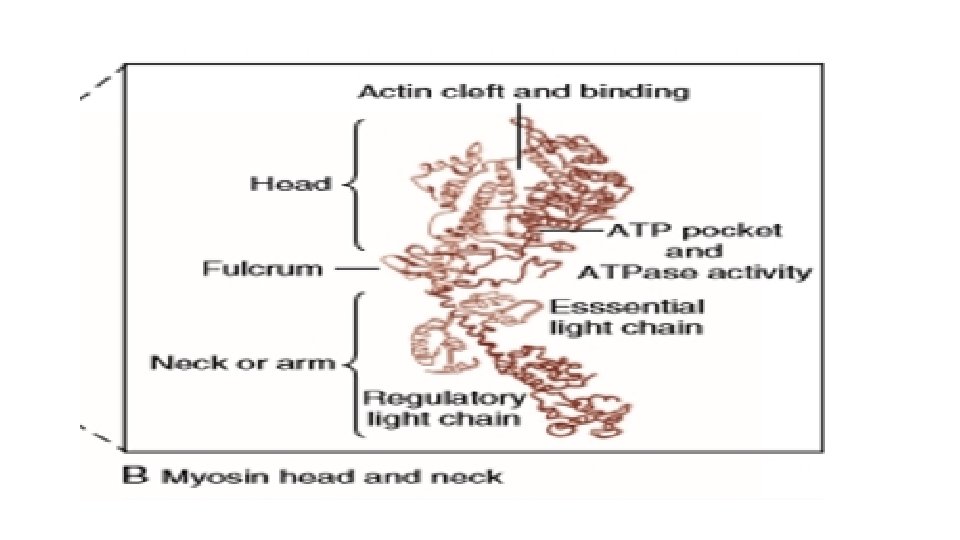
MYOSIN STRUCTURE AND FUNCTION • Each myosin head is the terminal part of the myosin heavy chain held via a short neck and the other end of 2 myosin molecules coil and form the thick filament. • Each head has an ATP binding pocket and a narrow cleft • According to the Rayment model, the base of the head, or the neck, changes configuration in the contractile cycle. • During isometric ( or isovolumic) contraction the cross bridges rotate but cannot fully move the actin filament, and the stretched strong-binding cross bridges bear force. During shortening (ejection) the actin filament moves during the power stroke. • Each cycle of cross bridge consumes 1 molecule of ATP and this myosin ATPase activity is the major site of ATP consumption in the beating heart. Thus when the heart is more strongly activated , the level of ATP consumption is similarly increased

MYOSIN ISOFORMS ALPHA FORM BETA FORM This has slower ATPase rate and is the predominant form in adult humans Each myosin molecule neck also has two light chains MCL 1 (Essential) This limits the contractile process by interaction with Actin. MCL 2 (regulatory) This is the potential site for phosphorylation on beta stimulatn and promotes cross-bridging.

EFFECTS OF CALCIUM • Calcium is the central regulator of cardiac contraction and relaxation • The higher the calcium, the more fully saturated are the calcium binding sites on troponin C and hence more sites are available for cross-bridges to form. • Ca 2+ bound to a single troponin C encourages local crossbridge formation, and both Ca 2+ binding and cross-bridge formation directly enhance the likelihood of cross-bridge formation in the seven actin molecules controlled by one tropomyosin molecule. This cooperativity means that a small change in [Ca 2+]i can have a large effect on the strength of contraction. • Relatively small amounts of calcium (trigger calcium) enter and leave the cardiomyocytes during each cardiac cycle, with larger amounts being released and taken up by the SR • In this calcium induced calcium release mechanism, a small amount of calcium entering via the calcium current triggers the release of a larger amount of calcium into the cytosol. • In human ventricles, SR Ca 2+ release is three to four times higher than calcium influx by ICa

• Besides calcium, sarcomere length at the end of diastole also influences the strength of contraction so More the diastolic filling of the heart increase sarcomere length sensing mechanism Greater the strength of the heart beat • When changes in diastolic length (or preload) are the cause of altered contractile strength, it is said to be a Frank-Starling (or sometimes just Starling) effect. Conditions in which contraction is strengthened (or weakened) independent of sarcomere length (e. g. , increased Ca 2+ transient amplitude) are referred to as positive (or negative) inotropic states or enhanced (or reduced) contractility.

FORCE TRANSMISSION • Volume and pressure overload may owe their different effects on myocardial growth to different patterns of force transmission. • Genetic-based hypertrophic and dilated cardiomyopathies are in general linked to mutant genes that cause abnormalities in the force generating system, such as β-MHC, troponin (T, I and C), MLCs, myosin-binding protein C, and alpha-tropomyosin. One hypothesis is that mutations that increase contractile performance and/or energy demand result in concentric hypertrophy whereas mutations that either reduce force generation or result in non–force-generating cytoskeletal proteins (e. g. , dystrophin, nuclear lamin, cytoplasmic actin, and titin) lead to a dilated cardiomyopathy.

CALCIUM RELEASE AND UPTAKE BY SARCOPLASMIC RETICULUM • SR is a continuous network surrounding the myofilaments with connections across Z-lines and transversely between myofibrils. • The total SR Ca 2+ content is the sum of [Ca 2+]SR plus Ca 2+ bound to the intra-SR Ca 2+ buffers (esp calsequestrin). • SR Ca 2+ content is critical to both normal cardiac function and electrophysiology, and its abnormalities contribute to systolic and diastolic dysfunction and arrhythmias. [Ca 2+]SR dictates the SR Ca 2+ content, the driving force for release of Ca 2+, and regulates Ry. R release channel gating.

JUNCTIONAL SR AND RYANODINE RECEPTORS • The ryanodine receptor that mediate SR calcium release are mainly located in the j. SR membrane at the junction with the T-tubule. • Each junction has 50 to 250 Ry. R channels that are directly under a cluster of 20 to 40 sarcolemmal L-type Ca 2+ channels across a 15 -nm junctional gap. • Function of Ry. R 2 is as a calcium channel and also as a scaffolding protein that localizes numerous key regulatory protein to the j. SR. • On the large cytosolic side, these include proteins that can stabilize Ry. R gating (e. g. , calmodulin [Ca. M]; FK-506 binding protein ; kinases that can regulate Ry. R gating by phosphorylation (e. g. , protein kinase A [PKA] and Ca 2+/ Ca. Mdependent protein kinase II [Ca. MKII]); and the protein phosphatases PP 1 and PP 2 A, which dephosphorylate the Ry. R.

• Inside the SR the Ry. R also couples to several proteins (e. g. , junctin, triadin, and via them calsequestrin) that similarly regulate Ry. R gating and, in the case of calsequestrin, provides a local reservoir of buffered Ca 2+ close to the release channel. The actual Ry. R channel is made up of a symmetric tetramer of Ry. R molecules, each of which may have the aforementioned regulatory proteins associated with it. Thus the Ry. R receptor complex is very large (>7000 k. Da) • When T-tubule is depolarised, one or more L-type calcium channels open and local cleft calcium increases sufficiently to activate atleast one local j. SR Ry. R This recruits additional Ry. R in the junction through calcium induced calcium release to amplify release of calcium into the junctional space activates contraction

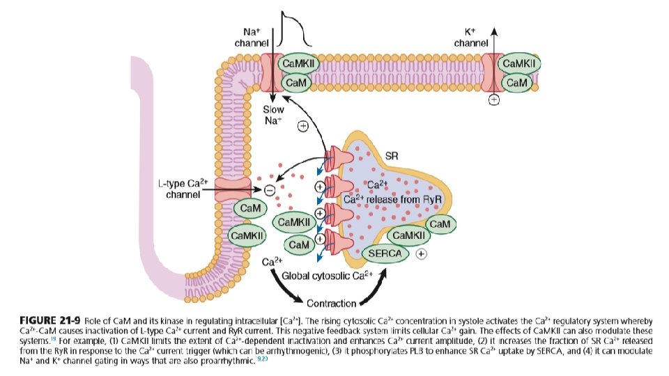
TURNING OFF CALCIUM RELEASE • SR calcium release turns off when Ca 2+ in the SR drops by approx. 50% • Ca 2+ in cytoplasm binds to calmodulin which is associated with ICa channel and it alters channel conformation such that ICa inactivation is favoured. • Binding of calcium to CAM that is pre-bound to Ry. R 2 favours closure of Ry. R channels and inhibits reopening • Moreover, Ry. R 2 gating is also sensitive to luminal calcium in SR such that high calcium favours opening and low calcium in SR favours closure. • Ca. M has four Ca 2+ binding sites, resembles troponin C, and participates in many different cellular pathways from ion channels to transcriptional regulation. In many cases (e. g. , L-type Ca 2+, Na+, and some K+ channels and Ry. R ), Ca. M is already prebound or “dedicated” such that elevation of local [Ca 2+]i can rapidly induce Ca 2+-Ca. M effects on their targets. • Many myocyte Ca. M targets (e. g. , Ca. MKII, calcineurin, nitric oxide synthase [NOS]) compete for this limited pool of “promiscuous” Ca. M. Thus Ca. M signaling in myocytes is complex and is further complicated by the effects of Ca. MKII, which influences some of the same targets and processes as Ca. M itself does.

CALCIUM SPARKS AND WAVES • In addition to SR Ca 2+ release triggered by ICa during normal excitationcontraction coupling, there is a finite probability that a given Ry. R will open stochastically. • Because of local Ca 2+-induced Ca 2+ release in the junctional cleft, this can lead to spontaneous local SR Ca 2+ release events known as Ca 2+ sparks. • Ca 2+ sparks are very local events (within 2 µm in the cell). • However, the probability of Ca 2+ sparks is greatly enhanced when [Ca 2+]i or [Ca 2+]SR is elevated or under conditions in which the Ry. R is otherwise sensitized (e. g. , by oxidation or Ca. MKII). These conditions can greatly enhance the likelihood that SR Ca 2+ release from one junction will be sufficient to trigger neighboring junctions 1 to 2 µm away and result in propagating Ca 2+ waves throughout the whole myocyte. These Ca 2+ waves can be arrhythmogenic.

• Calcium uptake into SER is mainly by SERCA (90% of the SR protein) and its dominant form in cardiac myocyte is SERCA 2 a • For each molecule of ATP hydrolysed by this enzyme, 2 calcium ions are taken up into the SR. • SR calcium uptake is the primary driver of cardiac myocyte relaxation and reuptake starts as soon as calcium ion in the cytoplasm begins to rise. • A reduction in SERCA expression or function (as seen in heart failure or energetic limitations) can thus directly result in slower rates of cardiac relaxation. • The strength of the SR calcium ion uptake directly influences the diastolic SR calcium ion content and calcium ion in SR dictates both the sensitivity of the Ry. R and the flux rate of SR calcium ion release Thus SR calcium ion uptake and release are an integrated system • Phospholamban (PLB), a single transmembrane protein binds directly to SERCA and decreases its affinity for cytosolic calcium ion and thereby weaken the calcium uptake by SR.

• However phosphorylation of PLB by either PKA or Ca. MK 11 Inhibits the inhibitory effect Increases the rate of SR Ca ion uptake Lusitropic effect and moreover increased SR calcium ion content which leads to positive inotropic effect

SARCOLEMMAL CONTROL OF Ca 2+ and Na+ • Calcium channels

Calcium channels T-type • Opens at a more negative voltage , have short burst of opening and donot interact with conventional calcium antagonist drugs. • In adults ventricular myocytes, there doesn’t seem to be appreciable T-type Ica • They donot seem to target the regions where Ry. Rs are, and consequently they donot participate in excitationcontraction coupling per-se. L-type • Long lasting type calcium channels and inhibited by calcium channel blockers. • Concentrated in the T-tubules at j. SR junction and cause calcium induced calcium release from the Ry. R. • This is rapidly activated during the rising phase of action potential but the combination of calcium influx via Ica itself and local SR calcium release causes rapid calcium dependent inactivation of ICa

L-Type channel continuation • Voltage dependent inactivation also contributes to ICa decline during the action potential but ICa continues at low levels through out the action potential. • Inward ICa is an important contributor to the plateau phase of action potential and excess ICa or failure of inactivation can prolong the duration of action potential Beta adrenergic stimulation c. AMP and PKA activity increases phosphorylation of calcium channels

this increases the opening time of the channel increases ICa Increases both the fraction of SR calcium release and the calcium load of the cell and SR

Sodium Channels • Depolarisation activates INa • Voltage dependent inactivation of INa is very rapid and under normal conditions sodium channels inactivates within a few millisecs of depolarisation. • However a small number of sodium channels remain open, thereby creating a small but persistent influx of sodium throughout the plateau of action potential This is called late sodium current INa. L and is characterised by ultraslow, voltage independent inactivation and reactivation Under pathophysiologic conditions this can cause acquired long QT syndrome which is arrhythmogenic

• Ca 2+/ Calmodulin dependent protein kinase II alters gating of INa, ICa and other channels: Ca. MKII is upregulated in heart failure, ROS, reperfusion ischaemia. At lower heart rate Causes phosphorylation and Increase INa. L and leads to acq form of long QT 3 syndrome in patients with genetically normal Calcium channels. At higher heart rate Shifts sodium channel availability to more negative voltages enhances intermediate inactivation And slows recovery from inactivation leading to acquired Brugada syndrome like condition
 NCX (20 -25%) Sarcolemmal")
Ion Exchangers and Pumps • For Ca 2+ SERCA (75%) NCX (20 -25%) Sarcolemmal ATPase (1%) • For Na+ Na-K ATPase which pumps 3 Na ions for each ATP consumed.
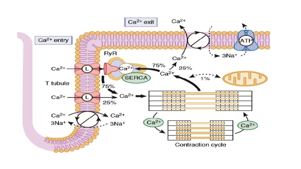

HEART RATE AND Na+/Ca 2+ EXCHANGE • NCX participates in the force-frequency relationship (Treppe or Bowditch phenomenon). An increasing heart rate (independent of sympathetic activation) increases the amount of Na+ and Ca 2+ entry per unit time and also diminishes the time available for extrusion of Na+ and Ca 2+. this will tend to increase the amount of Ca 2+ in the SR simply because of more frequent ICa pulses and less time for removal of Ca 2+ from the cell. Moreover, the elevation in [Na+]i also limits the ability of NCX to extrude Ca 2+ further increases the amount of Ca 2+ in the myocyte and SR • This NCX effect (once referred to as the “sodium pump lag” hypothesis) thus amplifies the intrinsic inotropic effect of an increase in heart rate.
 • During the normal heartbeat, Na+ enters the myocyte mainly")
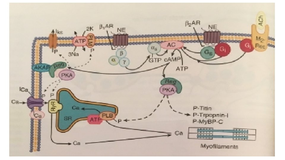
SODIUM PUMP (Na+/K+ ATPase) • During the normal heartbeat, Na+ enters the myocyte mainly via Na+ channels and NCX. Na+/H+ exchange also mediates significant Na+ influx, particularly when cells are acidotic. • In the steady state this Na+ influx is matched by an equal Na+ efflux, mediated mainly via sarcolemmal Na+/K+ATPase, or the Na+ pump. • The Na+ pump is activated by internal Na+ or external K+ and transports 3 Na+ ions out and 2 K+ ions in per ATP molecule used and is electrogenic and carries an outward current. • Na+ pump in the heart is modulated by the endogenous accessory protein phospholemman (PLM) • At baseline PLM reduces the intracellular Na+ affinity of Na+/K+-ATPase but when it is phosphorylated (by either PKA or protein kinase C [PKC]), that inhibitory effect is relieved. Thus during sympathetic activation, Na+/K+ATPase activity is increased at any given [Na+]i to better keep up with the higher rates of Na+ influx that occur under this condition.

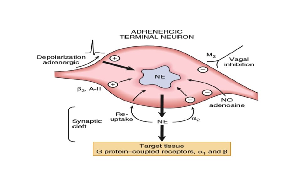
ADRENARGIC SIGNALING SYSTEM • During the classic adrenergic fight-or-flight response, cardiac myocyte betaadrenergic receptors are activated increased c. AMP production and PKA activation phosphorylation and altered function of numerous myocyte targets This results in an increase in the heart rate (positive chronotropy) increased contractility (positive inotropy) faster cardiac relaxation (positive lusitropy), enhanced conduction velocity (positive dromotropy) enhance cardiac output

• Increased HR and increased conduction is by acting on the SA node and conduction system. • Faster cardiac relaxation and increased contractility is by acting on the myocytes. • Increased alpha-adrenergic activity causes arteriolar constriction and increased resistance, although local metabolic control of arteriolar resistance is strong in the heart and dominates coronary resistance in arterioles. • Parasympathetic (vagal) innervation is strongest in the conduction system, where local release of acetylcholine (ACh) activates muscarinic receptors and tends to slow the heart rate and conduction velocity. In these conditions the heart rate and blood pressure fall. • Both alpha- and beta-adrenergic receptors are part of the family of seven– transmembrane domain G protein–coupled receptors (GPCRs).
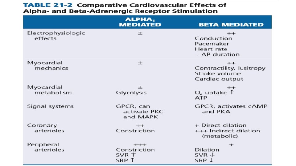

Beta-Adrenergic Receptor Subtypes • Cardiac beta-adrenergic receptors are chiefly the beta 1 subtype. • Beta 1 receptors are linked to the stimulatory G protein Gs, a component of the G protein–adenylyl cyclase system, beta 2 receptors are linked to both Gs and the inhibitory protein Gi, so their signaling pathway bifurcates at the very first postreceptor step. • In humans, the positive inotropic response to beta 2 stimulation by salbutamol occurs, at least in part, through beta 2 receptors on the terminal neurons of cardiac sympathetic nerves, thereby releasing norepinephrine, which in turn exerts dominant beta 1 effects. • Indirect evidence suggests that the Gi path is relatively augmented in heart failure whereas the strength of the Gs path is lessened because of uncoupling of Gs from the beta 2 receptor. • There also appear to be a small number of beta 3 -adrenergic receptors in cardiac myocytes that seem to produce more Gi-mediated negative inotropic signaling, mediated in part by NO. • The transmembrane domains are the site of agonist and antagonist binding, whereas the cytoplasmic domains interact with G proteins.

• Beta 1 stimulation: isoproterenol > epinephrine = nor-epinephrine • Beta 2 stimulation: isoproterenol > epinephrine > nor-epinephrine Alpha-Adrenergic Receptor Subtypes • There are two types of alpha-adrenergic receptors (alpha 1 and alpha 2). Those on the sarcolemma of vascular smooth muscle are vasoconstrictor alpha 1 receptors, whereas those situated on the terminal varicosities are alpha 2 -adrenergic receptors that feed back • Alpha 1 stimulation: nor-epinephrine > isoproterenol

G PROTEINS • G proteins are a superfamily of proteins that bind guanine triphosphate (GTP) and other guanine nucleotides. • The combination of the beta receptor, G protein complex, and adenylyl cyclase is the crux of beta-adrenergic signaling. • The G protein itself is a heterotrimer composed of Gα, Gβ, and Gγ, which on receptor stimulation splits into the alpha subunit that is bound to GTP and the beta -gamma subunit. • Either of these subunits may regulate different effectors such as adenylyl cyclase, phospholipase C, and ion channels. • The activity of adenylyl cyclase is controlled by two different G protein complexes, namely, Gs, which stimulates, and Gi, which inhibits. • The alpha subunit of Gs (αs) combines with GTP and then separates from the other two subunits to enhance the activity of adenylyl cyclase. The beta and gamma subunits (beta-gamma) appear to be linked structurally and functionally.

• The Inhibitory G Protein Gi in contrast is a second trimeric GTP-binding protein, Gi, is responsible for inhibition of adenylyl cyclase. • During stimulation of muscarinic and some beta 2 -adrenergic receptors, GTP binds to the inhibitory alpha subunit αi. The latter then dissociates from the other two components of the G protein complex, which are, as in the case of Gs, the combined beta-gamma subunits. • By stimulating the enzyme guanosine triphosphatase (GTPase), they break down the active αs subunit (αs-GTP) such that less activation of adenylyl cyclase occurs in response to alpha stimulation. • Furthermore, the beta-gamma subunit activates the KACh channel, which in turn can inhibit the SA node and thereby contribute to the bradycardic effect of cholinergic stimulation. • The major physiologic stimulus for Gi is thought to be vagal muscarinic receptor stimulation (although beta 2 adrenergic receptors may contribute as well).

• Adenosine interacts with A 1 receptors couples to Gi to inhibit contraction and the heart rate. • The adenosine A 2 receptor paradoxically increases c. AMP ( this effect, only of ancillary significance in the myocardium, is of major importance in vascular smooth muscle, where it induces vasorelaxation) • Pathologically, Gi is increased in experimental postinfarct heart failure and in donor hearts before cardiac transplantation.

Cyclic Adenosine Monophosphate and Protein Kinase A • Adenylyl cyclase catalyzes formation of the second messenger c. AMP. • Several isoforms exist, but AC 5 and AC 6 are most prominent in cardiac myocytes. • Adenylyl cyclase is the only enzyme system that produces c. AMP, and it requires just low concentrations of ATP and Mg 2+ as substrate. • It is a transmembrane enzyme, with most mass on the cytoplasmic side where G proteins interact. Cyclic guanosine monophosphate (c. GMP) is a related second messenger that often antagonizes c. AMP effects. • A number of hormones or peptides like glucagon, thyroid hormone, prostacyclin, and calcitonin gene–related peptide can couple to myocardial adenylyl cyclase independently of the beta-adrenergic receptor.
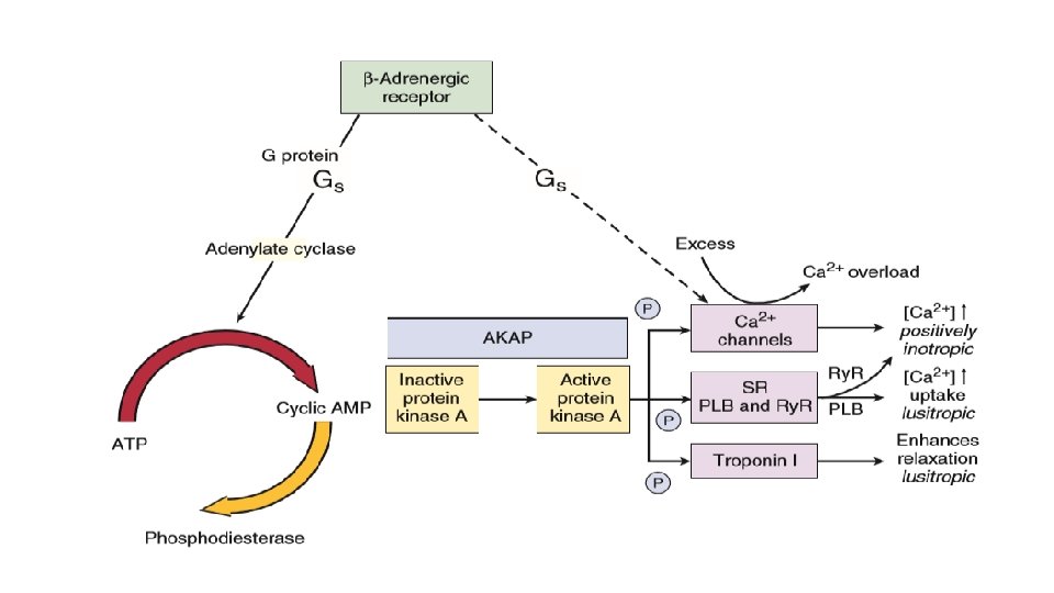

• In addition, myosin binding protein C is also a target for PKA and its phosphorylation appears to be responsible for accelerating the crossbridge turnover rate. • There is also a GTP exchange protein directly activated by c. AMP (Epac) that is activated in parallel to c. AMP dependent PKA activation. This allows additional parallel signalling downstream of betaadrenergic activation. For example, beta- adrenergic activation of SR Ca 2+ release is mediated by c. AMP-Epac-dependent signalling to Ca. MKII and consequent Ry. R 2 phosphorylation.

BETA- ADRENARGIC RECEPTOR DESENSITIZATION

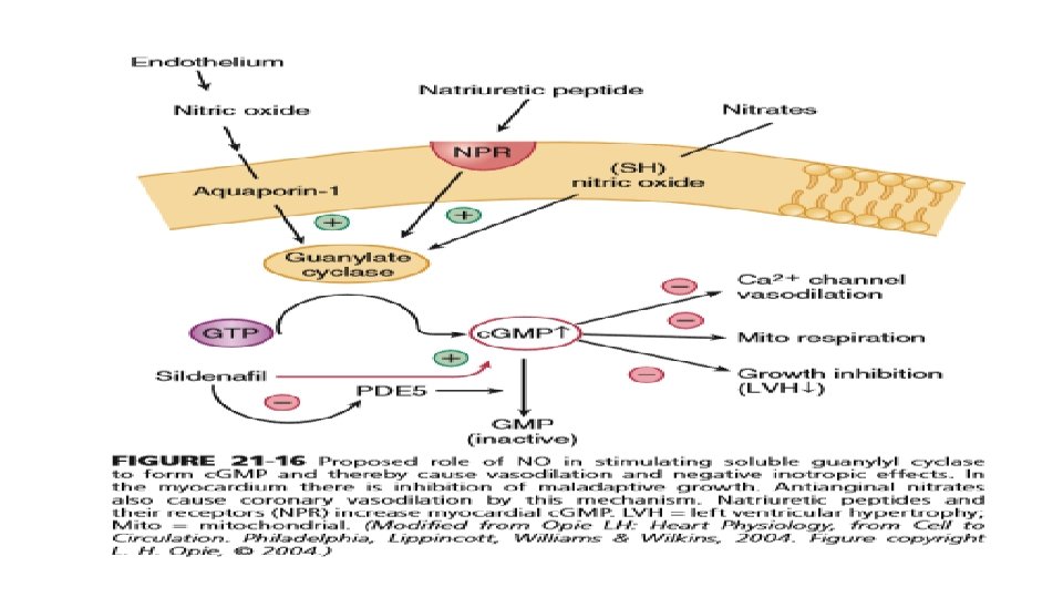
CHOLINERGIC AND NITRIC OXIDE SIGNALING • Parasympathetic stimulation reduces the heart rate and is negatively inotropic. • As in adrenergic signaling, there is an extracellular messenger (ACh), a GPCR (the cholinergic muscarinic receptor), and a sarcolemmal signaling system (G protein system, specifically Gi). The myocardial muscarinic receptor (M 2) is a GPCR associated with the activity of vagal nerve endings. • Receptor stimulation produces a negative chronotropic response that is inhibited by atropine. NO, also formed by beta 3 signaling facilitates cholinergic signaling at two levels, the nerve terminal and the activity of the enzyme system that produces the second messenger c. GMP. Neuregulins are growth factors that maintain the activity of this muscarinic receptor.

• Muscarinic Gi activation also inhibits adenylyl cyclase and thereby reduces the resultant c. AMP being produced by the ambient sympathetic tone and hence results in slowing of the HR. • Vagal innervation in the heart is highest in the SA and atrioventricular (AV) nodes, with lower density in atrial myocardium and the lowest density in ventricular myocardium. • Activation of M 2 muscarinic receptors results in activation of the coupled Gi and consequent activation of the ACh-activated K+ current (IK[ACh]). This increased K+ conductance causes more negative diastolic potential in pacemaker cells and also hinders the rate of diastolic depolarization. These factors slow the SA node pacemaker firing rate down and hence the heart rate.

THANK YOU
- Slides: 57