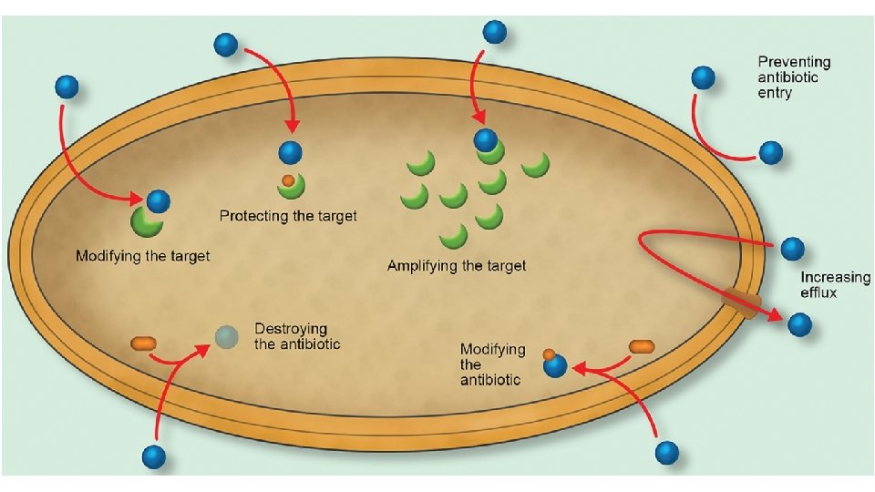
Mechanisms of antibiotic resistance The main mechanisms of

Mechanisms of antibiotic resistance The main mechanisms of resistance: • 1. Impermeability: Where the antibiotic fails to get into the bacterial cell. • 2. Efflux pumps: Where the antibiotic is removed from the bacterial cell faster then it can enter. • 3. Destruction: Where the antibiotic is destroyed so that an active drug is no longer intact and thus cannot attack its normal target. • 4. Modification: Where the antibiotic is modified so that an active drug is no longer intact and thus cannot attack its normal target.

• 5. Alteration of target: When the target of the antibiotic is modified so that the drug can no longer bind to it. • 6. Additional target: The production of a second target, which is usually less sensitive to binding by the antibiotic and thus provides a by-pass to the original inhibition. • 7. Hyper-production of target: When the target is produced in larger quantities than normal, thus the excess target molecules mop-up the available antibiotic. • This mechanism is often used in conjunction with one of the others.

 By prevention of the antimicrobial from reaching its target by reducing")
• (1) By prevention of the antimicrobial from reaching its target by reducing its ability to penetrate into the cell • Antimicrobial compounds almost always require access into the bacterial cell to reach their target site where they can interfere with the normal function of the bacterial organism. • Porin channels are the passageways by which these antibiotics would normally cross the bacterial outer membrane. • Some bacteria protect themselves by prohibiting these antimicrobial compounds from entering past their cell walls.

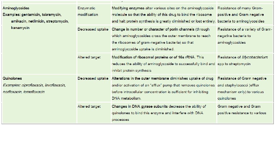
• For example, a variety of Gram-negative bacteria reduce the uptake of certain antibiotics, such as aminoglycosides and beta lactams, by modifying the cell membrane porin channel frequency, size, and selectivity. • Prohibiting entry in this manner will prevent these antimicrobials from reaching their intended targets that, for aminoglycosides and beta lactams, are the ribosomes and the penicillin-binding proteins (PBPs), respectively. • This strategy have been observed in: • Pseudomonas aeruginosa against imipenem (a beta-lactam antibiotic) • Enterobacter aerogenes and Klebsiella spp. against imipenem • Vancomycin intermediate-resistant S. aureus or VISA strains with thickened cell wall trapping vancomycin • Many Gram-negative bacteria against aminoglycosides • Many Gram-negative bacteria against quinolones
 By expulsion of the antimicrobial agents from the cell via general")
• 2) By expulsion of the antimicrobial agents from the cell via general or specific efflux pumps • To be effective, antimicrobial agents must also be present at a sufficiently high concentration within the bacterial cell. • Some bacteria possess membrane proteins that act as an export or efflux pump for certain antimicrobials, extruding the antibiotic out of the cell as fast as it can enter. • This results in low intracellular concentrations that are insufficient to elicit an effect.

• Efflux pumps are variants of membrane pumps possessed by all bacteria, both pathogenic and non-pathogenic, to move lipophilic or amphipathic molecules in and out of the cells. • Some are used by antibiotic producers to pump antibiotics out of the cells as fast as they are made, and so constitute an immunity protective mechanism for the bacteria to prevent being killed by their own chemical weapons

• Some efflux pumps selectively extrude specific antibiotics such as macrolides, lincosamides, streptogramins and tetracyclines, whereas others (referred to as multiple drug resistance pumps) expel a variety of structurally diverse anti-infectives with different modes of action. • This strategy has been observed in: • E. coli and other Enterobacteriaceae against tetracyclines • Enterobacteriaceae against chloramphenicol • Staphylococci against macrolides and streptogramins • Staphylococcus aureus and Streptococcus pneumoniae against fluoroquinolones
 By inactivation of antimicrobial agents via degradation • Another means by which bacteria")
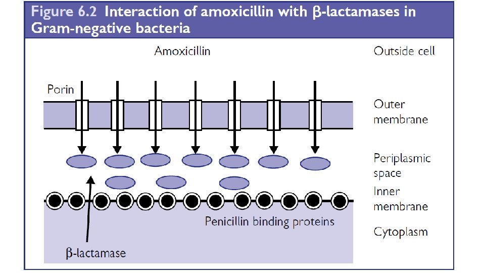
(3) By inactivation of antimicrobial agents via degradation • Another means by which bacteria preserve themselves is by destroying the active component of the antimicrobial agent. • A classic example is the hydrolytic deactivation of the beta-lactam ring in penicillins and cephalosporins by the bacterial enzyme called beta lactamase. • The inactivated penicilloic acid will then be ineffective in binding to PBPs (penicllin binding proteins), thereby protecting the process of cell wall synthesis.
")
• This strategy has also been observed in: • Enterobacteriaceae against chloramphenicol (acetylation) • Gram negative and Gram-positive bacteria against aminoglycosides (phosphorylation, adenylation, and acetylation) • Note: The first evidence of antimicrobial resistance • The first antibiotic resistance mechanism described was that of penicillinase. Its presence and activity was first reported by Abraham and Chain in 1940 shortly after its discovery (Abraham, E. P. and E. Chain. 1940. An enzyme from bacteria able to destroy penicillin. Nature 146: 837)

Figure: Example of Beta-lactamases for destruction of beta-lactam antibiotics: βlactamases are enzymes that can hydrolyse and inactivate various members of β-lactam drugs (penicillins, cephalosporins, and carbapenems). The β-lactamase hydrolyses the carbon-nitrogen bond of the β-lactam ring. The integrity of this ring is crucial to the activity of the antibiotic.

• In Gram-positive bacteria, the β-lactamase is produced within the cytoplasm of the cell and is exported through the cell membrane into the surrounding medium. • It provides a drug-free blanket around the cell. • It also provides protection for other microorganisms in close proximity. • In Gram-negative bacteria, the antibiotic has more difficulty in penetrating. • The β-lactamase is still produced in the cytoplasm; however, most of it is exported only as far as the periplasmic space, between the two membranes. • It is here that the β-lactamase intercepts the incoming β-lactam drug and destroys it. • It has recently been suggested that there are over 750 β-lactamases found in clinical bacteria. • They have conveniently been classified by their molecular structure into four groups (Classes A, B, C, and D).

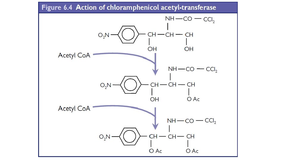
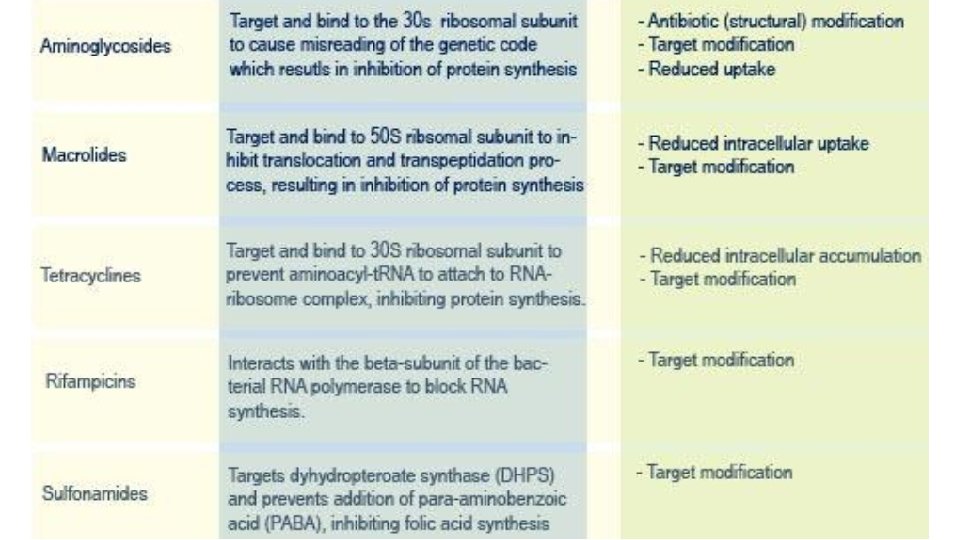
4. Modification of antibiotic • The consequence of modification on an antibiotic has the same effect as destruction, an inactive drug is created and it can no longer inhibit its target. • Essentially the plasmids encode a gene that adds a functional • group to the antibiotic. • The enzyme can have one of three actions: • 1. Acetyl transferase: The enzyme adds an acetyl group. • 2. Adenyl-tranferase: The enzyme adds an adenyl group. • 3. Phospho-transferase: The enzyme adds a phosphate group. • The main antibiotics that are modified in this manner are chloramphenicol and the aminoglycosides.
 By modification of the antimicrobial target within the bacteria • Some resistant bacteria")
(4) By modification of the antimicrobial target within the bacteria • Some resistant bacteria evade antimicrobials by reprogramming or camouflaging critical target sites to avoid recognition. • Therefore, in spite of the presence of an intact and active antimicrobial compound, no subsequent binding or inhibition will take place.

This strategy has been observed in: • Staphylococci against methicillin and other beta-lactams (Changes or acquisition of different PBPs that do not sufficiently bind betalactams to inhibit cell wall synthesis. ) • Enterococci against vancomycin (alteration in cell wall precursor components to decrease binding of vancomycin) • Mycobacterium spp. : • against streptomycin (modification of ribosomal proteins or of 16 s r. RNA); Mutations in RNA polymerase resulting in resistance to the rifamycins; Mutations in DNA gyrase resulting in resistance to quinolones

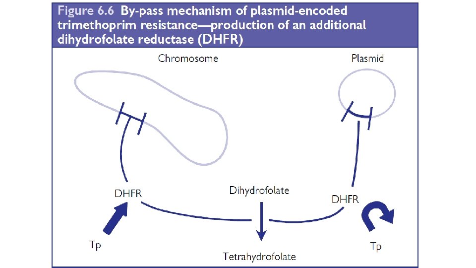
5. Additional target • This is usually a plasmid-mediated mechanism of resistance. • The antibiotic binds to its normal target but the plasmid produces an • additional target, which is less susceptible to the antibiotic. • This mechanism of resistance can only work if the quantity required of the product of the inhibited step is low. • This is the case for the co-factor tetrahydrofolate as trimethoprim selectively inhibits bacterial dihydrofolate reductase.

• The plasmid produces an additional dihydrofolate reductase that cannot readily bind the drug but can still reduce dihydrofolate to tetrahydrofolate. • Often the plasmid-encoded enzyme binds the drug around 10, 000 fold less effectively than the chromosomal enzyme and there is a corresponding increase in resistance on the host bacterium

6. Hyper-production of target • Chromosomal dihydrofolate reductase can be hyperproduced 100 fold so that it is able to bind many trimethoprim molecules. • Even if 99% of the produced dihydrofolate reductase molecules are inhibited by trimethoprim, there are enough enzyme molecules to reduce dihydrofolate to provide sufficient tetrahydrofolate for the cell’s needs. • It is a highly expensive mechanism of resistance in energy so host cell has a selective disadvantage once the antibiotic has been removed.

• Mechanisms of Resistance Against Different Antimicrobial Classes

• ANTIBIOTIC MODES OF ACTION AND BACTERIAL MECHANISMS OF RESISTANCE
- Slides: 26