Mechanism of Hormone Action Dr Jehad AlShuneigat The


 2 - β-subunit 3")


")


 • G proteins also uses the phosphoinositide cascade")
 (IP 3) Phosphatidylinositols in cellular regulation. Phosphatidylinositol 4, 5 -bisphosphate in the")


 to an")


- Slides: 47
Mechanism of Hormone Action Dr Jehad Al-Shuneigat
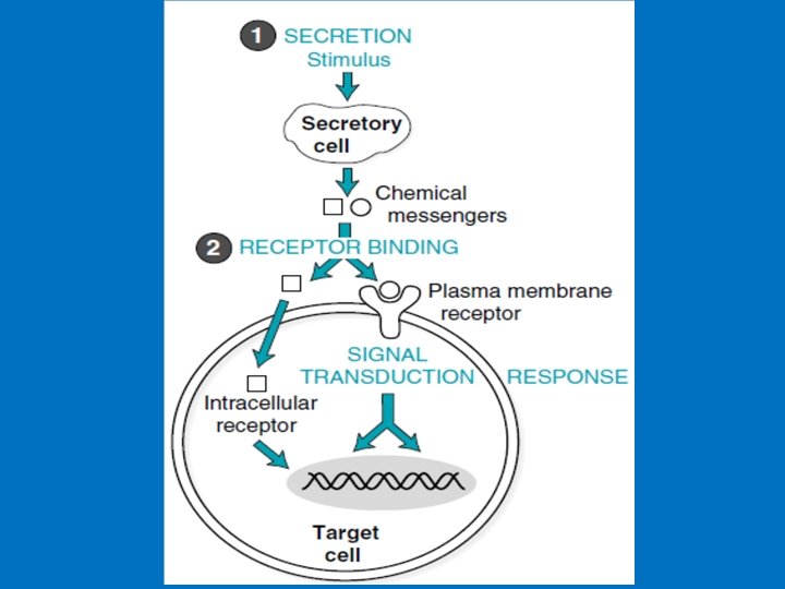
• The first step of hormone action is binding of the hormone to its appropriate receptor protein. • The hormone receptors have the following characteristics: • (a) high specificity, so that the receptors can recognize their specific ligand discriminate between the incoming signals; • (b) high affinity for their ligand. • The physical binding of hormone to receptors is dependent on several weak chemical forces including: hydrogen bond, van der waals forces, ionic bond.
• Receptors are proteins containing a binding site specific for a single chemical messenger and another binding site involved in transmitting the message. • Classification of receptors based on their location 1 - Plasma-membrane receptors: The peptide and protein hormones and the catecholamine hormones are hydrophilic and can’t inter inside cells thus their receptors are found on cell surface. Plasma-membrane receptors transmit their signals through second messenger that are generated intracellularly 2 -Intracellular receptors: found in the cytoplasm or inside nucleus and function in the nucleus as transcription factors to alter the rate of transcription of particular genes. • The steroid hormones and thyroid hormones are hydrophobic and diffuse freely across the plasma membrane and thus their receptors are intracellular receptors. Plasma membrane receptors categories A- G-proteins receptors B- Ion channel receptors, C- Tyrosine kinase receptors, tyrosine-kinase associated receptors (JAK-STAT receptors), or serine-threonine kinase receptors.
• 1 - Plasma-membrane receptors • Structure of Plasma-membrane receptors: 1 - Extracellular component (N-terminal) which binds the hormone. 2 - One or more than one membrane spanning region that are α -helix, this varies in structure from a simple linear hydrophobic region to a more complex eg G-receptors. 3 - Cytosolic domain (C-terminal) which initiate the intracellular signaling cascade (Signal transduction). • Mechanisms of signal transduction that follow the binding of signaling molecules to plasma membrane receptors include: 1 - Phosphorylation of receptors at tyrosine residues (receptor tyrosine kinase activity), 2 - Conformational changes in signal transducer proteins (e. g. , proteins with SH 2 domains, the monomeric G protein Ras, heterotrimeric G proteins) 3 - Increases in the levels of intracellular second messengers Examples: c. AMP, inositol trisphosphate (IP 3), and diacylglycerol (DAG).
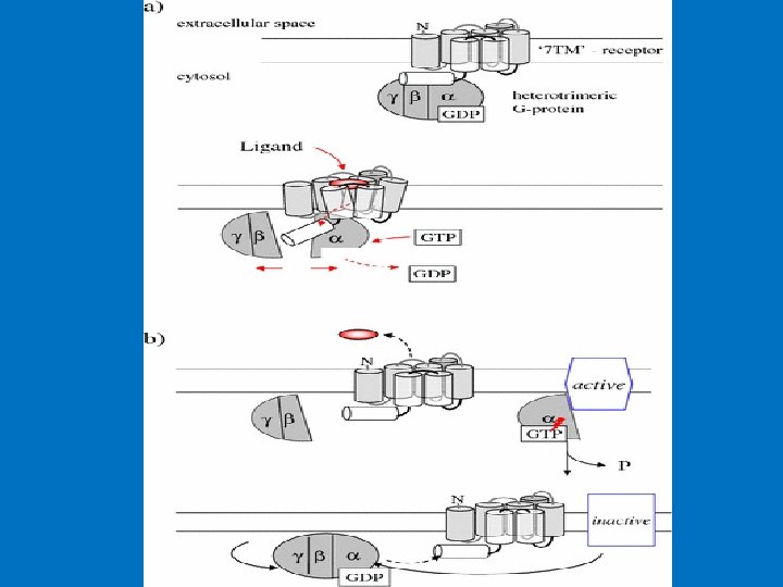
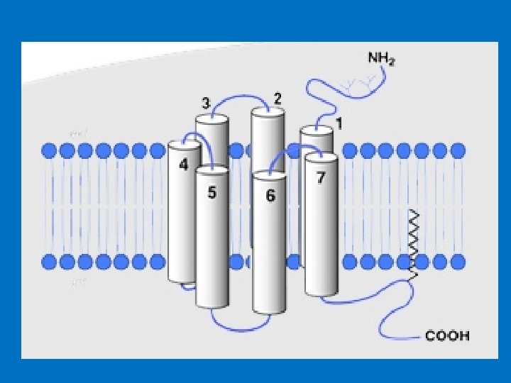
• We will study two major groups of cell surface receptors: • 1 - G-protein coupled receptors (GPCRs); 2 - Tyrosine kinase receptors • G-protein coupled receptors (GPCRs) • They are the largest family of cell-surface molecules involved in signal transmission. • They are activated by a wide variety of ligands, including hormones, neurotransmitters, growth factors, odorant molecules and light, • G-protein-coupled receptors (GPCRs) function is to transduce extracellular stimuli into intracellular signals. • G proteins are so named because they bind to and are regulated by nucleotides with a guanine base: GDP and GTP. When G protein is bound to GDP it is inactive while once activated it binds GTP. • G protein has two important binding sites: one link it to receptor and the other link it to the nucleotide GDP/GTP. • The G protein–coupled receptors consist of 1 - an extracellular N-terminal domain; 2 - seven membrane-spanning α-helix domains, each made up of 20– 30 amino acids, connected by three extracellular (E 1 -E 3) and three intracellular (C 1 -C 3) polypeptide loops; a disulfide bridge connects E-II and E-III loops 3 - intracellular, C-terminal cytoplasmic tail that interact with the G-protein.
structure of G protein–coupled receptors. All receptors of this type have the same orientation in the membrane and contain seven transmembrane -helical regions (H 1–H 7)
• Structural of G proteins 1 - α-subunit (Large) 2 - β-subunit 3 - γ-subunit. The β- and γ-subunits exist as a tightly associated complex and are active in this form. The α- and γ-subunits are associated with the cell membrane via lipid anchors. • The α-subunit has a binding site for GTP or GDP and carries the GTPase activity. • Most G-protein activity is determined by the α-subunit that interacts with the effector molecules. • The βγ-complex is also involved in signal transmission to the effector proteins. • Since different G proteins interact with different hormones, there are significant differences in the structure of the α-subunits. • Based on comparison of the amino acid sequences, the Gα proteins are divided into four families, the Gs, Gi, Gq and G 12 families.
Structural of G proteins
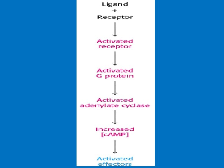
Activation of G protein–coupled receptors Signal transduction 1 - Hormone binding to its receptor 2 - Change in the conformation of the receptor that may involve disruption of a strong ionic interaction between the third and sixth transmembrane helices 3 - This facilitates activation of the G-protein and the release of GDP and the binding of GTP to α subunits which result in decreased affinity of Gα for Gβγ. 4 - GTP-bound α subunit & separate from βγ subunits, and diffusing in the plane of the lipid bilayer until it encounters an effector protein to which it can bind. 5 - An effector protein causes a specific effect in the cell through activation or inhibition of a variety of second messengers. Example of effectors: adenylyl and guanylyl cyclases (produces second messenger c. AMP from ATP and c. GMP from GTP), phospholipases, phosphodiesterases, and ion channels for calcium or potassium.
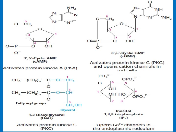
• Second messenger • They are nonprotein molecules generated inside the cell in response to hormone binding that continue transmission of the message. • Examples include 3', 5'-cyclic AMP (c. AMP), 3', 5'-cyclic GMP (c. GMP), inositol 1, 4, 5 trisphosphate (IP 3), diacylglycerol (DAG) and Ca 2+ • The second-messengers are the ones that bring out the cellular responses. • The use of second messengers has several consequences. • First, second messengers are often free to diffuse to other compartments of the cell where they can influence gene expression and other processes. • Second, the signal may be amplified significantly in the generation of second messengers. Thus, a low concentration of signal in the environment, even as little as a single molecule, can yield a large intracellular signal and response. • We will study: • 1 - c. AMP; 2 - c. GMP; 3 - IP 3 4 - DAG 5 - Calcium ions
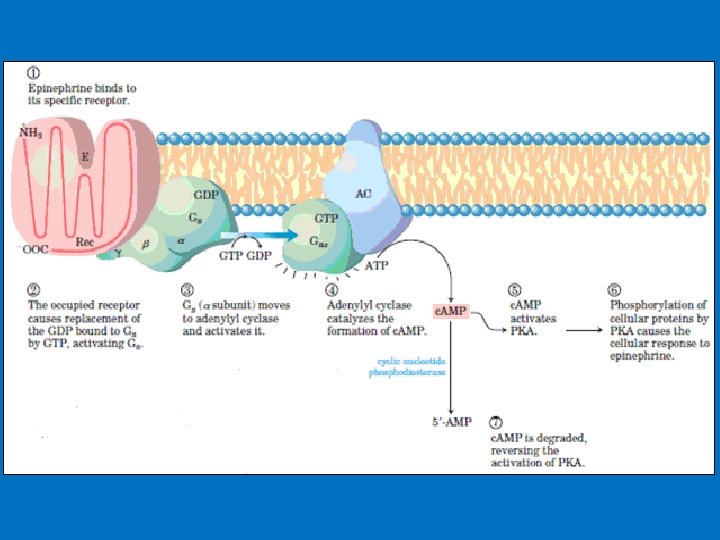
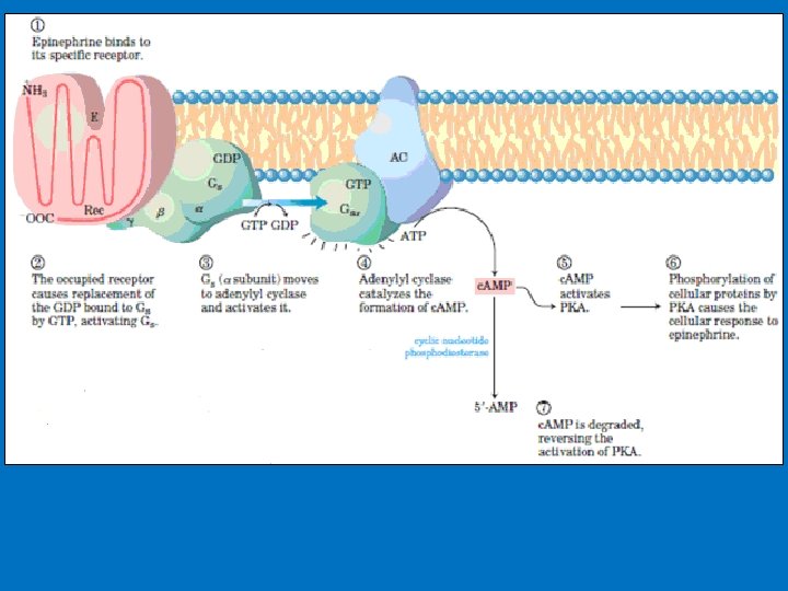
• 1 - c. AMP production and Adenylyl Cyclase • Adenylyl cyclase is a membrane-bound enzyme that converts ATP to c. AMP. • Different peptide hormones can either stimulate (s) or inhibit (i) the production of c. AMP from adenylyl cyclase. • Two parallel systems, a stimulatory (s) one and an inhibitory (i) one, converge upon a single catalytic molecule (C). Each consists of a receptor (Rs or Ri) and a regulatory complex, Gs and Gi. c. AMP effects in cells including 1 -Activation of protein kinase A which is a serine/threonine protein kinase that phosphorylates a large number of metabolic enzymes 2 - Enter the nucleus and phosphorylate a gene-specific transcription factor thus affecting gene transcription. 3 - Directly activates ligand-gated channels.
Hormone-induced activation and inhibition of adenylyl cyclase in adipose cells (prostaglandins)
• Phosphorylation • c. AMP activate enzymes called c. AMP-dependent protein kinases, example protein kinase A (PKA). • Phosphorylation reaction involve the transfer of a phosphate from the phosphate donor (ATP) to the acceptor protein. • In mammalian proteins serine, threonine, and tyrosine residue are the phosphate acceptors residues. • Serine/threonine protein kinases transfer a phosphate from ATP to the hydroxyl group of a specific serine (and sometimes threonine) on the target enzyme; tyrosine kinases transfer a phosphate to the hydroxyl group of a specific tyrosine residue. • Phosphate is a bulky, negatively charged residue that interacts with other nearby amino acid residues of the protein to create a conformational change at the catalytic site. The conformational change makes certain enzymes more active and other enzymes less active. The effect is reversed by a specific protein phosphatase that removes the phosphate by hydrolysis.
2 - c. GMP is also an intracellular signal • Cyclic GMP (guanosine 3', 5'-cyclic monophosphate) is made from GTP by the enzyme guanylyl cyclase • Cyclic GMP is a second messenger that carries different messages in different tissues. • In the kidney and intestine it triggers changes in ion transport and water retention; in cardiac muscle it signals relaxation; in the brain it may be involved both in development and in adult brain function. • Guanylyl cyclases in the kidney is activated by the hormone atrial natriuretic factor (ANF), which is released by cells in the atrium of the heart when the heart is stretched by increased blood volume. Carried in the blood to the kidney, ANF activates guanylyl cyclase in cells of the collecting ducts. The resulting rise in (c. GMP) triggers increased renal excretion of Na+ and, consequently, of water, driven by the change in osmotic pressure. Water loss reduces the blood volume, countering the stimulus that initially led to ANF secretion. • Vascular smooth muscle also has an ANF receptor—guanylyl cyclase; on binding to this receptor, ANF causes relaxation (vasodilation) of the blood vessel, which increases blood flow while decreasing blood pressure.
• Phosphodiesterases • Phosphodiesterase converts c. AMP to AMP, and c. GMP to GMP thereby decreasing c. AMP and c. GMP levels and inactivating protein kinase A. • Phosphodiesterase resides in the plasma membrane. • The presence of these enzymes ensures a rapid turnover of the signal (c. AMP) and hence a rapid termination of the biologic process once the hormonal stimulus is removed. • Some hormones change the concentration of c. AMP by targeting the phosphodiesterase enzyme rather than adenylyl cyclase. For example, insulin lowers c. AMP levels by causing phosphodiesterase activation.
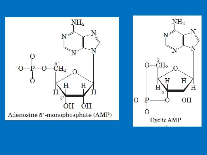
Formation and cleavage of the cyclic phosphodiester bond in c. AMP. When activated, adenyl cyclase converts ATP to 3, 5 - cyclic AMP PPi. c. AMP phosphodiesterase hydrolyzes c. AMP to AMP.
Example: c. AMP production due to adrenalin • Epinephrine and norepinephrine hormones prepares our body for fight or flight. • These hormones increase fuel mobilization, cardiac output, blood flow, etc. , which enable us to meet these stresses. They bind to adrenergic receptors. • There are nine different types of adrenergic receptors: α 1 A, α 1 B, α 1 D, α 2 A, α 2 B, α 2 C, β 1, β 2, and β 3. • adrenergic receptors are class of GPCR for catecholamine hormones. • The three β receptors work through the Adenylyl cyclase–c. AMP system and eventually protein kinase A.
• The β 1 receptor is the major adrenergic receptor in the human heart. • Contraction is initiated by the release of calcium from intracellular stores, whereas relaxation occurs as the calcium is re-sequestered in the sarcoplasmic reticulum, which in part mediated by the SERCA 2 a pump protein. • The SERCA 2 a pump is regulated, in part, by its association with the protein phospholamban (PLN). • PLN reduces SERCA 2 a pumping activity. • Epinephrine binds to its receptor, activating a G protein, which leads to adenylyl cyclase activation, elevation of c. AMP levels, and activation of protein kinase A. PKA phosphorylates PLN, thereby reducing its association with SERCA 2 a and relieving the inhibition of pumping activity. • The β 2 receptor is present in liver, skeletal muscle, and other tissues and is involved in the mobilization of fuels (such as the release of glucose through glycogenolysis). It also mediates vascular, bronchial, and uterine smooth muscle contraction. • The β 3 receptor is found predominantly in adipose tissue and to a lesser extent in skeletal muscle. Activation of this receptor stimulates fatty acid oxidation and thermogenesis, and agonists for this receptor may prove to be beneficial weight loss agents.
Examples continue • Epinephrine hormone causes glucose mobilization for energy and muscle contraction through binding to its G protein-linked receptor that leads to activation of protein kinase A (PKA) which: 1 - Phosphorylate and thereby activate an enzyme that activates glycogen phosphorylase which in turn breaks down glycogen into glucose-l-phosphate molecules. 2 - It phosphorylates glycogen synthase, and in this way turns it off, thereby preventing the reconversion of the released glucose to glycogen. These two changes together ensure the mobilization of glucose through the breakdown of glycogen stored in the liver.
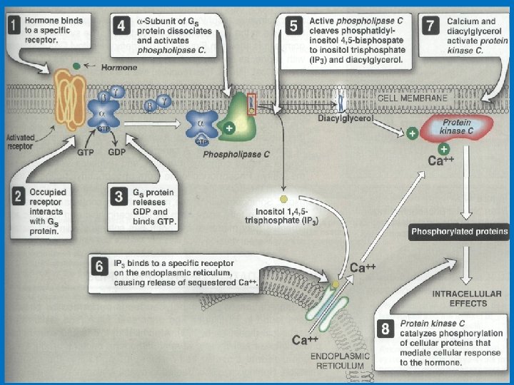
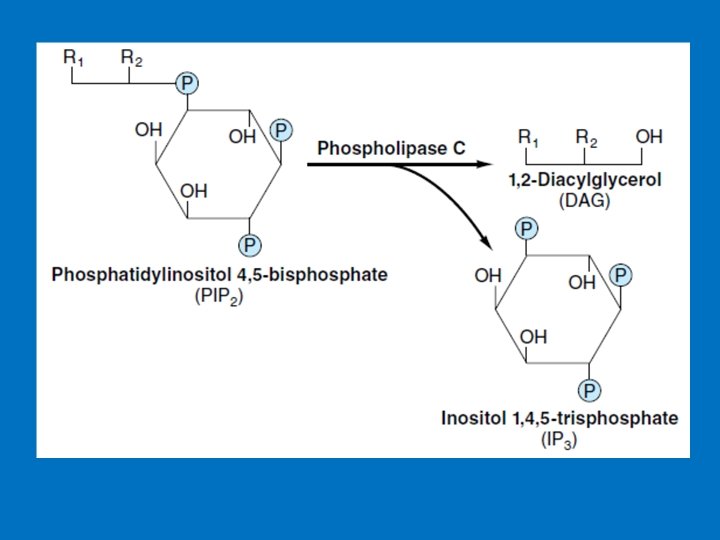
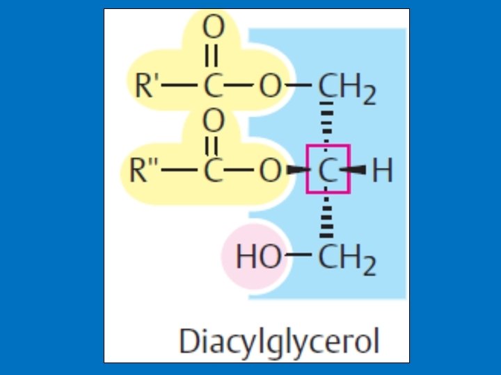
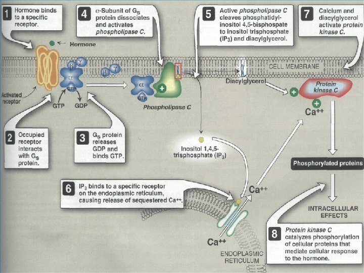
• 3 - Phosphatidylinositol (PI) • G proteins also uses the phosphoinositide cascade as a second messenger. • Phosphatidylinositol (PI) is a phospholipid in cell membrane its phosphorylation produces polyphosphoinositides, for example, phosphatidylinositol 4, 5 -bisphosphate (PIP 2). • The cleavage of PIP 2 by phospholipase C produces inositol 1, 4, 5 -trisphosphate (IP 3) and diacylglycero (DAG). • IP 3 and DAG work as a second messenger • IP 3 is a soluble molecule therefore diffuse from the membrane to cytoplasm • Inositol is a carbohydrate that has about half sweetness of classical sugar • DAG is hydrophobic and therefore stays in the membrane. • IP 3 stimulates the release of calcium ions from the smooth endoplasmic reticulum, whereas DAG activates cyclicnucleotide-independent kinase (protein kinase C ‘PKC’). PKC can be activated by Ca 2+ and DAG.
(PIP 2) (IP 3) Phosphatidylinositols in cellular regulation. Phosphatidylinositol 4, 5 -bisphosphate in the plasma membrane is hydrolyzed by a specific phospholipase C in response to hormonal signals. Both products of hydrolysis act as intracellular messengers.
• IP 3 and Calcium • IP 3 causes the rapid release of Ca 2+ from intracellular stores in endoplasmic reticulum and, in smooth muscle cells. Ca 2+ act as intracellular messenger of hormone action. The elevated level of Ca 2+ in the cytosol triggers processes such as smooth muscle contraction, glycogen breakdown, and vesicle release. • IP 3 is able to increase Ca 2+ concentration by associating with a membrane protein called the IP 3 gated channel or IP 3 receptor. This receptor, which is composed of four large, identical subunits, forms an ion channel. At least three molecules of IP 3 must bind to sites on the cytosolic side of the membrane protein to open the channel and release Ca 2+.
IP 3 signaling calcium release from the endoplasmic reticulum
• Ca 2+ is a signalling substance • The concentration of Ca 2+ ions in the cytoplasm is normally very low (10– 100 n. M), as it is kept down by ATP driven Ca 2+ pumps and Na+/Ca 2+ exchangers. In addition, many proteins in the cytoplasm and organelles bind calcium and thus act as Ca 2+ buffers. • Specific signals, from second messenger such as IP 3 or c. AMP, can trigger a sudden increase in the cytoplasmic Ca 2+ level to 500– 1000 n. M by opening Ca 2+ channels in the plasma membrane or in the membranes of the endoplasmic or sarcoplasmic reticulum. In the cytoplasm, the Ca 2+ level always only rises very briefly (Ca 2+ “spikes”), as prolonged high concentrations in the cytoplasm have cytotoxic effects.
• 3 a-Calcium effects. • The biochemical effects of Ca 2+ in the cytoplasm are mediated by special Ca 2+ -binding proteins (“calcium sensors”) eg; calmodulin. • Calmodulin the binding of four Ca 2+ ions activate it into a regulatory element. • The Ca 2+-calmodulin complex can activate specific kinases which in turn regulate the activity of enzymes, ion pumps, and components of the cytoskeleton. • There are three ways of changing cytosolic Ca 2+: • (1) Certain hormones by binding to receptors that are themselves Ca 2+ channels, enhance membrane permeability to Ca 2+ and thereby increase Ca 2+ influx. • (2) Hormones also indirectly promote Ca 2+ influx by modulating the membrane potential at the plasma membrane. Membrane depolarization opens voltage-gated Ca 2+ channels and allows for Ca 2+ influx. • (3) Ca 2+ can be mobilized from the endoplasmic reticulum, and possibly from mitochondrial pools. An important observation linking Ca 2+ to hormone action involved the definition of the intracellular targets of Ca 2+ action.
• How is the IP 3 -initiated signal turned off? • IP 3 is a short-lived just a few seconds. 1 - Degraded to inositol by phosphatases 2 - Phosphorylated to inositol.
• 3 b- Diacylglycerol as a Signal Molecule • DAG is made up of two fatty acids and a glycerol. Diacylglycerol can be produced by different pathways, and it has at least two functions. 1 - It is an important source for the release of arachidonic acid (found in membrane), from which biosynthesis of prostaglandins takes place. 2 - Stimulation of protein kinase C. • Protein kinase C is a protein kinase occurring in almost all cells and has a regulating effect on many reactions of the cell. • Characteristic for protein kinase C is its stimulation by Ca 2+, diacylglycerol and phosphatidyl serine.
• Receptor Tyrosine Kinase • RTKs are the second major type of cell-surface receptors. • RTK signalling pathways have a wide spectrum of functions including regulation of cell proliferation and differentiation, promotion of cell survival, and modulation of cellular metabolism. • Misregulation of these networks results in a variety of human diseases, including cancer, diabetes, and immune deficiency. • Some RTKs have been identified in studies on human cancers associated with mutant forms of growth-factor receptors, which send a proliferative signal to cells even in the absence of growth factor. • One such mutant receptor, encoded at the neu locus, contributes to the uncontrolled proliferation of certain human breast cancers.
Ligand Binding Leads to Autophosphorylation of RTKs • RTKs structure: A- Extracellular domain containing a ligand-binding site, B- Single hydrophobic transmembrane α helix, C- Cytosolic domain that includes a region with protein-tyrosine kinase activity. There are two types of RTK receptors 1 - Single transmembrane spanning proteins which dimerise when ligand binds eg; platelet derived growth factor (PDGF) receptor 2 - Covalently linked dimers (eg; insulin receptor) • The binding of ligand causes most RTKs to dimerize which lead to protein kinase activation • Then each receptor monomer phosphorylates a distinct set of tyrosine residues in the cytosolic domain of its dimer partner, a process termed autophosphorylation.
Tyrosine kinases receptor
• The phosphotyrosine residues in activated RTKs interact with adapter proteins containing SH 2 or PTB domains which then catalyzes phosphorylation of other target proteins. • SH 2 (Src homology region 2) and PTB (phosphotyrosine-binding) domains are small protein modules that mediate protein-protein interactions involved in many signal transduction pathways. Both domains were initially identified as modules that recognize phosphorylated tyrosines in receptor tyrosine kinases and other signaling proteins.
Activation of Ras following binding of a hormone (e. g. , EGF) to an RTK The adapter protein GRB 2 binds to a specific phosphotyrosine on the activated RTK and to Sos, which in turn interacts with the inactive Ras · GDP. The guanine nucleotide – exchange factor (GEF) activity of Sos then promotes formation of the active Ras · GTP. Note that Ras is contected to the membrane by anchor.
Signal Termination 1 - The first level of termination is the chemical messenger itself. When the stimulus is no longer applied to the secreting cell, the messenger is no longer secreted, and existing messenger is catabolized. For example, many polypeptide hormones such as insulin are taken up into the liver and degraded. 2 - The receptor might be desensitized to the messenger by phosphorylation. G proteins automatically terminate messages as they hydrolyze GTP. • Termination also can be achieved through degradation of the second messenger (e. g. phosphodiesterase cleavage of c. AMP). Each of these terminating processes is also highly regulated. 3 - Protein phosphatases, enzymes that reverse the action of kinases by removing phosphate groups from proteins. Specific tyrosine or serine/threonine phosphatases (enzymes that remove the phosphate group from specific proteins) exist for all of the sites phosphorylated by signal transduction kinases. Some receptors are even protein phosphatases.
Second Messenger Examples of Hormones Which Utilize This System Cyclic AMP Epinephrine and norepinephrine, glucagon, luteinizing hormone, follicle stimulating hormone, thyroid-stimulating hormone, calcitonin, parathyroid hormone, antidiuretic hormone Insulin, growth hormone, prolactin, Protein kinase activity oxytocin, erythropoietin, several growth factors Epinephrine and norepinephrine, Calcium and/or angiotensin II, antidiuretic hormone, phosphoinositides gonadotropin-releasing hormone, thyroidreleasing hormone. Cyclic GMP Atrial naturetic hormone, nitric oxide