
Mechanical Ventilation for Nursing Melissa Dearing BS RRTNPS
Mechanical Ventilation for Nursing Melissa Dearing, BS, RRT-NPS, RCP Associate Professor of Respiratory Care Curtis Shelley, BS, RRT-NPS, RCP Respiratory Educator – Hermann Children’s Hospital
Indications for Mechanical Ventilation ü Airway Compromise – airway patency is in doubt or patient may be at risk of losing patency
Indications for Mechanical Ventilation Respiratory Failure – 2 Types üHypoxemic Respiratory Failure üHypercapnic Respiratory Failure
Hypoxemic Respiratory Failure üPa. O 2 < 60 mm. Hg in an otherwise healthy individual
Hypercapnic Respiratory Failure üPa. CO 2 > 50 mm. Hg in an otherwise healthy individual • AKA “Ventilatory Failure” • Caused by increased WOB, ↓ventilatory drive, or muscle fatigue
Indications for Mechanical Ventilation üNeed to Protect the Airway For some reason the patient’s ability to sneeze, gag or cough has been dulled and aspiration is possible.
Contraindications for an Artificial Airway When a pt’s desire to not be resuscitated has been expressed and is documented in the pt’s chart
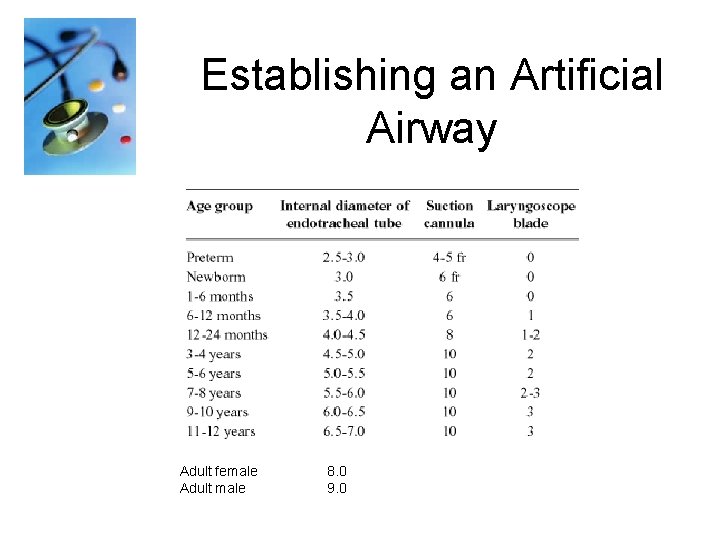
Establishing an Artificial Airway Adult female Adult male 8. 0 9. 0
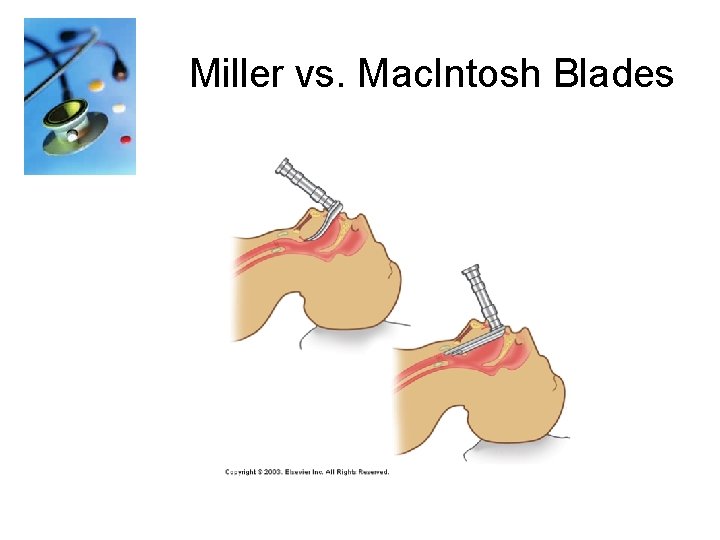
Miller vs. Mac. Intosh Blades
Intubation Procedure Check and Assemble Equipment: üOxygen flowmeter and O 2 tubing üSuction apparatus and tubing üSuction catheter or yankauer üAmbu bag and mask üLaryngoscope with assorted blades ü 3 sizes of ET tubes üStylet üStethoscope üTape üSyringe üMagill forceps üTowels for positioning
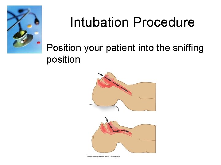
Intubation Procedure Position your patient into the sniffing position
Intubation Procedure Preoxygenate with 100% oxygen to provide apneic or distressed patient with reserve while attempting to intubate. Do not allow more than 30 seconds to any intubation attempt. If intubation is unsuccessful, ventilate with 100% oxygen for 3 -5 minutes before a reattempt.
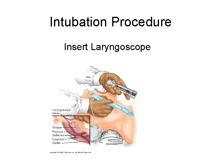
Intubation Procedure Insert Laryngoscope
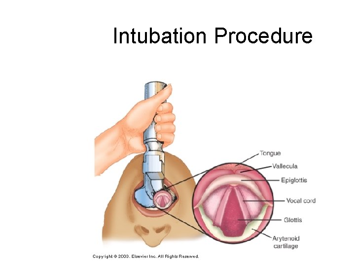
Intubation Procedure
Intubation Procedure After displacing the epiglottis insert the ETT. The depth of the tube for a male patient on average is 21 -23 cm at teeth The depth of the tube on average for a female patient is 19 -21 at teeth.
Intubation Procedure Confirm tube position: üBy auscultation of the chest üBilateral chest rise üTube location at teeth üCO 2 detector – (esophageal detection device)
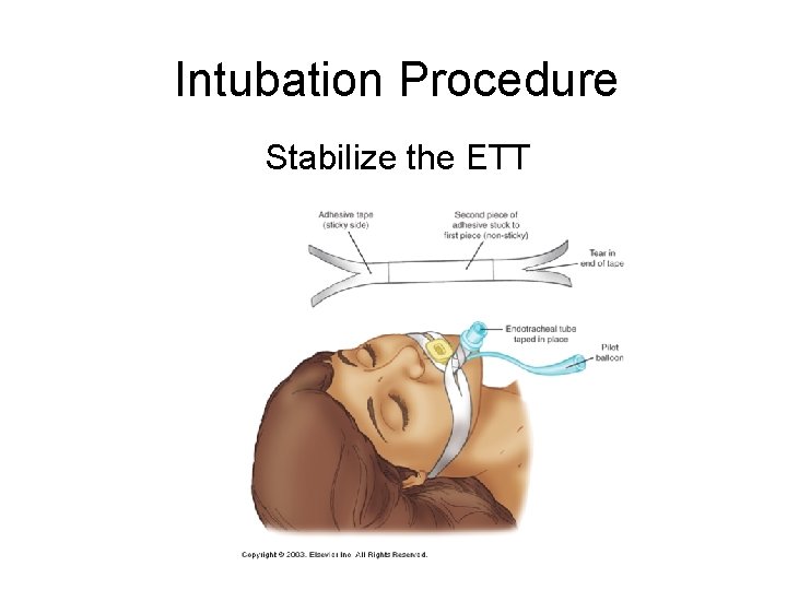
Intubation Procedure Stabilize the ETT
Intubation Procedure Video on Intubation: http: //youtube. com/watch? v=e. Rkley. IJi 9 U&fe ature=related
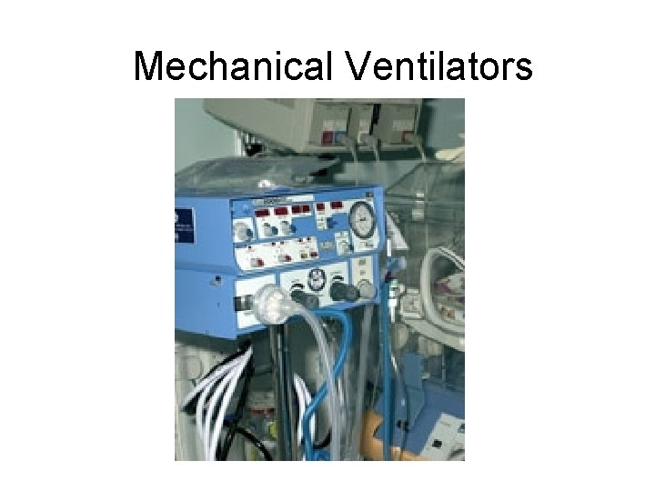
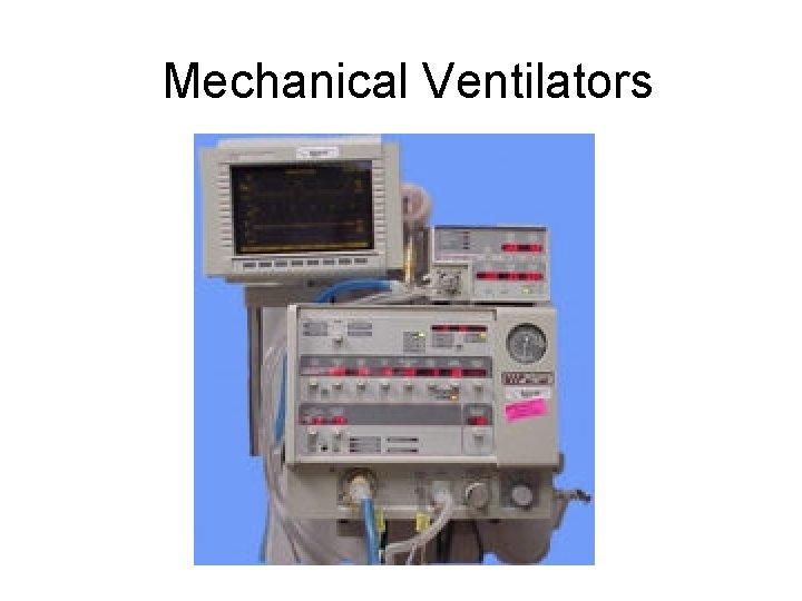
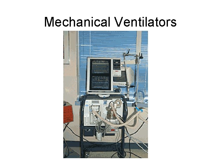
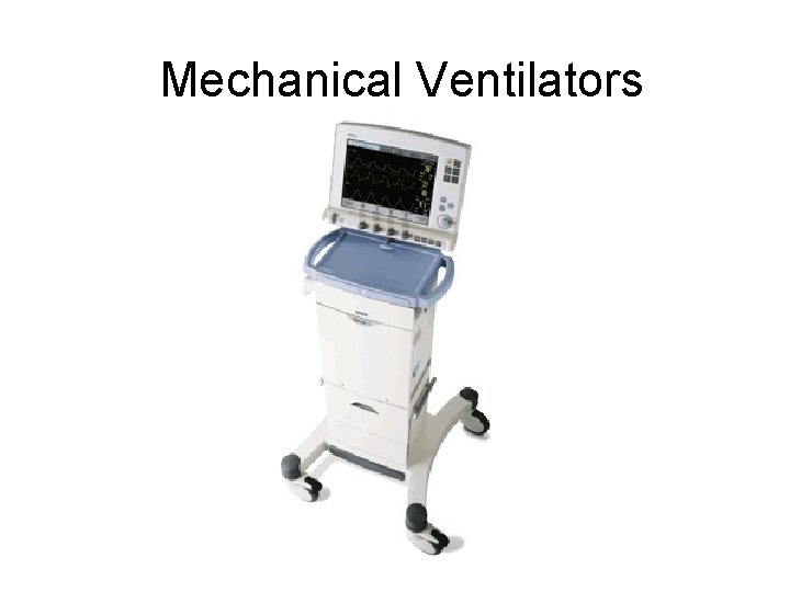
Mechanical Ventilators Different Types of Ventilators Available: Will depend on you place of employment
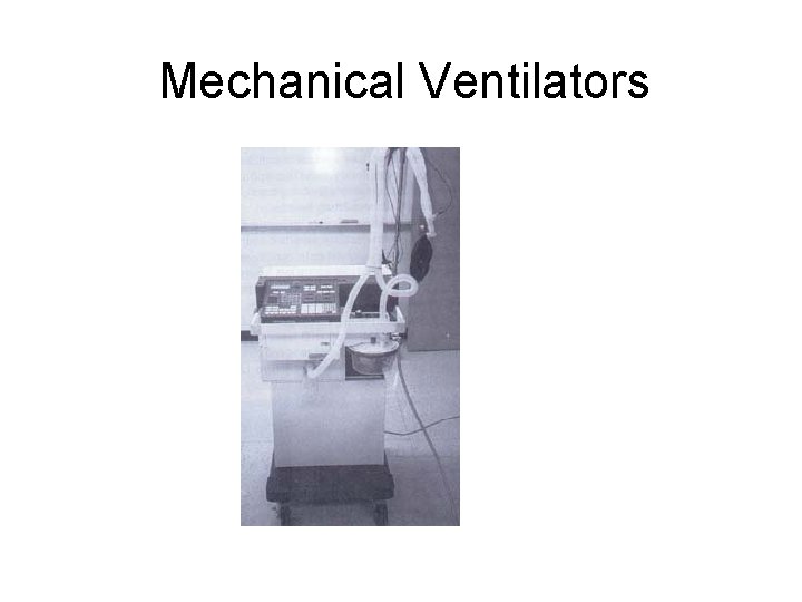
Mechanical Ventilators
Mechanical Ventilators
Mechanical Ventilators
Mechanical Ventilators
Mechanical Ventilators
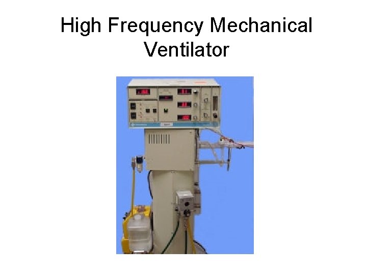
High Frequency Mechanical Ventilator
Ventilator Settings Terminology • A/C: Assist-Control • IMV: Intermittent Mandatory Ventilation • SIMV: Synchronized Intermittent Mandatory Ventilation • Bi-level/Biphasic: Non-inversed Pressure Ventilation with Pressure Support (consists of 2 levels of pressure)
Ventilator Settings Terminology (con’t) • PRVC: Pressure Regulated Volume Control • PEEP: Positive End Expiratory Pressure • CPAP: Continuous Positive Airway Pressure • PSV: Pressure Support Ventilation • NIPPV: Non-Invasive Positive Pressure Ventilation
VOLUME vs. PRESSURE VENTILATION ØVolume ventilation: Volume is constant and pressure will vary with patient’s lung compliance. ØPressure ventilation: Pressure is constant and volume will vary with patient’s lung compliance.
MODES of VENTILATION
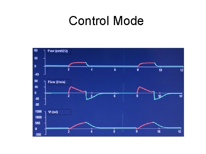
Control Mode Delivers pre-set volumes at a pre-set rate and a pre-set flow rate. The patient CANNOT generate spontaneous breaths, volumes, or flow rates in this mode.
Control Mode
Assist/Control Mode • Delivers pre-set volumes at a preset rate and a pre-set flow rate. • The patient CANNOT generate spontaneous volumes, or flow rates in this mode. • Each patient generated respiratory effort over and above the set rate are delivered at the set volume and flow rate.
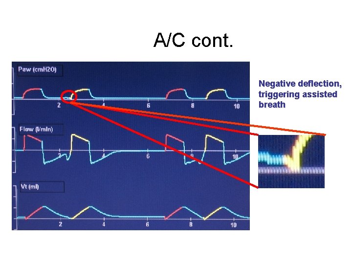
A/C cont. Negative deflection, triggering assisted breath
SYCHRONIZED INTERMITTENT MANDATORY VENTILATION (SIMV): üDelivers a pre-set number of breaths at a set volume and flow rate. üAllows the patient to generate spontaneous breaths, volumes, and flow rates between the set breaths. üDetects a patient’s spontaneous breath attempt and doesn’t initiate a ventilatory breath – prevents breath stacking
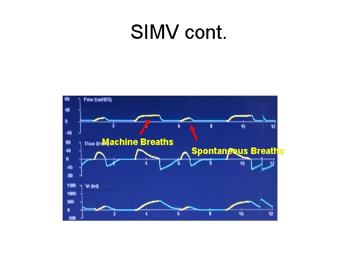
SIMV cont. Machine Breaths Spontaneous Breaths
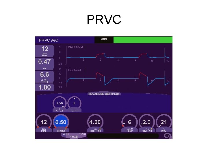
PRESSURE REGULATED VOLUME CONTROL (PRVC): • This is a volume targeted, pressure limited mode. (available in SIMV or AC) • Each breath is delivered at a set volume with a variable flow rate and an absolute pressure limit. • The vent delivers this pre-set volume at the LOWEST required peak pressure and adjust with each breath.
PRVC
POSITIVE END EXPIRATORY PRESSURE (PEEP): • This is NOT a specific mode, but is rather an adjunct to any of the vent modes. • PEEP is the amount of pressure remaining in the lung at the END of the expiratory phase. • Utilized to keep otherwise collapsing lung units open while hopefully also improving oxygenation.
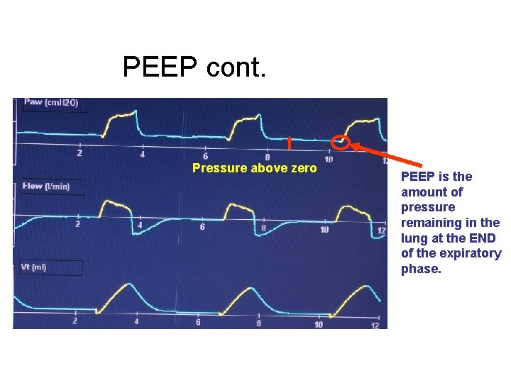
PEEP cont. Pressure above zero PEEP is the amount of pressure remaining in the lung at the END of the expiratory phase.
Demonstration of PEEP • http: //youtube. com/watch? v=o. KH 7 Cts. Eg. H w
Continuous Positive Airway Pressure (CPAP): • This IS a mode and simply means that a preset pressure is present in the circuit and lungs throughout both the inspiratory and expiratory phases of the breath. • CPAP serves to keep alveoli from collapsing, resulting in better oxygenation and less WOB. • The CPAP mode is very commonly used as a mode to evaluate the patients readiness for extubation.
HIGH FREQUENCY VENTILATION
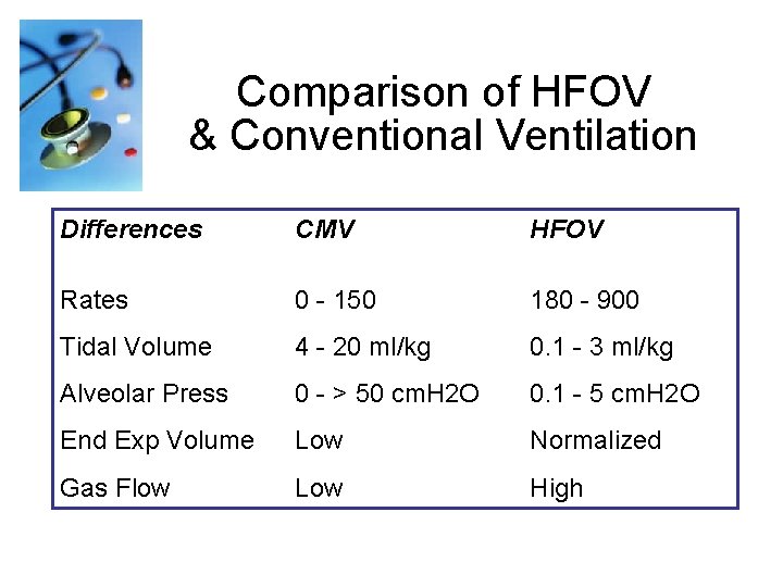
Comparison of HFOV & Conventional Ventilation Differences CMV HFOV Rates 0 - 150 180 - 900 Tidal Volume 4 - 20 ml/kg 0. 1 - 3 ml/kg Alveolar Press 0 - > 50 cm. H 2 O 0. 1 - 5 cm. H 2 O End Exp Volume Low Normalized Gas Flow Low High
Oxygenation • Oxygenation is primarily controlled by the Mean Airway Pressure (Paw) and the Fi. O 2. • Mean Airway Pressure is a constant pressure used to inflate the lung and hold the alveoli open. • Since the Paw is constant, it reduces the injury that results from cycling the lung open for each breath

Video on HFOV http: //youtube. com/watch? v=j. Lro. OPo. Plig
Initial Settings • • Select your mode of ventilation Set sensitivity at Flow trigger mode Set Tidal Volume Set Rate Set Inspiratory Flow (if necessary) Set PEEP Set Pressure Limit Humidification
Post Initial Settings • Obtain an ABG (arterial blood gas) about 30 minutes after you set your patient up on the ventilator. • An ABG will give you information about any changes that may need to be made to keep the patient’s oxygenation and ventilation status within a physiological range.
ABG • Goal: • Keep patient’s acid/base balance within normal range: • p. H • PCO 2 • PO 2 7. 35 – 7. 45 35 -45 mm. Hg 80 -100 mm. Hg
TROUBLESHOOTING • Anxious Patient – Can be due to a malfunction of the ventilator – Patient may need to be suctioned – Frequently the patient needs medication for anxiety or sedation to help them relax • Attempt to fix the problem • Call your RT
Low Pressure Alarm • Usually due to a leak in the circuit. – Attempt to quickly find the problem – Bag the patient and call your RT.
High Pressure Alarm • Usually caused by: – A blockage in the circuit (water condensation) – Patient biting his ETT – Mucus plug in the ETT – You can attempt to quickly fix the problem – Bag the patient and call for your RT.
Low Minute Volume Alarm • Usually caused by: – Apnea of your patient (CPAP) – Disconnection of the patient from the ventilator – You can attempt to quickly fix the problem – Bag the patient and call for your RT.
Accidental Extubation • Role of the Nurse: – Ensure the Ambu bag is attached to the oxygen flowmeter and it is on! – Attach the face mask to the Ambu bag and after ensuring a good seal on the patient’s face; supply the patient with ventilation. – Bag the patient and call for your RT.
OTHER • Anytime you have concerns, alarms, ventilator changes or any other problem with your ventilated patient. – Call for your RT – NEVER hit the silence button!
- Slides: 54