
MDS Anomalie morfologiche Emopoiesi inefficace Midollo pieno e
- Slides: 42
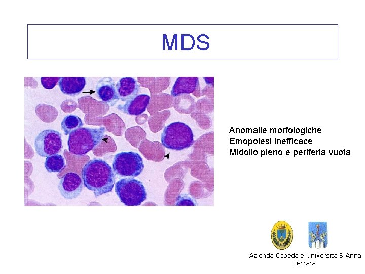
MDS Anomalie morfologiche Emopoiesi inefficace Midollo pieno e periferia vuota Azienda Ospedale-Università S. Anna Ferrara
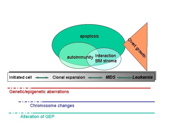
apoptosis ve O g rt Initiated cell Clonal expansion Genetic/epigenetic aberrations Chromosome changes Alteration of GEP MDS h wt ro autoimmunity Interaction BM stroma Leukemia
Lesioni citogenetiche nelle MDS • Meccanismi • Descrizione delle anomalie e di alcune entità • Relazione con la patogenesi e con la clinica
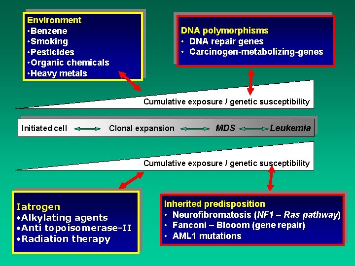
Environment • Benzene • Smoking • Pesticides • Organic chemicals • Heavy metals DNA polymorphisms • DNA repair genes • Carcinogen-metabolizing-genes Cumulative exposure / genetic susceptibility Initiated cell Clonal expansion MDS Leukemia Cumulative exposure / genetic susceptibility Iatrogen • Alkylating agents • Anti topoisomerase-II • Radiation therapy Inherited predisposition • Neurofibromatosis (NF 1 – Ras pathway) • Fanconi – Blooom (gene repair) • AML 1 mutations
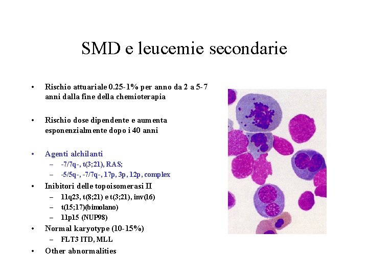
SMD e leucemie secondarie • Rischio attuariale 0. 25 -1% per anno da 2 a 5 -7 anni dalla fine della chemioterapia • Rischio dose dipendente e aumenta esponenzialmente dopo i 40 anni • Agenti alchilanti – – • Inibitori delle topoisomerasi II – – – • 11 q 23, t(8; 21) e t(3; 21), inv(16) t(15; 17)(bimolano) 11 p 15 (NUP 98) Normal karyotype (10 -15%) – • -7/7 q-, t(3; 21), RAS; -5/5 q-, -7/7 q-, 17 p, 3 p, 12 p, complex FLT 3 ITD, MLL Other abnormalities
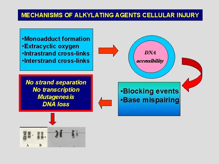
MECHANISMS OF ALKYLATING AGENTS CELLULAR INJURY • Monoadduct formation • Extracyclic oxygen • Intrastrand cross-links • Interstrand cross-links No strand separation No transcription Mutagenesis DNA loss DNA accessibility • Blocking events • Base mispairing
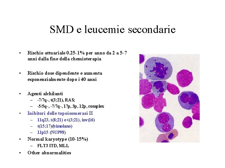
SMD e leucemie secondarie • Rischio attuariale 0. 25 -1% per anno da 2 a 5 -7 anni dalla fine della chemioterapia • Rischio dose dipendente e aumenta esponenzialmente dopo i 40 anni • Agenti alchilanti – – • Inibitori delle topoisomerasi II – – – • 11 q 23, t(8; 21) e t(3; 21), inv(16) t(15; 17)(bimolano) 11 p 15 (NUP 98) Normal karyotype (10 -15%) – • -7/7 q-, t(3; 21), RAS; -5/5 q-, -7/7 q-, 17 p, 3 p, 12 p, complex FLT 3 ITD, MLL Other abnormalities
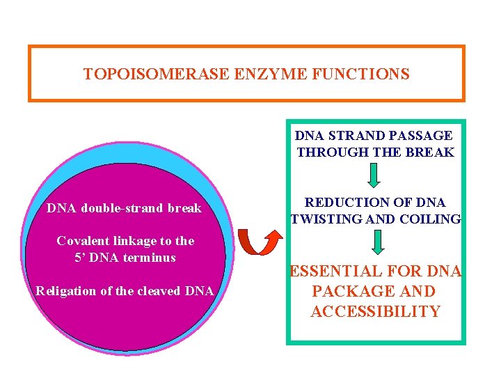
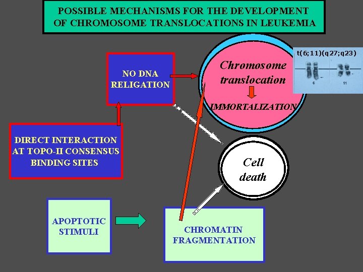
TOPOISOMERASE ENZYME FUNCTIONS DNA STRAND PASSAGE THROUGH THE BREAK DNA double-strand break Covalent linkage to the 5’ DNA terminus Religation of the cleaved DNA REDUCTION OF DNA TWISTING AND COILING ESSENTIAL FOR DNA PACKAGE AND ACCESSIBILITY
POSSIBLE MECHANISMS FOR THE DEVELOPMENT OF CHROMOSOME TRANSLOCATIONS IN LEUKEMIA t(6; 11)(q 27; q 23) NO DNA RELIGATION Chromosome translocation IMMORTALIZATION DIRECT INTERACTION AT TOPO-II CONSENSUS BINDING SITES APOPTOTIC STIMULI Cell death CHROMATIN FRAGMENTATION
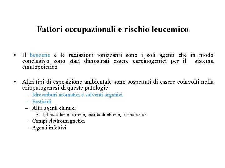
Fattori occupazionali e rischio leucemico • Il benzene e le radiazionizzanti sono i soli agenti che in modo conclusivo sono stati dimostrati essere carcinogenici per il sistema ematopoietico • Altri tipi di esposizione ambientale sono sospettati di essere coinvolti nella eziopatogenesi di queste patologie: – Idrocarburi aromatici e solventi organici – Pesticidi – Altri agenti chimici • 1, 3 -butadiene, stirene, ossido di etilene, formaldeide – Campi elettromagnetici – Agenti infettivi
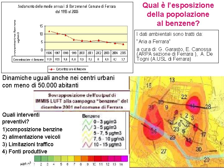
Qual è l’esposizione della popolazione al benzene? I dati ambientali sono tratti da: “Aria a Ferrara” a cura di: G. Garasto, E. Canossa (ARPA sezione di Ferrara ), A. De Togni (A. USL di Ferrara) Dinamiche uguali anche nei centri urbani con meno di 50. 000 abitanti Quali interventi preventivi? 1)composizione benzine 2) alimentazione veicoli 3) Limitazioni traffico 4) Fonti produttive
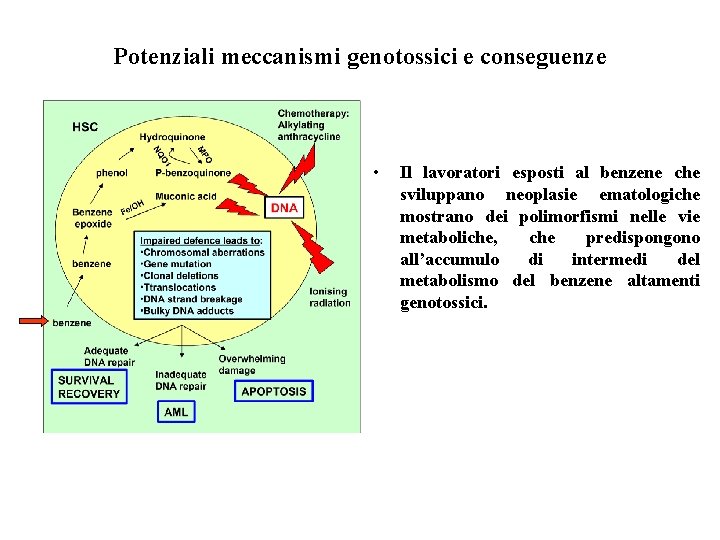
Potenziali meccanismi genotossici e conseguenze • Il lavoratori esposti al benzene che sviluppano neoplasie ematologiche mostrano dei polimorfismi nelle vie metaboliche, che predispongono all’accumulo di intermedi del metabolismo del benzene altamenti genotossici.
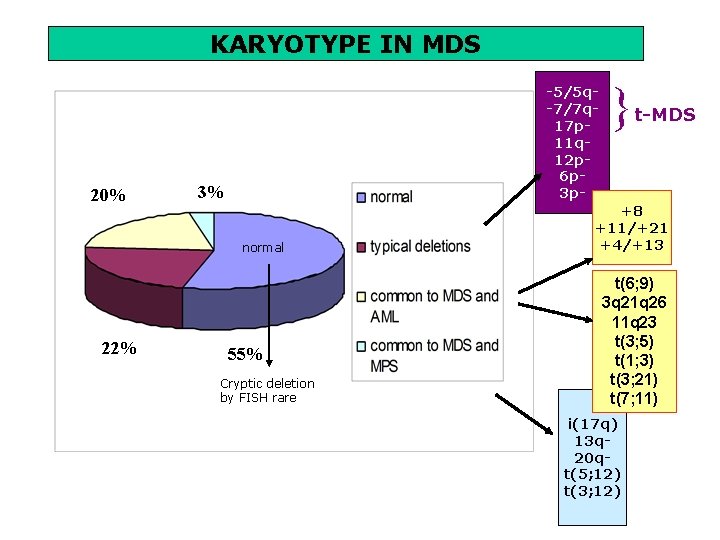
KARYOTYPE IN MDS 20% -5/5 q-7/7 q 17 p 11 q 12 p 6 p 3 p- 3% normal 22% 55% Cryptic deletion by FISH rare } t-MDS +8 +11/+21 +4/+13 t(6; 9) 3 q 21 q 26 11 q 23 t(3; 5) t(1; 3) t(3; 21) t(7; 11) i(17 q) 13 q 20 qt(5; 12) t(3; 12)
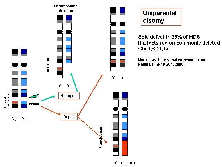
Chromosome deletion Uniparental disomy deletion Sole defect in 33% of MDS It affects region commonly deleted Chr 1, 6, 11, 13 Maciejewski, personal communication Naples, june 19 -20° , 2006 5* Abnormal oncosuppressor 5* 5 5 q- No repair break Repair translocation 5♂ 5♀ 5* der(5 q)
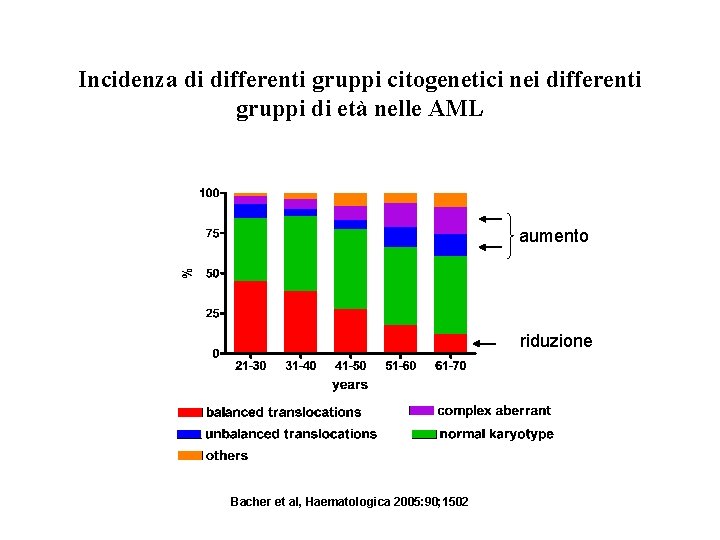
Incidenza di differenti gruppi citogenetici nei differenti gruppi di età nelle AML aumento riduzione Bacher et al, Haematologica 2005: 90; 1502
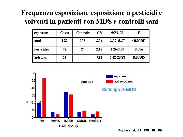
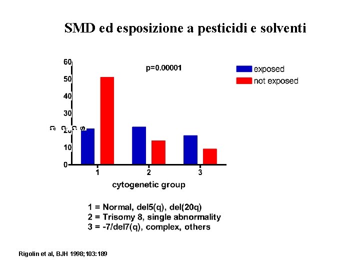
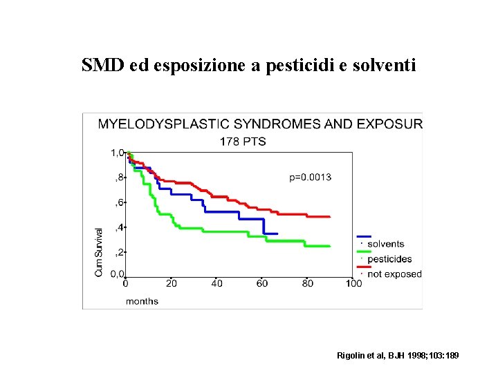
Frequenza esposizione a pesticidi e solventi in pazienti con MDS e controlli sani exposure Cases Controls OR 95% CI P total 178 3. 74 2. 02 -. 5. 37 <0. 00001 Pesticides 48 27 2. 12 1. 26 -3. 59 0. 006 Solvents 25 4 7. 11 2. 42 -20. 88 0. 00005 Sottotipo di MDS Rigolin et al, BJH 1998; 103: 189
SMD ed esposizione a pesticidi e solventi Rigolin et al, BJH 1998; 103: 189
SMD ed esposizione a pesticidi e solventi Rigolin et al, BJH 1998; 103: 189
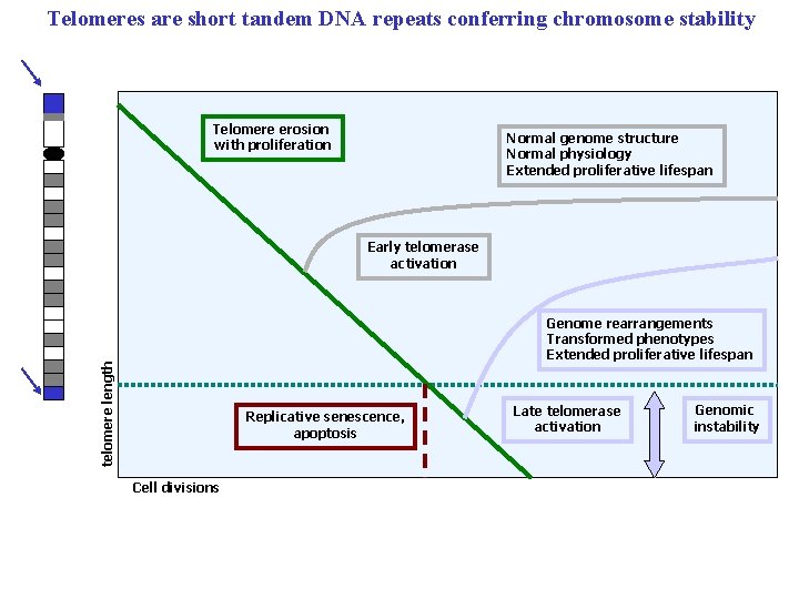
Telomeres are short tandem DNA repeats conferring chromosome stability Telomere erosion with proliferation Normal genome structure Normal physiology Extended proliferative lifespan Early telomerase activation telomere length Genome rearrangements Transformed phenotypes Extended proliferative lifespan Replicative senescence, apoptosis Cell divisions Late telomerase activation Genomic instability
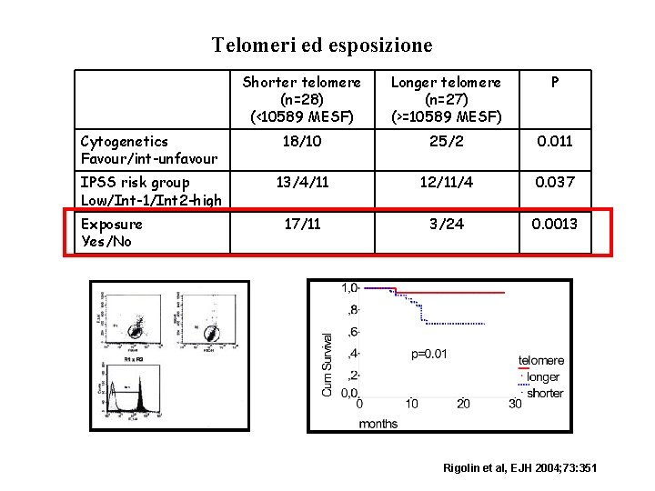
Telomeri ed esposizione Shorter telomere (n=28) (<10589 MESF) Longer telomere (n=27) (>=10589 MESF) P Cytogenetics Favour/int-unfavour 18/10 25/2 0. 011 IPSS risk group Low/Int-1/Int 2 -high 13/4/11 12/11/4 0. 037 17/11 3/24 0. 0013 Exposure Yes/No Rigolin et al, EJH 2004; 73: 351
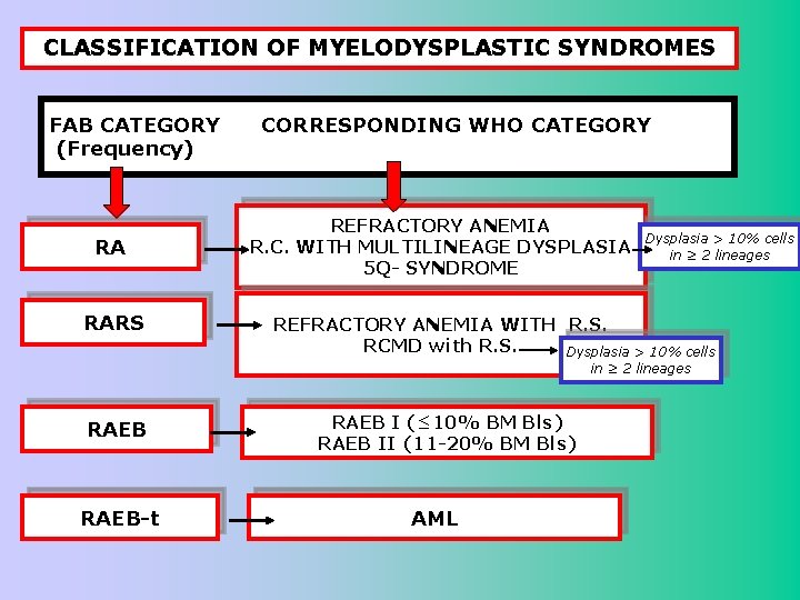
CLASSIFICATION OF MYELODYSPLASTIC SYNDROMES FAB CATEGORY CORRESPONDING WHO CATEGORY (Frequency) RA RARS REFRACTORY ANEMIA R. C. WITH MULTILINEAGE DYSPLASIA 5 Q- SYNDROME Dysplasia > 10% cells in ≥ 2 lineages REFRACTORY ANEMIA WITH R. S. RCMD with R. S. Dysplasia > 10% cells in ≥ 2 lineages RAEB-t RAEB I (≤ 10% BM Bls) RAEB II (11 -20% BM Bls) AML
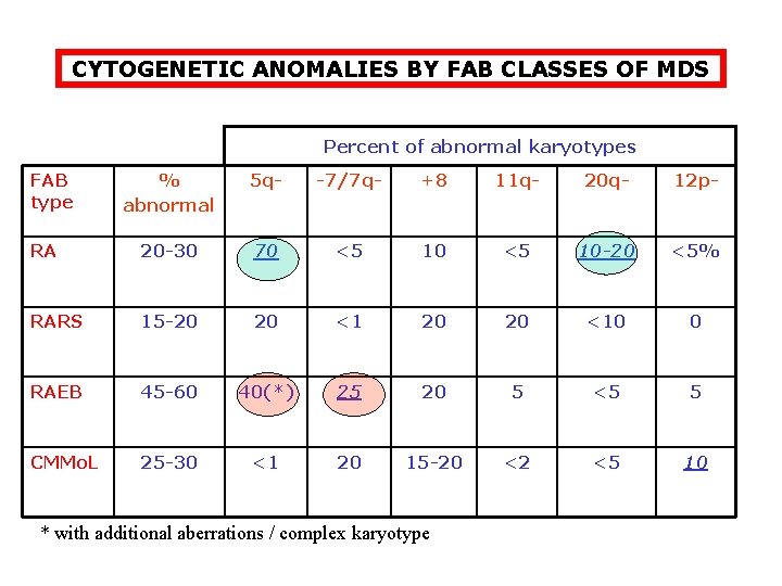
CYTOGENETIC ANOMALIES BY FAB CLASSES OF MDS Percent of abnormal karyotypes FAB type % abnormal 5 q- -7/7 q- +8 11 q- 20 q- 12 p- RA 20 -30 70 <5 10 -20 <5% RARS 15 -20 20 <10 0 RAEB 45 -60 40(*) 25 20 5 <5 5 CMMo. L 25 -30 <1 20 15 -20 <2 <5 10 * with additional aberrations / complex karyotype
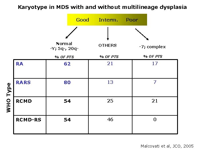
Karyotype in MDS with and without multilineage dysplasia Good Interm. Poor Normal -Y; 5 q-, 20 q- WHO Type % OF PTS OTHERS % OF PTS -7; complex % OF PTS RA 62 21 17 RARS 80 13 7 RCMD 54 25 21 RCMD-RS 54 46 0 Malcovati et al, JCO, 2005
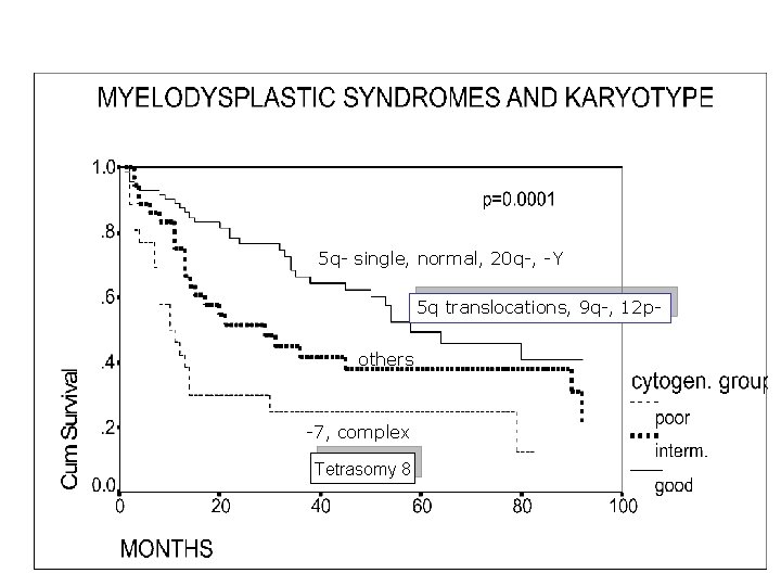
5 q- single, normal, 20 q-, -Y 5 q translocations, 9 q-, 12 pothers -7, complex Tetrasomy 8
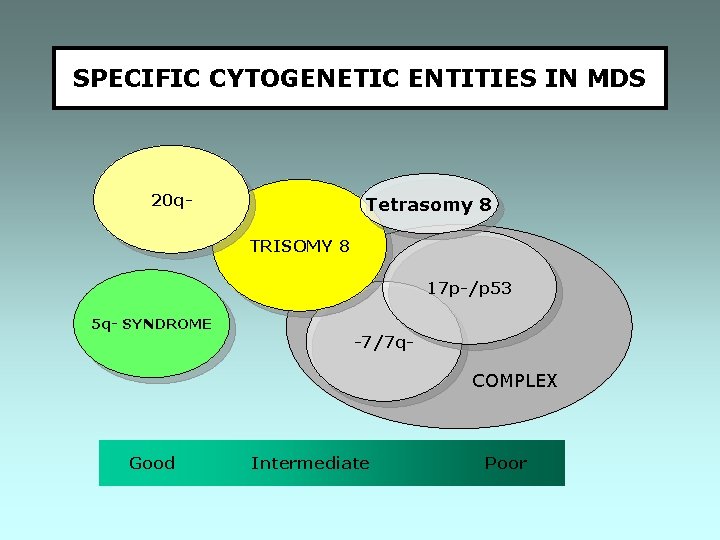
SPECIFIC CYTOGENETIC ENTITIES IN MDS 20 q- Tetrasomy 8 TRISOMY 8 17 p-/p 53 5 q- SYNDROME -7/7 q- COMPLEX Good Intermediate Poor
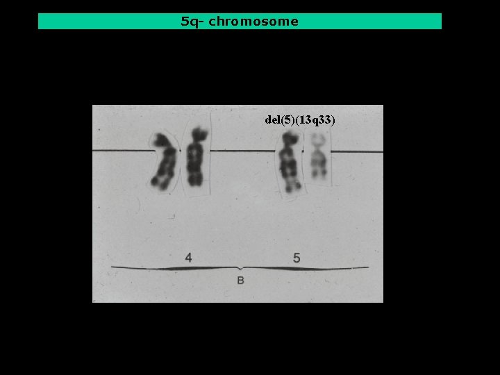
5 q- chromosome del(5)(13 q 33)
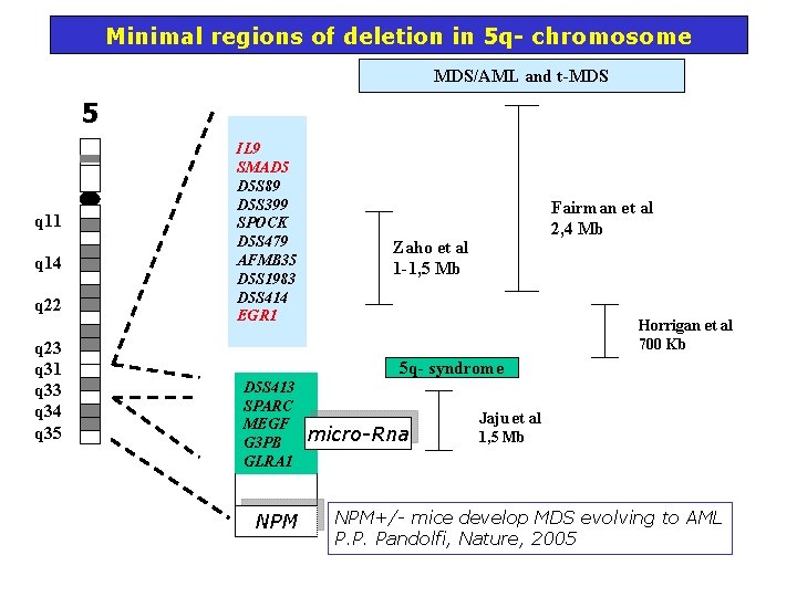
Minimal regions of deletion in 5 q- chromosome MDS/AML and t-MDS 5 q 11 q 14 q 22 q 23 q 31 q 33 q 34 q 35 IL 9 SMAD 5 D 5 S 89 D 5 S 399 SPOCK D 5 S 479 AFMB 35 D 5 S 1983 D 5 S 414 EGR 1 Fairman et al 2, 4 Mb Zaho et al 1 -1, 5 Mb Horrigan et al 700 Kb 5 q- syndrome D 5 S 413 SPARC MEGF G 3 PB GLRA 1 NPM micro-Rna Jaju et al 1, 5 Mb NPM+/- mice develop MDS evolving to AML P. P. Pandolfi, Nature, 2005
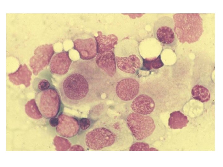
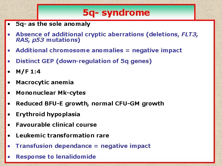
5 q- syndrome • 5 q- as the sole anomaly • Absence of additional cryptic aberrations (deletions, FLT 3, RAS, p 53 mutations) • Additional chromosome anomalies = negative impact • Distinct GEP (down-regulation of 5 q genes) • M/F 1: 4 • Macrocytic anemia • Mononuclear Mk-cytes • Reduced BFU-E growth, normal CFU-GM growth • Erythroid hypoplasia • Favourable clinical course • Leukemic transformation rare • Transfusion dependance = negative impact • Response to lenalidomide
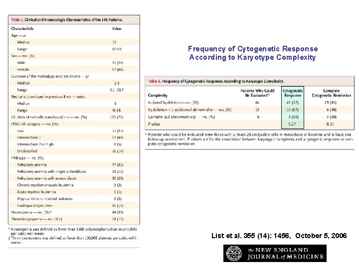
Frequency of Cytogenetic Response According to Karyotype Complexity List et al. 355 (14): 1456, October 5, 2006
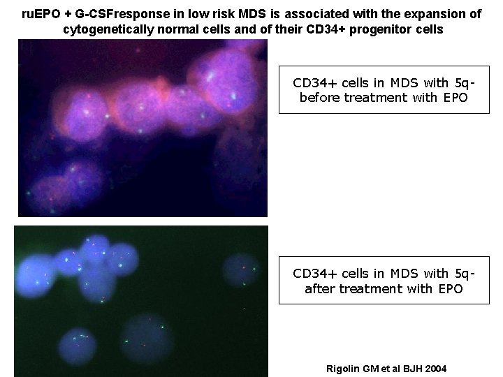
ru. EPO + G-CSFresponse in low risk MDS is associated with the expansion of cytogenetically normal cells and of their CD 34+ progenitor cells CD 34+ cells in MDS with 5 qbefore treatment with EPO CD 34+ cells in MDS with 5 qafter treatment with EPO Rigolin GM et al BJH 2004
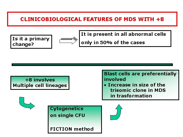
CLINICOBIOLOGICAL FEATURES OF MDS WITH +8 It is present in all abnormal cells Is it a primary change? only in 50% of the cases +8 involves Multiple cell lineages Cytogenetics on single CFU FICTION method Blast cells are preferentially involved • Increase in size of the trisomic clone in MDS in trasformation
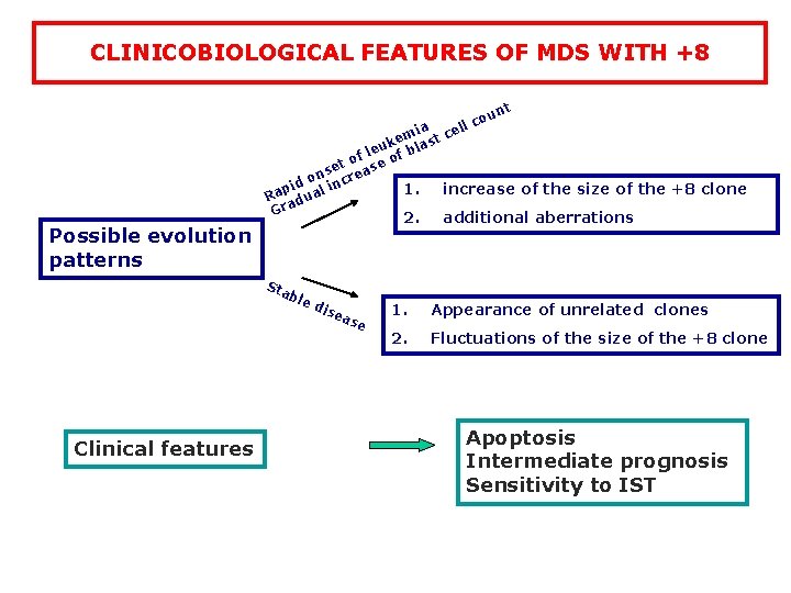
CLINICOBIOLOGICAL FEATURES OF MDS WITH +8 nt u co Possible evolution patterns ll a mi st ce e k leu f bla f o t o se ease n r o id l inc p 1. increase of the size of the +8 clone Ra dua a Gr 2. additional aberrations Sta ble Clinical features di sea se 1. Appearance of unrelated clones 2. Fluctuations of the size of the +8 clone Apoptosis Intermediate prognosis Sensitivity to IST
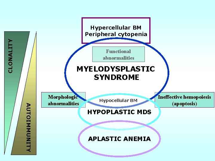
CLONALITY Hypercellular BM Peripheral cytopenia Functional abnormalities MYELODYSPLASTIC SYNDROME AUTOIMMUNITY Morphologic abnormalities Hypocellular BM HYPOPLASTIC MDS APLASTIC ANEMIA Ineffective hemopoiesis (apoptosis)
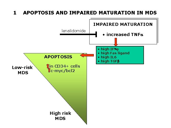
1 APOPTOSIS AND IMPAIRED MATURATION IN MDS IMPAIRED MATURATION lenalidomide APOPTOSIS Low-risk MDS in CD 34+ cells c-myc/bcl 2 High risk MDS • increased TNFa • high IFNg • high Fas ligand • high IL 6 • high TGFb
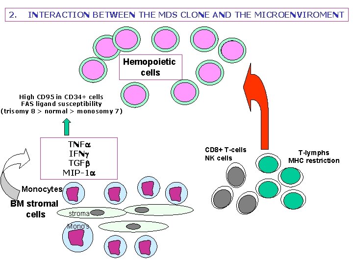
2. INTERACTION BETWEEN THE MDS CLONE AND THE MICROENVIROMENT Hemopoietic cells High CD 95 in CD 34+ cells FAS ligand susceptibility (trisomy 8 > normal > monosomy 7) TNFa IFNg TGFb MIP-1 a Monocytes BM stromal cells stroma Mono’s CD 8+ T-cells NK cells T-lymphs MHC restriction
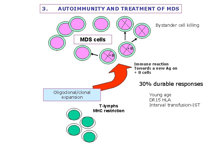
3. AUTOIMMUNITY AND TREATMENT OF MDS Bystander cell killing MDS cells +8 +8 Immune reaction Towards a new Ag on + 8 cells 30% durable responses Oligoclonal/clonal expansion T-lymphs MHC restriction Young age DR 15 HLA Interval transfusion-IST
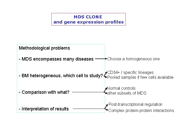
MDS CLONE and gene expression profiles Methodological problems - MDS encompasses many diseases Choose a homogeneous one CD 34+ / specific lineages - BM heterogeneous, which cell to study? Pooled samples if few cells available - Comparison with what? - Interpretation of results Normal controls other subsets of MDS Post-transcriptional regulation Complex protein-protein interactions
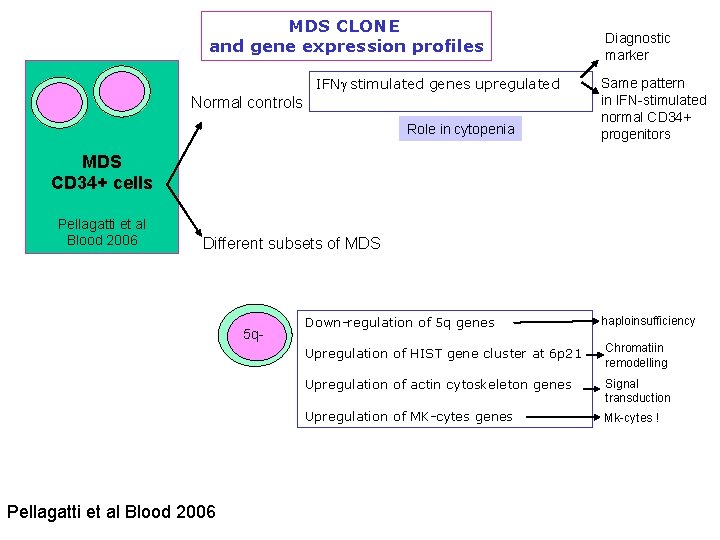
MDS CLONE and gene expression profiles IFNg stimulated genes upregulated Normal controls Role in cytopenia Diagnostic marker Same pattern in IFN-stimulated normal CD 34+ progenitors MDS CD 34+ cells Pellagatti et al Blood 2006 Different subsets of MDS 5 q- Pellagatti et al Blood 2006 Down-regulation of 5 q genes haploinsufficiency Upregulation of HIST gene cluster at 6 p 21 Chromatiin remodelling Upregulation of actin cytoskeleton genes Signal transduction Upregulation of MK-cytes genes Mk-cytes !
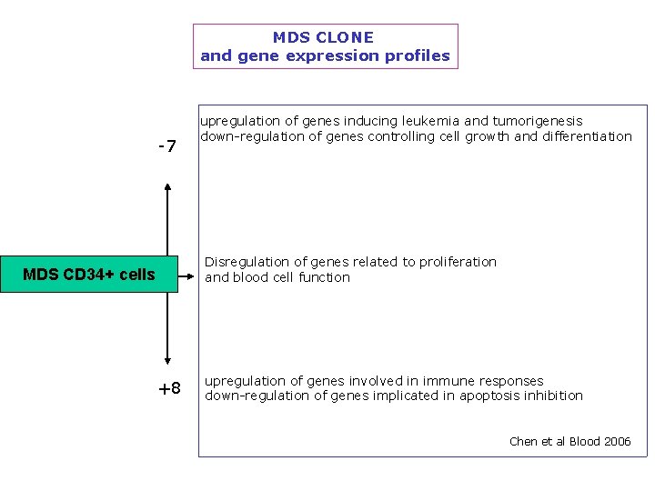
MDS CLONE and gene expression profiles -7 upregulation of genes inducing leukemia and tumorigenesis down-regulation of genes controlling cell growth and differentiation Disregulation of genes related to proliferation and blood cell function MDS CD 34+ cells +8 upregulation of genes involved in immune responses down-regulation of genes implicated in apoptosis inhibition Chen et al Blood 2006
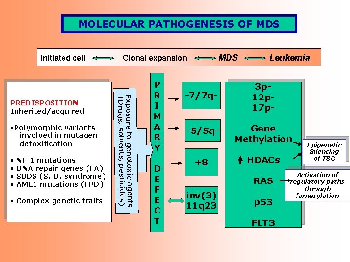
MOLECULAR PATHOGENESIS OF MDS Initiated cell • Polymorphic variants involved in mutagen detoxification • NF-1 mutations • DNA repair genes (FA) • SBDS (S. -D. syndrome) • AML 1 mutations (FPD) • Complex genetic traits Exposure to genotoxic agents (Drugs, solvents, pesticides) PREDISPOSITION Inherited/acquired MDS Clonal expansion P R I M A R Y D E F E C T Leukemia -7/7 q- 3 p 12 p 17 p- -5/5 q- Gene Methylation +8 HDACs RAS inv(3) 11 q 23 p 53 FLT 3 Epigenetic Silencing of TSG Activation of regulatory paths through farnesylation
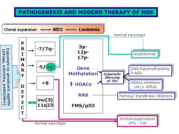
PATHOGENESIS AND MODERN THERAPY OF MDS Clonal expansion Leukemia Normal karyotype Exposure to genotoxic agents (Drugs, solvents, pesticides) P R I M A R Y D E F E C T -7/7 q- -5/5 q 5 q+8 inv(3) 11 q 23 Normal karyotype 3 p 12 p 17 p. Gene Methylation HDACs RAS lenalidomide Epigenetic Silencing of TSG DNA-hypomethylating 5 -AZA HDACs inhibitors VA (+ ATRA) Farnesyl transferase inhibitors FMS/p 53 Immunosuppression ATG - Cs. A