MCB 101 Introductory Microbiology Laboratory Lecture 2 January

MCB 101 Introductory Microbiology Laboratory Lecture 2 January 25, 2019 Today’s Topics - Experiment 3: Gram Stain - Experiment 4: - Schaeffer-Fulton Endospore Stain - Capsule Stain - Flagella Stain

Things in MCB 101 Next Week: Mon. and Tues: Experiment 3 - Gram Stain Wed. and Thurs: Experiment 4 – Special Stains LON-CAPA: Pre-lab 2 is due Wednesday, 1/30/19 at 8: 00 AM Quiz 2 opens today, due Friday, 2/1/19 at 8: 30 AM Pre-lab 3 opens, due Monday, 2/4/19 at 8: 00 AM
 Still used in")
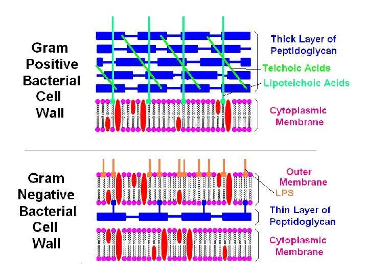
Ex. 3 The Gram Stain (Invented by Christian Gram in 1884) Still used in clinical labs Divides bacteria into two groups Bacteria with thick cell walls are purple, Gram + Bacteria with thin cell walls are pink, Gram – Cell wall refers to the layer of peptidoglycan Gram – bacteria have an outer membrane, the space between the inner and outer membranes is called the periplasmic space. Gram + bacteria do not have an outer membrane or periplasmic space. • Most Gram – bacteria are more resistant to Penicillin, bile salts and dyes. • • •
 Streptococcus pneumonia – causes lung infection, used in")
Examples of Gram Positive Bacteria 1) Streptococcus pneumonia – causes lung infection, used in MCB 101 as an example of -hemolytic bacteria 2) Streptococcus pyogenes – causes strep throat, scarlet fever, used as an example of -hemolytic bacteria 3) Enterococcus faecalis – found in intestines, used in 101 4) Staphylococcus aureus – found on skin, causes boils, wound infections & food poisoning, used in MCB 101 5) Staphylococcus epidermidis – is on skin, used in 101 6) Bacillus subtilis – found on hay, makes heat resistant endospores, aerobic, used in MCB 101 7) Bacillus cereus – endospores, aerobic, food poisoning 8) Clostridium botulinum – makes endospores, grows anaerobically, can cause food poisoning 9) Lactobacillus bulgaricus – used to make yogurt 10) Corynebacterium diphtheria – seen on day 1
 Esherichia coli – found in intestines, some")
Examples of Gram Negative Bacteria 1) Esherichia coli – found in intestines, some strains cause diarrhea, used a lot in MCB 101, can grow using fermentative metabolism, makes acid from lactose 2) Salmonella typhi – found in intestines, causes typhoid fever, can be deadly, not used in MCB 101 3) Salmonella arizonae – found in intestines, used in 101 ferments glucose but not lactose 4) Enterobacter aerogenes – intestinal, used in MCB 101 ferments lactose, called a “coliform” 5) Proteus vulgaris – bladder infections, used in MCB 101 6) Pseudomonas aeruginosa – found on rotting plants, can cause burn infections, does not ferment glucose, used in MCB 101 7) Vibrio cholera - causes cholera, not used in MCB 101 8) Yersinia Pestis - causes bubonic plague, not used here

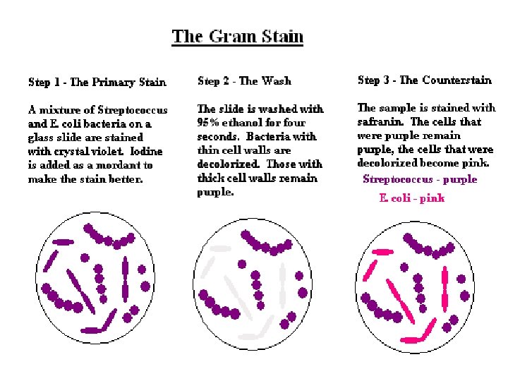
Gram Stain Procedure 1. Crystal Violet, 2 minutes, Primary Stain Both Gram + and Gram – cells are stained purple. 2. Iodine, 1 minute, Mordant (helps dye stick) Both Gram + and Gram – cells are still purple. 3. 95% Ethanol, 4 seconds, Decolorizer, washes out dye Removes color from Gram - cell wall but Gram + cells are still purple. 4. Safranin, 30 seconds, Counter Stain 1. Gram – cells get stained pink, Gram + cells stay purple.

The Gram Stain

The Gram Stain – Expected Results Choose the following statement about the Gram stain that is TRUE. A. After a good Gram stain Streptococcus pyogenes cells will be colored red-pink. B. After a successful Gram stain Escherichia coli bacterial cells will be colored red-pink. C. After a good Gram stain Staphylococcus aureus cells will be colored green. D. After a successful Gram stain Salmonella typhi cells will be colored purple-blue. E. After a successful Gram stain Clostridium botulinum cells will be colored red-pink.

Gram positive cocci Gram negative rods examples: Staphylococcus aureus A Escherichia coli B

What is the purpose of using iodine in the Gram stain? What is the purpose of using iodine in the A. It helps the crystal violet stick to the Gram stain? bacterial cell wall. A. It helps the crystal violet stick to the B. It degrades the outer membrane of bacterial cell wall. B. It degrades the outer membrane of Gram-negative bacteria. C. It colors the bacterial cells a reddish C. It colors the bacterial cells a reddishbrown color. D. It kills the bacterial cells. brown color. E. It helps the bacterial cells to stick to the slide. D. It kills the bacterial cells. E. It helps the bacterial cells to stick to the slide.

Common Problems With the Gram Stain - Gram positive cells come out lavender or pink instead of purple. - Gram negative cells come out lavender or purple instead of pink. - Decolorized too long. - Iodine solution old. - Cells old. - Stopped ethanol wash too soon. - Cells too crowded. - Ethanol solution diluted.

Experiment 4 A: The Schaeffer-Fulton Spore Stain - The spore stain is used to help identify bacteria in the genera Bacillus and Clostridium. - Bacillus and Clostridium are both Gram positive rods. - Bacillus is aerobic. Clostridium is anaerobic.

Some endospores are located terminally, others in the middle of the cell, others just slightly off-center. The endospore is a tough metabolic resting state that is very resistant to heat, drying, freezing, chemicals and radiation. The endospore has a specialized, extra-thick cell wall.

Endospores are produced when conditions become unfavorable for growth and reproduction. Vegetative cells are actively absorbing nutrients and growing but endospores make and use very little ATP. Endospores do not grow or divide. Frozen endospores can survive for years. Sporulation: when a vegetative cell makes an endospore Germination: when an endospore breaks open and a small vegetative cell emerges
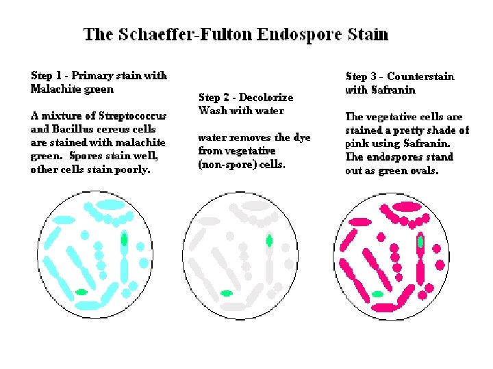

What will you actually see? Carolina Biological Supply Company used a modified Schaeffer. Fulton endospore staining technique to make the slides that you will examine. Vegetative (growing) cells are a red color but the endospores are also a red color. You should be able to see the difference.

Reagents Used in the Schaeffer. Fulton Endospore Stain What reagent is used as the primary stain in the Schaeffer-Fulton Endospore stain. A. 95% ethanol B. water C. malachite green D. safranin E. crystal violet

Experiment 4 B: The Capsule Stain The capsule stain is a negative stain that uses an acidic dye to stain the glass background around the bacteria. Usually the cells are counterstained with a basic dye such as safranin. In the prepared slides we will look at, the negative stain is Congo red and the cells are stained with safranin. The capsules are seen as a clear zone around the cells.

Negative Stains – Acidic Dyes For an acidic dye the chromophore is a proton donor when the dye molecule is dissolved in water. This makes the colored reagent negatively charged. Acidic dyes usually don’t stain microorganisms but they can stain glass. The negative stain is a procedure that is used to visualize the bacterial capsule. A capsule is a slimy layer of polysaccharides that coats the outside surface of some bacteria and protects the bacterium from the host immune system. Acidic Dyes Nigrosin India Ink Congo Red Eosin

Capsule Stain Which one of the following reagents is an ACIDIC DYE that can be used to prepare a negative stain such as a capsule stain. A. Methylene Blue B. Safranin C. Congo Red D. Crystal Violet E. Carbol Fuchsin

Experiment 4 C: The Flagella Stain Flagella are hard to see with light microscopy because they are very thin. The diameter of a bacterial flagellum is less than the limit of resolution for visible light. To see the flagella the sample must be treated with a mordant that acts as a thickening agent. Usually tannic acid is used. The tannic acid binds to the flagella making them thick enough to see when stained with a basic dye such as crystal violet.

Bacterial Flagellar Arrangements

Flagella Stain Why is it difficult to see bacterial flagella when using light microscopy? A. Bacterial flagella fall off very easily so most of the bacteria in a smear loose them. B. The width of a bacterial flagellum is less than the limit of resolution with a light microscope. C. The bacteria with flagella move too fast to get the microscope focused on them. D. There are no dyes that will stick to and stain a bacterial flagellum.
 The slides are made by")
What will you see in the flagella stain? 1) The slides are made by an experienced technician for commercial sale. But even so, flagella stain slides tend to be a bit messy. Any little bit of debris or dust will take up the mordant and end up larger, stained and visible. 2) You will look at two slides: Spirillum volutans is a large helical shaped cell with amphitrichous flagella, while Proteus vulgaris has peritrichous flagella. 3) Proteus vulgaris is a cause of bladder infection. Proteus cells can be seen in different forms: swarming (running mode) and sessile (not moving). Also look for large cells with many peritrichous flagella in all directions (a sessile stage).

Chemotaxis Run and Tumble Motion Chemotaxis is purposeful movement of a cell in a chemical concentration gradient towards an attractant or away from a repellant. Run and Tumble Motion is a semi-random form of movement. Progress in the right direction occurs when the length of runs in the correct direction are longer than the length of runs in a wrong direction. The direction of flagellar rotation (clockwise vs. counterclockwise) determines if the cell will run or tumble.


Vegetative Cells vs. Endospores Choose all of the following statements abut the differences between vegetative cells and endospores that are TRUE. 1) vegetative cells absorb nutrients but endospores don’t 2) all pathogenic bacteria can form endospores 3) vegetative cells are more resistant to killing by heat or drying out than endospores 4) tetanus and botulism are diseases caused by endospore-forming bacteria 5) vegetative cells grow and divide but endospores don’t A. 2, 3, 4 B. 1, 3, 5 C. 1, 2, 3 D. 1, 4, 5 E. 2, 4, 5
- Slides: 29