MAXILLARY FRACTURES Midface Fractures LEFORT AP VIEW Le


 Unilateral/ bilateral �")


 Separation of NF suture,")

 Separation of")


 Halo-frame Plaster of paries")
 Interosseous Wire Fixation Bone Grafts")

- Slides: 40
MAXILLARY FRACTURES
Midface Fractures LEFORT - AP VIEW
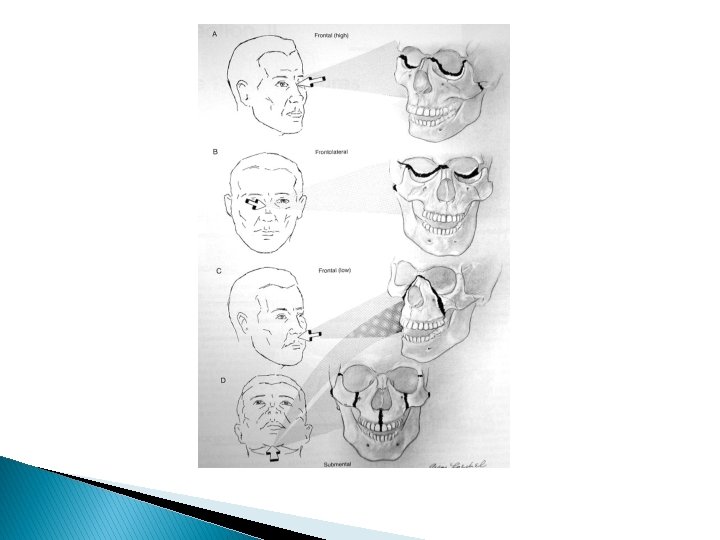
Le Fort’s fractures Le Fort I (low level or Guerian fracture) Unilateral/ bilateral � � Horizontal fracture through the maxilla above the level of the nasasl floor and alveolar process Piriform rims Anterior maxilla Zygomatic buttresses Ptrygoid laminae � � 4
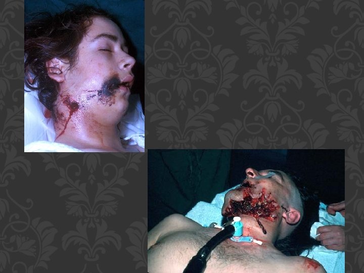
Signs and symptoms � Slight swelling of upper lip � Ecchymosis in upper lip sulcus � Hematoma intra-orally over zygoma and in palate � Disturbed occlusion � Mobility of teeth of the involved segment of maxilla � Combination of soft tissue laceration � Impacted type of fracture is oftenly not mobile and teeth cusps may be damaged 5
Lé Fort I
Le Fort’s fractures � Le Fort II (pyramidal or subzygomatic) Separation of NF suture, medial orbital walls (lacrimal bone), inferior orbital floor and rim (adjacent to infrorbital canal and foramen), anterior maxilla below zygomatic buttress and ptrygoid laminae about halfway up. Separation of the block from the base of skull is completed via the nasal septum and may involve the floor of the anterior cranial fossa 7
Lé Fort II
Le. Fort’s fractures � Le. Fort III (cranifacial dysjunction, high transverse, suprazygomatic) Separation of NF suture, medial orbital walls (involve the depth of the ethmoid bone and cribriform plate, pass below optic foramen and cross the inferior orbital fissur), inferior orbital floor, lateral orbital wall, ZF suture, zygomatic arch, suprazygomatic to the root of ptrygoid plate. 9
Le Fort 3 and mastication problem
Signs and symptoms although it is possible to distinguish between le fort II and III, the signs and symptoms are almost similar � � � Gross edema of soft tissue Bilateral circumorbital ecchymosis Bilateral subconjunctival hemorrahge Obvious deformity of the nose Nasal bleeding and obstruction CSF leak rhinorrhea Dish-face deformity Limitation of ocular movement Possible diplopia and enophthalmous Retropostioning of the maxilla with anterior open bite Lengthening of the face � � � � � Difficulty in mouth opening Mobility of the upper jaw Occusional hematoma of the palate Cracked-pot sound on percussion Step deformity at infra-orbiatal margin Anasthesia of midface Nasal bone moves with mid-face as a whole Tenderness and sepration at FZ suture Tenderness and deformity of zygomatic arch Depression of occular level and pseudoptosis 11
DIAGNOSIS OF MAXILLOFACIAL INJURIES Inspection Palpation Diagnostic Imaging Plain films CT
INSPECTION Sublingual ecchymosis Step defects, ridge discontinuity, malocclusion
DIAGNOSIS OF MAXILLOFACIAL INJURIES PALPATION “Step” Defect Crepitus Bony segments Subcutaneous emphysema Mobility
FACIAL EXAMINATION PALPATION OF MIDFACE/BRIDGE OF NOSE
FACIAL EXAMINATION ORBITS EVALUATION
FACIAL EXAMINATION Orbits evaluated Periorbital edema and ecchymosis Gross visual acuity determined Diplopia Pupillary size & shape Subconjunctival hemorrhage
FACIAL EXAMINATION Orbits evaluated Lid lacerations Attachment of medial canthal tendon Rounding of lacrimal lake Increased intercanthal distance Epiphora Prompt Ophthamology consult
FACIAL EXAMINATION Evaluate mandibular opening Palpation of buccal vestibule Crepitus of lateral antral wall Occlusion evaluated Absence and quality of dentition noted Ecchymosis common finding Pharynx evaluated for laceration & bleeding
Indications for treatment � Physical signs of a fracture of the maxilla. � Evidence of a fractured maxilla on imaging. � Disruption of the occlusion of the teeth. � Displacement of the maxilla. � Post traumatic facial deformity. 24
Indications for treatment � Fractured or displaced teeth. � Cerebrospinal fluid leak. � Abnormal eye movement or restriction of eye movement. � Occlusion of the nasolacrimal duct. � Sensory or motor nerve deficit. � Other evidence of loss of function 25
Aims of treatment � Relieve pain � Restore function. � Restore bone anatomy. � Prevent infection � Restore the dental occlusion � Restore jaw movement at the earliest possible stage � Restore normal nerve function 26
Factors affecting the risk � Association with multiple injuries. � Presence of uncontrolled haemorrhage � Impairment of the airway. � Association with a dural tear. � Association with a base of skull fracture. 27
Factors affecting the risk � Presence of a pre-existing dentofacial deformity. � Time elapsed since the injury. � � � Presence of a medical or surgical factor which would delay general anesthesia Presence of any factor which would delay healing. (eg nutritional deficiency or alcoholism) Stage of dental development (deciduous, mixed or permanent dentition) 28
Factors affecting the risk � Presence of fractured teeth. � Total absence of teeth (edentulous) � � Inability of the patient to co-operate with treatment. Association with fractures of the mandible especially bilateral fractures of the condyles. 29
Principles of treatment Closed reduction may be appropriate in cases � Simple uncomplicated fractures � Complex or comminuted fractures � Medical or surgical contraindications to open reduction �Maxillary fractures in children 31
Open reduction may be appropriate where �Immediate or early jaw function is desirable �Difficulty is encountered in reducing the fracture by a closed method �The fracture is unstable 32
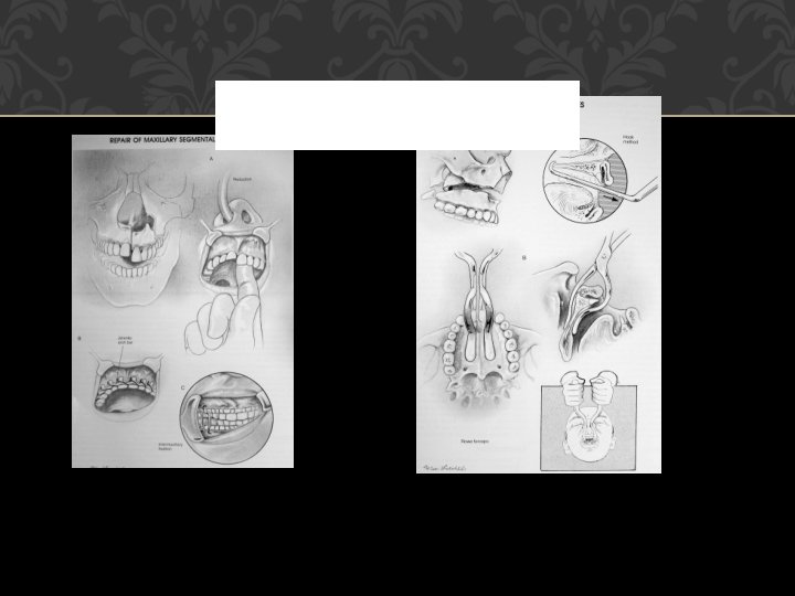
Definitive treatment � Reduction Manual manipulation Use of dis-impaction forceps 33
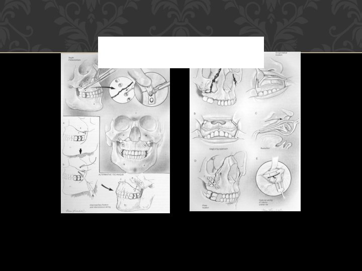
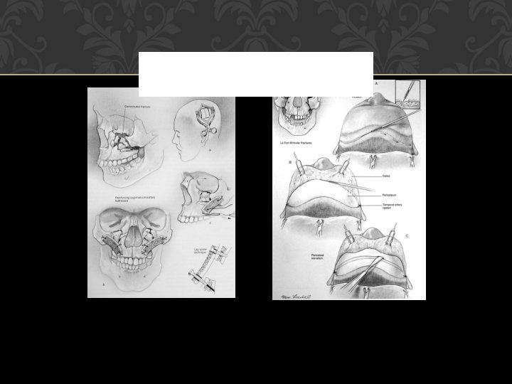
Fixation and immobilization Extraoral fixation Craniomandibular fixation Box-frame (pin fixation) Halo-frame Plaster of paries headcap Craniomaxillary fixation Supra-orbital pins Zygomatic pins Halo-frame 34
Cont: Management Techniques Ø Ø Ø Plate Fixation (Miniplates) Interosseous Wire Fixation Bone Grafts
Management by Le Fort Classification Ø Le Fort I: reduced digitally, MMF, fixation of ZM Ø Le Fort II: stabilization of the ZM buttress, MMF , nasofrontal process and inferior orbital rim. Ø Le Fort III: usually requires coronal flap for adequate exposure for exploration and miniplate fixation