Maternal Physiological Changes during Pregnancy Labor and Puerperium

Maternal Physiological Changes during Pregnancy, Labor and Puerperium ASSOCIATE PROFESSOR IOLANDA BLIDARU MD, Ph. D

Pregnancy is a period of adaptation for : • The needs of the fetus • Meeting the stress of pregnancy and labour

THE L A T I N GE S E G N CHA
 The whole uterus")
(A) The whole uterus

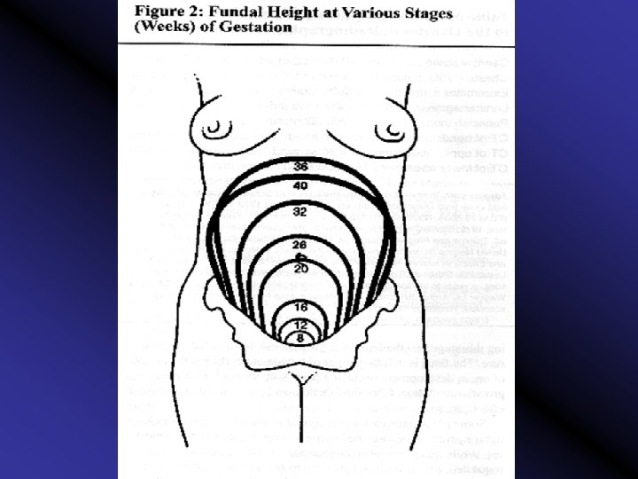
1 - Size increase from 7. 5 x 2. 5 cm in nonpregnant states to 36 x 25 x 20 cm at term i. e. the volume increase 1000 time

2 - Weight increases from 50 gm in nonpregnant state to 1000 gm at term

3 - Shape pyriform in the nonpregnant state , becomes globular at 8 th week, then ovoid by 16 th week till term.

4 - Position with ascent from the pelvis , the uterus usually undergoes rotation with tilting to the right (dextrorotation) due to the presence of the rectosegmoid colon on the left side.

5 - Consistency : becomes progressively softer due to : 1 Increased vascularity 2 Presence of amniotic fluid

6 - Contractility from the first trimester onwards , the uterus undergoes irregular painless contractions (Braxton Hicks contractions). They may cause some discomfort late in pregnancy and may account for false labour pain.

7 - Capacity increases from 4 ml in non-pregnant state to 4 -5000 ml at term
 Myometrial changes")
(B) Myometrial changes
 + hyperplasia (progesterone effect) 2 - the fetus exerts")
1 - Hypertrophy (estrogen effect) + hyperplasia (progesterone effect) 2 - the fetus exerts a direct stretch
 from the")
2 - Formation of the lower uterine segment (L. U. S. ) from the isthmus
 starts")
Formation of lower uterine segment After 12 weeks, the isthmus (0. 5 cm) starts to expand gradually to form the lower uterine segment which measures 10 -11 cm in length at term

Upper Uterine Segment • Peritoneum: Firmly-attached • Myometrium: 3 layers; outer longitudinal, middle oblique (interlacing network) and inner circular. • The middle layer forms 8 -shaped fibers around the blood vessels to control postpartum hemorrhage

Upper Uterine Segment • Decidua: Well-developed • Membranes: Firmly-attached • Activity: Active, contracts, retracts and becomes thicker during labour.

Lower Uterine Segment • Peritoneum: Looselyattached • Myometrium : 2 layers; outer longitudinal and inner circular.

Lower Uterine Segment • Decidua: Poorly-developed • Membranes: Looselyattached. • Activity: Passive, dilates, stretches and becomes thinner during labour
 which is thick")
The junction between the upper uterine segment (U. U. S. ) which is thick and the lower uterine segment which is thin is called the physiologic contraction ring at the level of the symphysis pubis (not seen or felt)
 Uterine blood vessels")
(C) Uterine blood vessels

Uterine and vaginal blood supply

1 - Uterine artery lumen: is doubled and its blood flow increases 5 times 2 - Myometrial and decidual arteries (spiral arteries) undergo fibrinoid degeneration due to 2 waves of trophoblastic migration , so they become dilated to be the uteroplacental arteries

Trophoblast invasion

• Uterine blood flow increases progressively and reaches about 500 ml / minute at term
 Changes in the cervix : 1 It becomes hypertrophied , soft and bluish")
(D) Changes in the cervix : 1 It becomes hypertrophied , soft and bluish in colour due to oedema and increased vascularity.

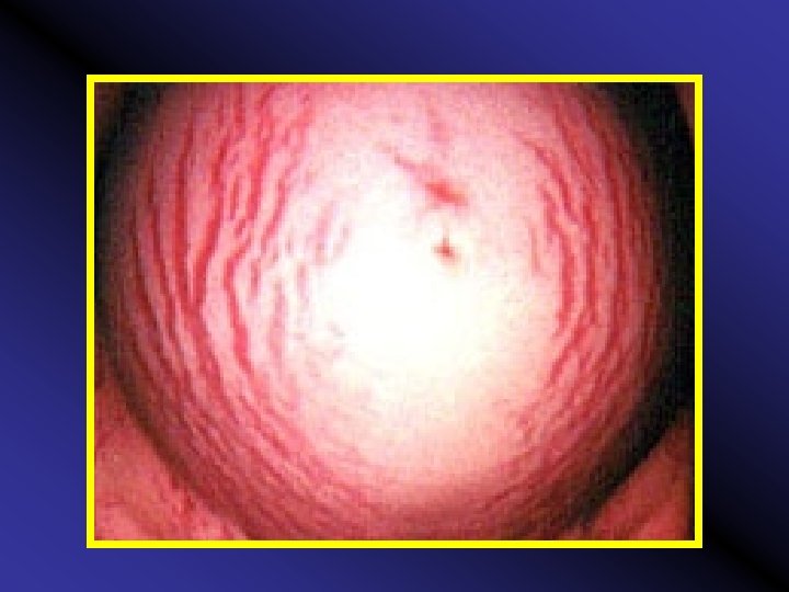
2 Soon after conception, a thick cervical secretion obstructs the cervical canal forming a mucous plug. 3 The endocervical epithelium proliferates and / or everts forming cervical ectopy (previously called erosion) (sometimes).

(Cervical eversion (colposcopy
,")
4 There are 3 principal structural components of the cervix: smooth muscle (only 10%), collagen and connective tissue or extracellular matrix (ground substance).
,")
5 The cervical ripening process changes in collagen (breakdown and rearrangement of the fibers), connective tissue and its ground substance (alterations of the various glycosaminoglycans). 6 - In the ripening of uterine cervix there are involved: PG E 2 and PG F 2 alpha, E, P and relaxin.
 Changes in fallopian tubes and round & broad ligaments : Inactive , elongated")
(E) Changes in fallopian tubes and round & broad ligaments : Inactive , elongated , marked increase in vascularity There may be broad ligament varicose veins
 Changes in the vagina : The vagina becomes soft , warm , moist")
(F) Changes in the vagina : The vagina becomes soft , warm , moist with increased secretion and violet in colour (Chadwick's sign) due to increased vascularity
 Changes in the vulva : • It becomes soft, violet in colour •")
(G) Changes in the vulva : • It becomes soft, violet in colour • Oedema and varicosities may develop
 Changes in the ovaries 1 Both ovaries are enlarged due to increased vascularity")
(H) Changes in the ovaries 1 Both ovaries are enlarged due to increased vascularity and oedema particularly the ovary which contains the corpus luteum.
 Changes in the ovaries 2 - Corpus luteum continues to grow till 7")
(H) Changes in the ovaries 2 - Corpus luteum continues to grow till 7 - 8 weeks, then it stops growing. It becomes inactive and starts degeneration at 12 weeks (degeneration is completed after labour)

Corpus luteum secretes 1. estrogen 2. progesterone 3. relaxin hormones
 Changes in the ovaries 3 Ovulation ceases during pregnancy due to pituitary inhibition")
(H) Changes in the ovaries 3 Ovulation ceases during pregnancy due to pituitary inhibition by the high levels of oestrogen and progesterone

• Relaxin is a protein hormone. • Its complete role in pregnancy is debated. • It may induce softness and effacement of the cervix.

II - Haematological Changes
 Blood volume The total blood volume increases steadily from early pregnancy to reach")
(A) Blood volume The total blood volume increases steadily from early pregnancy to reach a maximum of 35 45 % above the non pregnant level at 32 week.

Plasma volume : Increases from 2600 ml by ± 45 % (1200 in the 1 st pregnancy) and 1500 ml in subsequent pregnancies After delivery, it decreases rapidly by 600 800 ml.
 by 30 %")
Red blood cell mass : • Increases from 1400 ml (nonpregnant) by 30 % (± 450 ml) due to increased production resulting from erythropoietin action • The increase is steady till full term.

The increase in plasma volume is more than the increase in red blood cell mass (Hb mass) resulting in haemodilution (physiologic anemia)

However, the minimal Hb. accepted is 10 -11 gm%

Changes in the blood volume

Values of increased blood volume 1 - Meets increased demands for uterus, baby. . etc. 2 - Protects against supine hypotension syndrome. 3 - Protects against fluid loss at delivery.

Increased blood volume more than the increase in red cell mass, leads to decreased blood viscosity which leads to a decrease in peripheral resistance
 Blood indices")
(B) Blood indices

1 - Decreased Hb % and RBCs % : • Erythrocytes decrease from 4. 5 million / mm 3 to 3. 7 million / mm 3 (due to the relative increase in plasma volume more than red cell mass).

Hb concentrations falls from 14 g / dl To 12 g / dl.

2 - Reticulocytes : mild 3 - E. S. R : from 12 to 50 mm / hour (Erythrocyte Sedimentation Rate ) 4 – Fibrinogen: from 200 - 400 mg / dl to 400 - 600 mg / dl.
")
• All these changes are reversed after labour with RBCs production (not destruction) & the excess Fe is stored.

5 - White blood cells: (from 7. 000 / mm 3 to 10. 500 / mm 3 during pregnancy and up to 16. 000 / mm 3 during labour. 6 - PNL & its enzymes. (Polymorphonuclear Neutrophilic Leukocyte) 7 - Lymphocytes: no change.
 resulting in osmotic pressure.")
8 - Platelets: 9 -Total plasma proteins: slightly (mainly albumin) resulting in osmotic pressure. 10 - Increased plasma lipids
 Coagulation system")
(C) Coagulation system

• Platelets , unchanged adhesivity. • Fibrinogen doubled to 600 mg % • Factor VIII tripled. • Factor VII & factor X doubled • Factor XI & factor XIII slight • Fibrinolytic activity .

• Therefore pregnancy is a hypercoagulative state. • After delivery, fibrinogen and plasminogen decrease and the fibrinolytic activity in the plasma increases.

Ill - Cardiovascular system changes
 Changes in the heart")
(A) Changes in the heart

Position: As the diaphragm is elevated progressively during pregnancy the apex is displaced upwards and to the left so that it lies in the 4 th intercostal space outside the midclavicular line.

Pulse rate : - The resting pulse rate increases by 8 beats / min. at 8 weeks and 16 beats / min. at full term. Some episodes of ectopic beats

Heart sounds • The first heart sound become louder before midpregnancy and splitting of this sound may occur due to earlier closer of the mitral than the tricuspid valve • The intensity of the second heart sound may increase.

Heart sounds • The third sound becomes louder before mid-pregnancy and persists as such till one week post partum. • The fourth sound may be detectable by phonocardiography.

Murmurs Systolic functional murmurs develop in most of women, usually early systolic, but mid systolic murmurs may occur and heard over the left sternal edge, they are thought to be due to functional tricuspid regurgitation

ECG CHANGES • The main features of ECG may be attributed to the changes in the position of the heart. • The axis undergoes left shift by 15 28°. • The QRS complexes become of low voltage, and T wave becomes flattened.
 Haemodynamic changes")
(B) Haemodynamic changes
")
1 - Cardiac output (C. O. P. )

Cardiac output: increases mainly by increased stroke volume rather than increased heart rate reaching a maximum of 40% above the non pregnant level at 20 weeks to be maintained till term. Cardiac output = stroke volume x heart rate

Cardiac output Distribution : • • 400 ml to the uterus, 300 ml to the kidneys, 300 ml to skin, 300 ml to gastrointestinal tract, breast & heart

• Roles: Distributes extra 0 2 • During labour: Cardiac output increases more particularly during the second stage due to pain , uterine contractions, and expulsive efforts pushing the blood into the general circulation

• Postpartum : the increased CARDIAC OUTPUT is maintained for up to 4 days and then declines rapidly

2 - Arterial blood pressure

Although CARDIAC OUTPUT increase, yet Arterial Blood Pressure is decreased in midtrimester to increase again in 3 rd trimester

This is due to: 1 Decreased Peripheral resistance : (mainly affect diastolic B. P. ) due to : vasodilatation + increased metabolism + arterio venous shunt at the level of placenta.

2. Supine hypotensive syndrome: may develop in late pregnancy while lying supine compression on the Inferior Vena Cava by the large pregnant uterus decreased venous return decreased Cardiac Output and low B. P. to the extent that fainting may occur. Treatment: lateral recumbent position

Vena Cava Syndrome

3. Decreased sensitivity of blood vessels to angiotensin II which is vasoconstrictor

• The posture of the pregnant woman affects arterial blood pressure. • Typically, it is highest when she is sitting, lowest when lying in the lateral recumbent position and intermediate when supine.

Peripheral Vasodilatation

Peripheral Vasodilatation Regional blood flows the utero placental flow, the renal blood flow, the skin, particularly in the hands and feet + mammary gland

Peripheral Vasodilatation Renal blood flow → glomerular filtration increase favoring the oxygen supply and the amplification of some active processes. The increase of glomerular filtration contributes to the explanation of glycosuria, aminoaciduria and to the increase of water-soluble vitamin excretion.

Peripheral Vasodilatation blood flow to the skin, particularly in the hands and feet generally giving the pregnant women a feeling of warmth

Peripheral Vasodilatation Increases the congestion of nasal mucosa leading to a common complaint of nasal obstruction and bleeding (epistaxis).

3 - Venous pressure

Increased venous pressure in the lower limbs due to: 1. Back pressure from the compressed Inferior Vena Cava by the pregnant uterus. 2. Mechanical pressure of the uterus on pelvic veins. 3. Increased venous return from internal iliac veins → increased pressure in external iliac veins.

Increased venous pressure in the lower limbs Predisposes to: Oedema, varicose veins and piles

Oedema and varicose veins in the lower limbs & vulva are due to 1. Venous pressure. 2. Relaxation of the smooth muscles in the wall of the veins by progesterone 3. Osmotic pressure in blood. 4. Capillary permeability (due to progesterone and aldosterone). 5. Interstitial pressure (Na retention).

IV - Respiratory system
 Anatomically: The enlarged uterus displaces the diaphragm up to 4 cm.")
(A) Anatomically: The enlarged uterus displaces the diaphragm up to 4 cm.

This results in : 1. The diaphragmatic mobility is reduced and respiration becomes mainly thoracic. 2. Widen the subcostal angle and increases the transverse diameter of the chest.

Respiratory functions The respiratory rate does not increase during pregnancy from its normal rate of 14 - 15 / minute.
 occurs due to the effect of excess progesterone")
Overbreathing (deep respiration) occurs due to the effect of excess progesterone
 and dyspnea are")
Shortness of breath (the need to breath becomes a conscious one) and dyspnea are common complaints of the pregnant women which may be due to unfamiliarity with low C 02 tension in the alveolar capillaries.
 is decreased")
The vital capacity 1. The inspiratory capacity (Tidal volume + inspiratory volume) is decreased in late pregnancy

2. The expiratory reserve volume (maximum amount of air which can be expired after normal expiration) is reduced 3. The residual volume is reduced.

The reduction in: 1. The inspiratory capacity 2. The expiratory reserve volume 3. The residual volume is not significant .
")
4. The tidal volume : (amount of gas inspired or expired in each respiration) rises through out pregnancy by about 40 %.

Hyperventilation is due to increased tidal volume not respiratory rate ►a respiratory alkalosis takes place

V - Urinary system
 Kidney and kidney function tests • Renal blood flow and glomerular filtration rate")
(A) Kidney and kidney function tests • Renal blood flow and glomerular filtration rate increases by 50 %. This leads to increased excretion
 2.")
• Blood Tests: 1. There is serum creatinine (due to creatinine cleareance) 2. serum uric acid. 3. blood urea. 4. kidney excretion of glucose is due to filtration load and renal threshold leading to renal glucosuria.

Therefore , in interpretating the results of kidney function test you should take into consideration that the lowest normal values in non-pregnant state = the highest normal values in pregnancy
 Ureters Dilatation of the ureters and renal pelvis due to: 1. Relaxation of")
(B) Ureters Dilatation of the ureters and renal pelvis due to: 1. Relaxation of the ureters by the effect of progesterone. 2. Pressure against the pelvic brim by the uterus particularly on the right side due to dextroposed uterus and dilatation of the right ovarian vessels
 Bladder and urethra • Frequency of micturition in early pregnancy due to: -")
(C) Bladder and urethra • Frequency of micturition in early pregnancy due to: - Pressure on the bladder by the enlarged uterus. - Congestion of the bladder muscosa.

VI - Gastrointestinal tract & liver

1 - Gingivitis : There is increased vascularity and tendency for bleeding as well as hypertrophy of the interdental papillae

• The gums may become hyperemic and soft and may bleed when mildly traumatized, as with a tooth brush. • Epulis of pregnancy may develop. Treated by dental hygiene and cryosurgery for severe cases

2 - Ptyalism: • It is excessive salivation which is more common in association with oral sepsis. • It is due to failure to swallow saliva and not due to increase in amount. • Smoking is stopped anticholinergic drugs may help.
 and vomiting (emesis gravidarum) occur in")
3 - Nausea and vomiting Nausea (morning sickness) and vomiting (emesis gravidarum) occur in early months
")
4 - Appetite changes (longing or craving)

• The pregnant woman dislikes some foods and odours while desires others • Reduced sensitivity of the taste buds during pregnancy creates the desire for markedly sweet, sour, or salt foods.
 Deviation may be so extreme to the extent of eating blackboard chalk, coal")
(pica) Deviation may be so extreme to the extent of eating blackboard chalk, coal or mud

5 - Indigestion and flatulence

This is probably due to : 1. - Decreased gastric acidity caused by regurgitation of alkaline secretion from the intestine to the stomach. 2. - Decreased gastric motility (progesterone effect).
 burn Due to reflux of acidic gastric contents to")
6 - Pyrosis (Heart burn) burn Due to reflux of acidic gastric contents to the oesophagus
 small frequent meals to prevent overdistension of the stomach.")
The treatment includes : (a) small frequent meals to prevent overdistension of the stomach. The evening meal should be taken at least 3 hours before going to bed.
 avoid fatty foods, chocolate, and smoking, as these relax the lower esophageal sphincter.")
(b) avoid fatty foods, chocolate, and smoking, as these relax the lower esophageal sphincter. (c) the bed should be raised at the head (15 -20 cm), and an extra pillow is used.
 Antacid Preparations containing aluminium hydroxide are favoured.")
(d) Antacid Preparations containing aluminium hydroxide are favoured.
. 2.")
7 - Constipation Due to: 1. Reduced motility of large intestine (progesterone effect). 2. Increased water reabsorption from large intestine (aldosterone effect).

7 - Constipation 3. Pressure on the pelvic colon by the pregnant uterus. 4. Sedentary life during pregnancy.
 evacuation of the bowel at the same time each")
It is treated by (a) evacuation of the bowel at the same time each day (bowel training)
 diet rich in fiber in the form of vegetables, fruits, and bran (c)")
(b) diet rich in fiber in the form of vegetables, fruits, and bran (c) milk and avoid dehydration by increasing fluid intake.
 minimize coffee and tea as they are diuretics and cause dehydration. (e) increase")
(d) minimize coffee and tea as they are diuretics and cause dehydration. (e) increase physical activity and avoid sedentary life.
 a mild laxative may be needed. Liquid paraffin is better avoided as it")
(f) a mild laxative may be needed. Liquid paraffin is better avoided as it prevents absorption of fat soluble vitamins.

In some women iron supplementation may be the cause

8 - Gall stones More tendency to stone formation due to atony and delayed emptying of the gall bladder

9 - Haemorroids

due to : 1. Mechanical pressure on the pelvic veins. 2. Laxity of the walls of the veins by progesterone 3. Constipation.

10 - Appendix Is displaced upwards and laterally (pain and tenderness due to appendicitis is higher than in nonpregnant state)

Appendix

Liver 1. - Decreased albumin and increased globulin resulting in decreased A/G ratio 2. - Increased heat labile serum alkaline phosphatase.

Therefore both A/G ratio and heat labile alkaline phosphatase are not reliable as liver function tests during pregnancy The functional disorders are minimal.

VII - Metabolic changes
 Weight gain The average weight gain in pregnancy is 10 - 12 kg")
(A) Weight gain The average weight gain in pregnancy is 10 - 12 kg

The increase occurs mainly in the second and third trimester at a rate of 350 - 400 gm/ week

Out of the 11 kg weight gain 6 kg is composed of maternal tissues (breasts, fat, blood and uterine tissues), and 5 kg of fetal tissue, placenta and amniotic fluid

Maternal Tissues Increases during weeks of Pregnancy King JC. Am J Clin Nutr 71 (5(S)); 2000.

Products of Conception Increases during weeks of Pregnancy King JC. Am J Clin Nutr 71 (5(S)); 2000.

Out of the 11 kg weight gain: 7 kg are water, 3 kg fat and 1 kg protein
 Water metabolism There is tendency to water retention secondary to sodium retention")
(B) Water metabolism There is tendency to water retention secondary to sodium retention
 Protein metabolism There is tendency for nitrogen retention (+ nitrogen balance) for fetal")
(C) Protein metabolism There is tendency for nitrogen retention (+ nitrogen balance) for fetal and maternal tissue formation
 Carbohydrate metabolism Pregnancy is potentially diabetogenic - Alimentary glucosuria may occur in early")
(D) Carbohydrate metabolism Pregnancy is potentially diabetogenic - Alimentary glucosuria may occur in early pregnancy. - Renal glucosuria may occur in the middle of pregnancy.
 Fat metabolism There is increase of plasma lipids with tendency to acidosis (Human")
(E) Fat metabolism There is increase of plasma lipids with tendency to acidosis (Human Placental Lactogen action)
 Mineral metabolism There is increased demand for iron, calcium, phosphate and magnesium")
(F) Mineral metabolism There is increased demand for iron, calcium, phosphate and magnesium

IX - Endocrine system

1 - Anterior pituitary

1. Increase in size more than increase in vascularity This renders anterior pituitary liable for ischaemia.

2. - Prolactin level increases up to 150 ng /ml at term to ensure lactation. 3. The ACTH and MSH secretion increases.

2 - Posterior pituitary Does not hypertrophy, but increase its oxytocin release near term. OXT is synthesized in the hypothalamus (supraoptic and paraventricular nuclei).

3 - Thyroid gland There is diffuse slight enlargement of the gland

Gland activity is as evidenced by normal free T 4 (although total T 4 ) due to thyroid binding globulin (TBG) , Basal Metabolism Rate (20 %), total T 3, protein bound iodine and TSH

4 - Parathyroid gland Hypertrophy due to increased demand for Calcium
 and")
5 - Suprarenal gland Hypertrophy particularly the cortex resulting in increased glucocorticoids (cortisone) and increased mineralocorticoids (aldosterone)
 levels")
5 - Suprarenal gland • Under the influence of estrogens, transcortin (cortisol-binding globulin) levels rise and, together with cortisol, produce a slight hypercorticism. • As early as 15 weeks of normal pregnancy, the maternal adrenal secretes considerably increased amounts of aldosterone.
")
6 - Insulin increased mainly due to HPL (anti insulin hormone)

7 -Ovaries corpus luteum of pregnancy functions till 8 12 wks. when its function is taken by the placenta

4 - Pigmentation Due to melanocyte stimulating hormone

• In the face = chloasma gravidarum = mask of pregnancy a butterfly pigmentation on the cheeks and nose. It usually disappears few months after labour.

Linea nigra

• In abdomen: Linea Nigra= pigmentation in midline below the umbilicus

Stria gravidarum pigmentation in the lower abdomen , flanks , inner thighs , buttocks & breast and increase as pregnancy advances
 , then becomes pale to become white (stria albicans)")
It starts bluish (stria rubra) , then becomes pale to become white (stria albicans) after delivery , which persists (primigravida has stria rubra only, while multigravida has both S. R and S. A)

It is due mainly not to mechanical stretching but to increased glucocorticoids which results in rupture of the elastic fibres in the dermis and exposure of the vascular subcutaneous tissues

5 - Secretions increase in sweat and sebaceous glands activity
 Breast signs")
(B) Breast signs

Breast changes
, mastodynia may be")
1. – Early pregnancy : increased size & vascularity (dilated veins), mastodynia may be present which ranges from tingling to frank pain due to hormonal responses of the mammary ducts and alveolar system

2. – end of I trim. : increased pigmentation of the nipple & areola and prominence of Montgomery tubercles nonpigmented nodules around the primary areola (12 20) enlarged sebaceous glands
 which can be expressed from")
3. - Midpregnancy: secretion of colostrum (thick yellowish fluid) which can be expressed from the nipple

4. - Midpregnancy: a pigmented area appears around the primary areola called the secondary areola

XII. Neurologic System

1. Headache It is relatively common, and attributed to intracranial vasodilatation caused by oestrogen and progesterone

2. Fainting It results from lowering of blood pressure due to vasodilatation which occur in pregnancy

3. Insomnia During pregnancy some women are sleepy and depressed, others may be irritable and suffer insomnia

LEUCORRHEA The normal vaginal discharge increases during pregnancy because of excess oestrogen and may form a complaint

However, a pathological discharge, e. g. , monilial infections which is common in pregnancy must be excluded.
- Slides: 178