Mastication Contents Introduction Definition and importance of mastication


 Superficial layer – anterior 2/3 rd of lower border of")
 Superficial layer –lower part of lateral surface of ramus of mandible")

 Temporal fossa, excluding the zygomatic bone Insertion : a) Margins")
 Elevates the mandible, this movement requires both the upward pull")


 Upper head – infratemporal surface & crest of greater")


 Superficial head – maxillary tuberosity b) Deep head –")


 Failure to penetrate the intervening food particle even")


- Slides: 56
Mastication
Contents • Introduction • Definition and importance of mastication • Masticatory Force • Masticatory muscles • Masticatory Cycle • Neural Masticatory Receptors • Neural control of mastication • Masticatory Reflexes
Introduction • Definition: Rhythmic opposition & separation of jaws with the involvement of teeth, lips, cheeks & tongue for chewing of food in order to prepare it for swallowing & digestion. • Various parts involved: Teeth, Tongue, Palate, Lips / Cheeks
Importance of mastication: • Increases surface area for action of enzymes • Stimulates mucosal circulation & keratinisation of mucosa • Increases thickness of periodontal ligament • Stimulates alveolar bone growth • Psychological value – for emotional needs
• Mastication helps in deglutination by breaking the large food particles into smaller particles to form bolus which can be easily swallowed.
Masticatory Force • Force exerted during mastication – variations in region, persons, age, sex, food habits & race • The average maximum sustainable biting force is 756 N{170 pounds}. • Average masticatory force: • Males – 520 N; Females – 340 N • Normal dentition – 80 N; • Dentures – 64 N
• • • Molar region: 400 -890 N Premolar region: 222 -445 N Cuspid region: 133 -334 N Incisor region: 89 -111 N Masticatory force measured by Gnathodynamometer
Factors influencing Bite Force: • Particular tooth • Dietary consistency • Degree of chronic periodontal disease • Jaw separation • Tooth – cusp configuration • Natural / artificial teeth • Biting practice
Masticatory muscles & its action on mastication I. Mandibular Elevator muscles II. Mandibular Depressor muscles
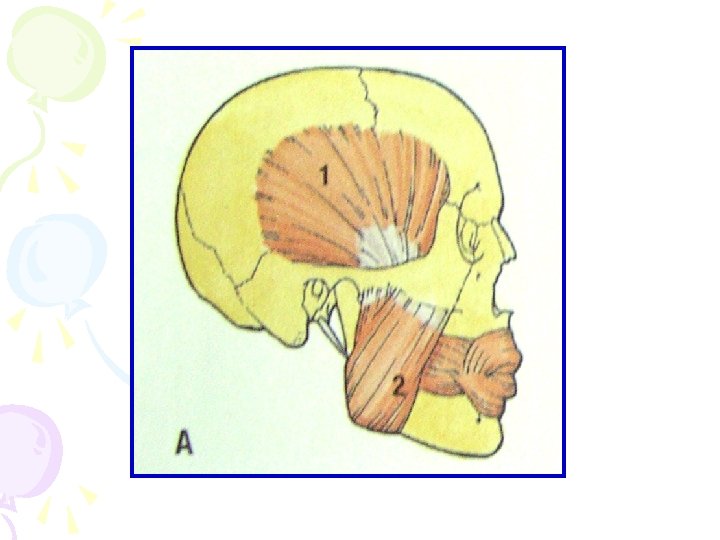
I. Mandibular Elevator muscles 1. Masseter 2. Temporalis 3. Medial Pterygoid 4. Lateral Pterygoid
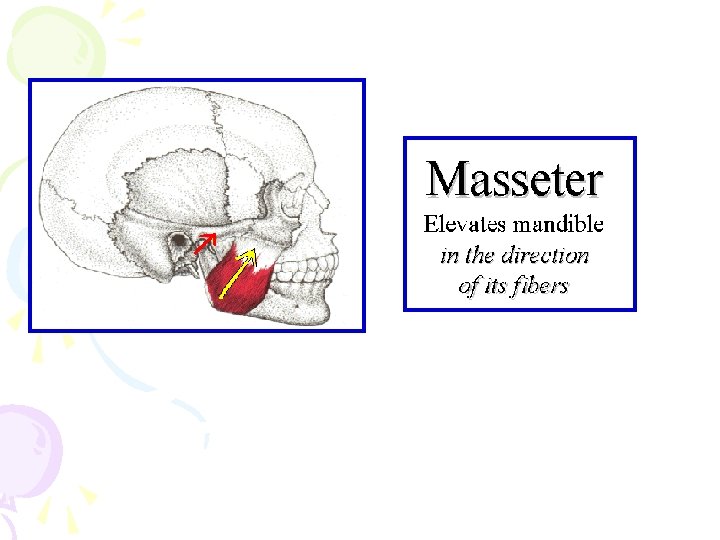
1. Masseter Origin: a) Superficial layer – anterior 2/3 rd of lower border of zygomatic arch & zygomatic processof maxilla b) Middle layer – anterior 2/3 rd of deep surface & posterior 1/3 rd of lower border of zygomatic arch c) Deep layer – deep surface of zygomatic arch
Insertion : a) Superficial layer –lower part of lateral surface of ramus of mandible b) Middle layer –middle part of ramus c) Deep layer – upper part of the ramus & coronoid process Nerve Supply: Massetric nerve Blood supply: Maxillary artery
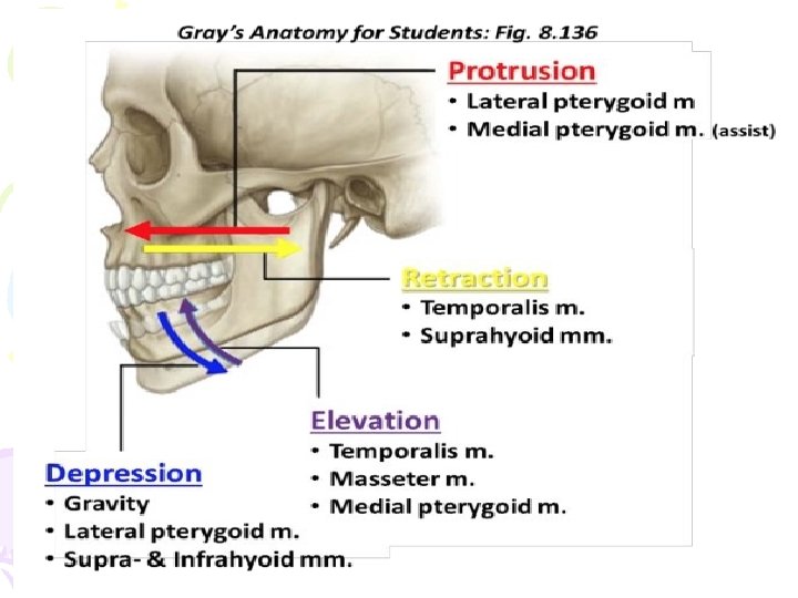
Actions: • Elevates the mandible to close the mouth and to occlude the teeth in mastication. • Its activity in the resting position is minimal. • It has a small effect in side-to-side movement, protraction and retraction.
2. Temporalis Origin: a) Temporal fossa, excluding the zygomatic bone Insertion : a) Margins & deep surface of coronoid process b) Anterior border of ramus of mandible Nerve Supply: Deep temporal nerves Blood Supply : Deep temporal part of maxillary artery
• Actions: a) Elevates the mandible, this movement requires both the upward pull of anterior fibers and backward pull of the posterior fibers. b) Posterior fibers draw the mandible backwards after it has been protruded. c) It is also a contributor to side grinding movement.
Elevation of Mandible
Posterior Fiber Draws Mandible Backwards
Side to Side Grinding Movement
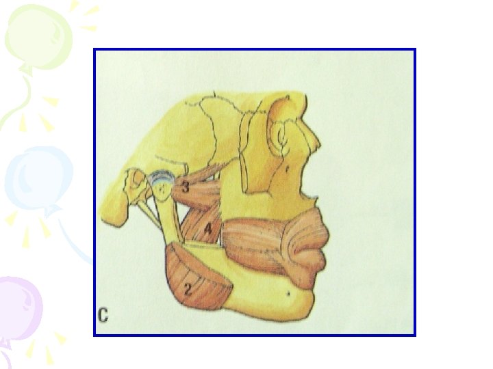
3. Lateral Pterygoid Origin: a) Upper head – infratemporal surface & crest of greater wing of sphenoid bone b) Lower head – lateral pterygoid plate Insertion : a) pterygoid fovea on the anterior surface of neck of mandible b) anterior margin of articular disc & capsule of TMJ
Nerve Supply: branch of anterior division of mandibular nerve Blood Supply: Pterygoid branch of 2 nd part of maxillary artery Action: a) Depresses the mandible b) protrudes the mandible c) side movements
The combined efforts of the Digastrics and Lateral Pterygoids provide for natural jaw opening.
Side To Side Grinding Movement
Medial and lateral pterygoid act together to protrude the mandible
4. Medial Pterygoid Origin: a) Superficial head – maxillary tuberosity b) Deep head – medial surface of lateral pterygoid plate Insertion : medial surface of angle & adjoining part of ramus of mandible
Nerve Supply: Nerve to medial pterygoid Blood Supply: Pterygoid branch of 2 nd part of maxillary artery Action: a) Depresses the mandible b) protrudes the mandible c) side movements
Mandibular Depressor Muscles I. Suprahyoid Muscles 1. Digastric 2. Stylohyoid 3. Mylohyoid 4. Geniohyoid
II. Infrahyoid muscles 1. Sternohyoid 2. Thyrohyoid 3. Omohyoid
Masticatory Cycle / Chewing cycle • The pathway of mandible during chewing is referred to as chewing cycle • Characterised by opening, closing, retrusive, protrusive & lateral jaw movements • 3 phases – 1) Opening phase (mandible is depressed) 2) Closing Phase (mandible is elevated) 3) Occlusal / Intercuspal phase
Jaw movements during masticatory cycle • Both condylar head and mandibular body move during masticatory cycle. • Mandibular and condylar movements are associated with mastication are the coordinated results of sequenced mandibular muscle contraction. • When movement of mandible is traced during masticatiory stroke “ a tear drop ” shaped tracing is observed.
Chewing
1. Opening phase: • Intake of bolus in the mouth – mouth opens by reflex , inhibition of elevators • Isotonic contraction : shorten to produce jaw movements against a constant load. • Isotonic contraction of depressors • Condyles move forward & downward • Divided into a) Slow opening b) Rapid opening
• In the opening phase mandible drops downward from intercuspal position to a point where incisal edge are about 16 to 18 mm apart. • Mandible moves 5 to 6 mm laterally from the midline as the closing movement begins. • In opening stage condyle on the working side moves laterally and on balancing side moves medially downward and forward make the mandible to shift on working side.
2. Closing Phase: • In the beginning of this phase – elevators show isotonic contraction • In the end of this phase – elevators (masseter) shows isometric contraction(muscle length do not change during contraction) • Gradual transition from isotonic contraction to isometric contraction • Condyles – On working side – moves back almost horizontally - On balancing side – moves upwards & backwards in reverse direction
• 2. Closing Phase: Crushing phase: it is the first phase in which food is trapped between the teeth Grinding phase: it is later phase which permit grinding of food.
• Crushing phase: first phase of the closure which traps food between the teeth. • As the crushing phase proceeds , mandible continues to close and trapped bolus will be grinded during grinding phase. • During grinding phase mandible is guided by occlusal surface of teeth, which has come back to the intercuspal position.
3. Intercuspal phase: • Tooth to tooth contact occurs • Path of mandibular closure determined by slide of mandibular teeth along the cuspal inclines of maxillary teeth • First cuspal contact B-B, then L-L, then cusps glide sideways
• 3 possibilities – a) Failure to penetrate the intervening food particle even after power stroke b) Slow penetration of food particle & tooth contact c) Sudden breakage of food particle leading to unloading reflex & separation of occluding surfaces
Jaw opening phase • In this chewing cycle there is first bilateral activation of- Mylohyoid - Anterior Belly of digastric - inferior head of lateral pterygoid muscle. • Degree of separation of occluding surfaces is variable even in one person because it depends on the size of food in the mouth. • Normally lower incisors move downwards by 10 -15 mm.
Slow jaw closing phase • Fasts closure phase ends when resistance is detected between the teeth. • Results into tooth-to-tooth contact. • There are three possible outcomes of the slow closure stroke: 1) failure to penetrate the intervening food particle even after a power stroke which aims to crush it. 2) slow penetration of the food particle and tooth contact. 3) sudden breakage of the food particle leading to the unloading reflex and separation of occluding surfaces. • In all these outcomes it is the stimulation of periodontal receptors that helps to initiate next cycle.
Neural Masticatory Receptors 1. 2. 3. 4. 5. Muscle spindles Golgi tendon organs Periodontal mechanoreceptors Mucous membrane receptors TMJ receptors
1. Muscle spindles: • Stretch sensitive specialized receptors that monitor any change in length of the muscle • Large number of spindles in mandibular elevators • Highest – Fingers • Intermediate – Masticatory muscles • Low – Limb muscles
• SENSORY RECEPTORS : Masticatory system uses four major types of sensory receptors to monitor the status of its structures : 1)Muscle spindle 2)Golgi tendon organs
2. Golgi Tendon organs: • Receptors primarily located at muscle – tendon junctions or TMJ capsule • Monitor tension • Masticatory muscles do not have golgi tendon organs 3. Periodontal mechanoreceptors: • Respond to forces applied to the teeth
PDL- RECEPTORS • There are conflicting reports of wheather the elevator muscles have golgi tendon organs. • It is however agreed that the role of the golgi tendon organ in protecting against the overdevelopment of muscle tension. • In oral cavity is performed by the “PDL receptors”, which limit the force and can be applied in mastication.
CONTROL OF MASTICATORY FORCE BY PDL RECEPTORS
4. Mucous membrane receptors: • Pressure receptors - palate • Touch receptors – tongue 5. TMJ receptors: • Free nerve endings (mainly) • Ruffini endings • Pacinian corpuscles • Golgi receptors
• Neural control of mastication
Control of mastication • Mastication may be controlled entirely voluntarily by the motor cortex. • Control automatically by the masticatory rhythm generator in the pons. • This is subjected to modifying influences from sensory receptors in oralmucosa, muscles of mastication, PDL of teeth and TMJ. • When food bolous is perceived to be appropriate consistancy the swallowing center is activated.
Control of masticaction
• Masticatory reflexes
Masticatory Reflexes / Jaw Reflexes • Reflex is a highly stereotyped automated response to a specific stimulus • Masticatory reflexes are type of stretch reflexes brought about by activation of muscle spindles in the muscles
• Types of Jaw Reflexes: 1. Jaw Closing Reflex / Jaw Jerk Reflex 2. Jaw Opening Reflex 3. Jaw Unloading Reflex 4. Tooth Contact Reflexes 5. Horizontal Jaw Reflexes