Mass Spectrometry Mass spectrometry MS differs from other
 differs from other common forms of organic spectral analysis")


- Slides: 27
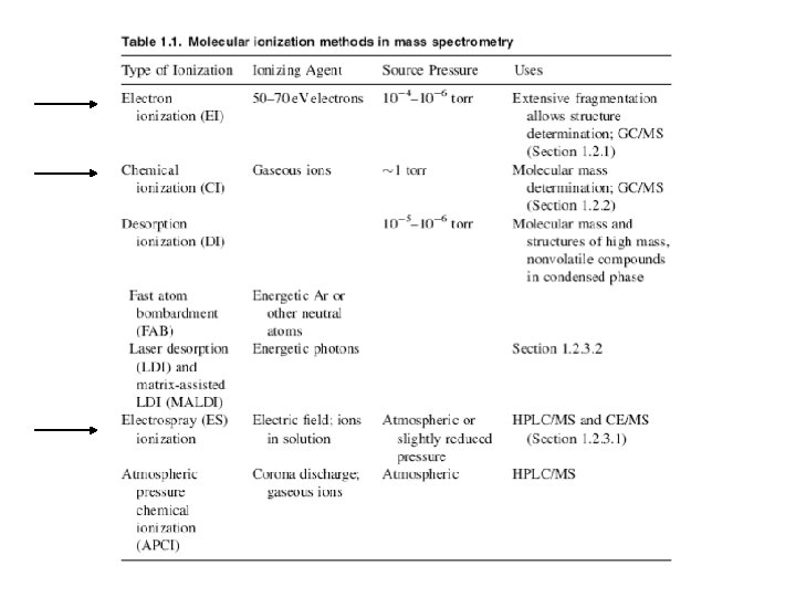
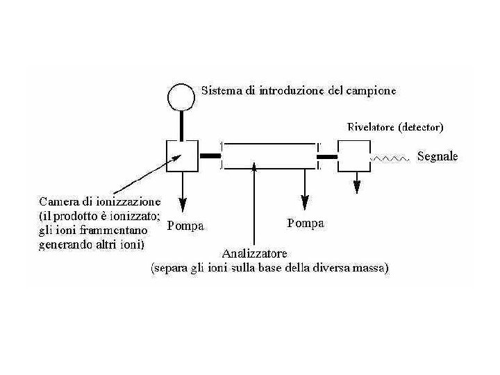
Mass Spectrometry Mass spectrometry (MS) differs from other common forms of organic spectral analysis in that the sample does not absorb radiation such as infrared, ultraviolet, or radio waves from the electromagnetic spectrum. In contrast to infrared (IR) or nuclear magnetic resonance (NMR) spectrometry, both of which identify compounds with specificity comparable to that of mass spectrometry, MS is a destructive method of analysis—that is, the sample cannot be recovered after mass spectral analysis. Mass spectrometers are typically not standalone instruments. Most often they are connected physically and electronically to a chromatograph: the chromatograph separates mixtures and introduces the sample into the mass spectrometer; the mass spectrometer ionizes analyte molecules, then separates and detects the resulting ions. In order to be analyzed by mass spectrometry, sample molecules must be ionized.
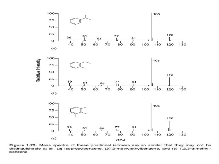
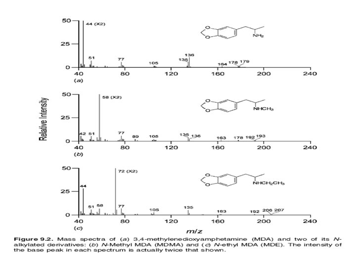
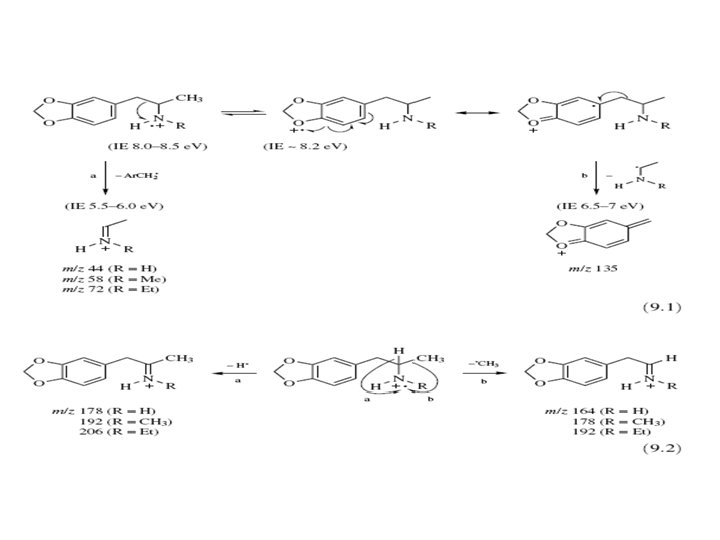
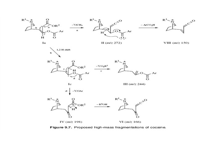
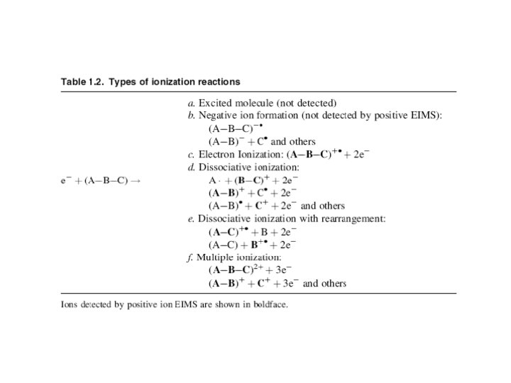
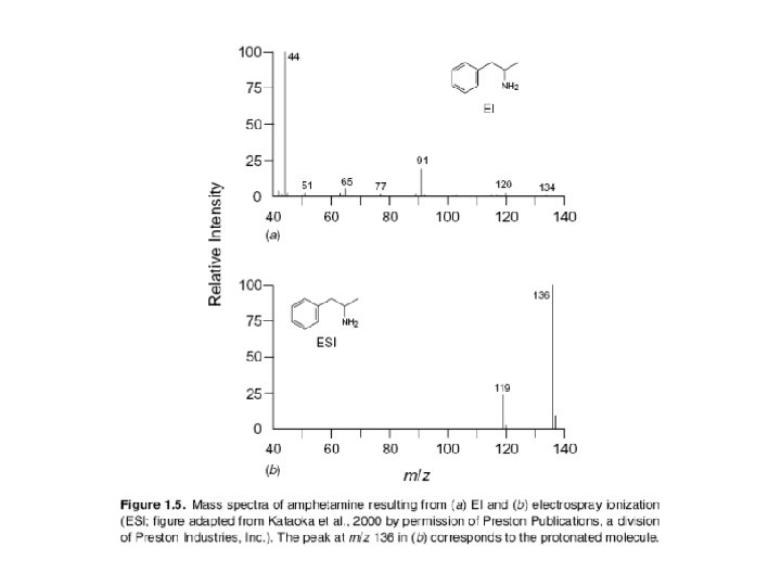
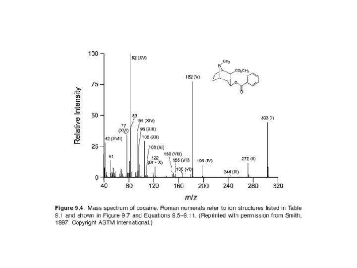
The ion that is formed first results directly from ejection of a single electron from the neutral molecule (product c). This molecular ion (M+) is very important because it has virtually the same mass as that of the analyte molecule (the small mass of the lost electron can be ignored). Since mass spectrometry actually measures the mass-tocharge ratio (m/z) of an ion, not its mass, an ion having a charge greater than +1 is found not at the m/z value corresponding to its mass (m), but rather at m/2, m/3, or m/4, depending on the number of charge states.
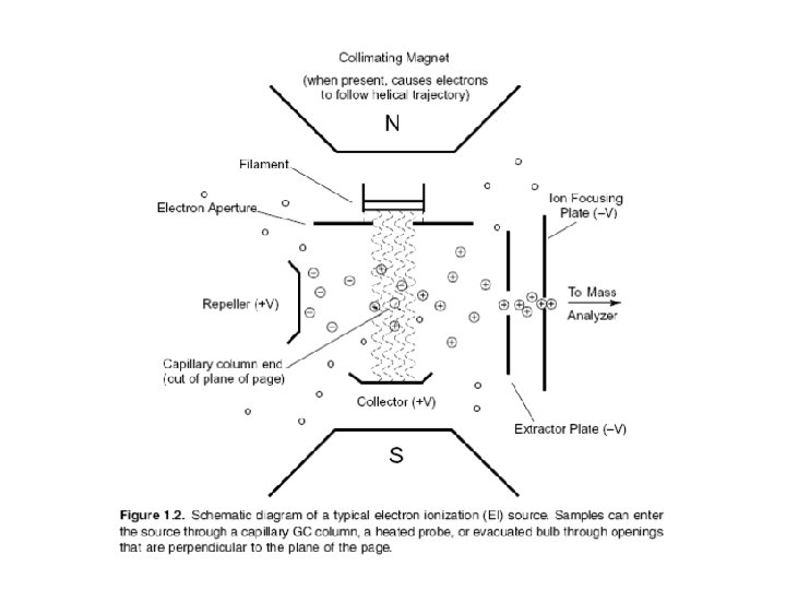
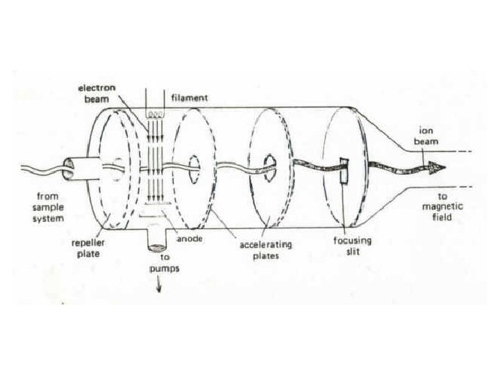
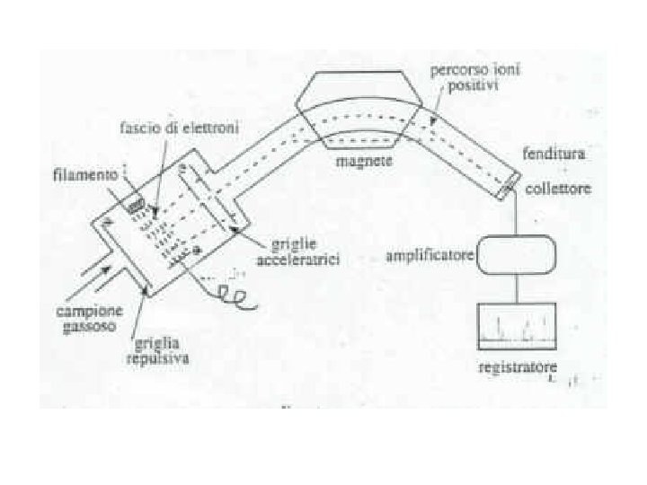
Electron Ionization Source The EI source is most commonly a small chamber about 1 cc in volume, in which analyte molecules interact with a beam of highly energetic electrons that have typically been accelerated through a potential difference of 50– 70 volts (V) across the volume of the ion source.
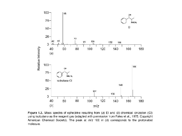
Chemical Ionization Unlike EIMS, in which molecules are ionized through interaction with high-energy electrons, ionization in chemical ionization mass spectrometry (CIMS) depends on collisions of ions and molecules. In positive ion CIMS the sample is ionized by reaction with ions generated within a large excess of a relatively low molecular mass reagent gas such as methane (as CH+5), isobutane [as (CH 3)3 C+], or ammonia.
In CIMS the concentration of analyte molecules is small compared to that of reagent gas molecules. Thus, the electron beam, which is more energetic than that used in EIMS (200 e. V), preferentially ionizes the reagent gas. Analyte molecules are ionized through reaction with reagent gas ions, rather than by the electron beam. Most reagent gas ions are strong proton donors and form protonated molecules (sometimes incorrectly called pseudomolecular ions) that have a mass 1 u greater than that of the molecular mass of the original compound: M + CH +5 MH+ + CH 4
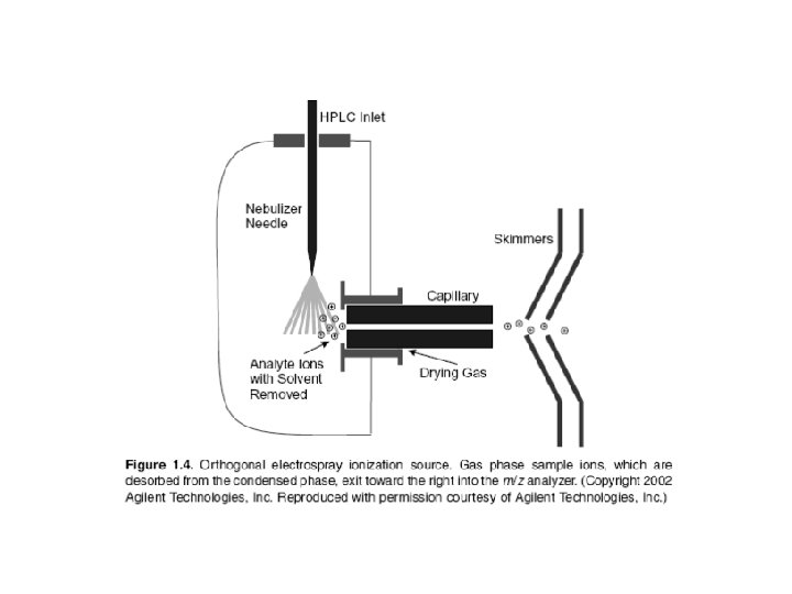
Electrospray Ionization Basically ESI works by converting the HPLC effluent, already containing the sample in ionic form, into an aerosol and subjecting the resulting spray to high voltage in a chamber held near atmospheric pressure. This process creates a mist of charged droplets that flow toward the orifice of the capillary. In the configuration shown, the nebulizing needle, which creates the aerosol, is orthogonal (perpendicular) to the eventual direction of ion flow toward the m/z analyzer. Other geometric configurations are possible and have been used. As the charged droplets travel toward the capillary opening, they are subjected to the counterflow of a drying gas, such as nitrogen (N 2), which causes evaporation of solvent molecules from the droplets. Evaporation, charge concentration, and droplet disintegration continue until the analyte ions are finally desorbed into the vapor phase, passed into the sampling capillary, then on into the high vacuum of the m/z analyzer.
Trappola Ionica: Può essere considerato una variante dell'analizzatore a quadrupolo. Anzichè permettere agli ioni di attraversare il campo quadrupolare, la trappola ionica trattiene tutti gli ioni al suo interno. Lo spettro di massa è generato variando il potenziale elettrico in modo da espellere in sequenza dalla trappola verso il rivelatore gli ioni secondo un valore m/z crescente.
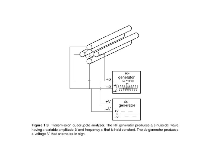
Analizzatore a quadrupolo: E’ costituito da quattro barre cilindriche metalliche, lunghe ca. 20 cm. , che delimitano il "cammino" percorso dagli ioni provenienti dalla camera di ionizzazione e diretti al detector. Le barre sono mantenute ad un potenziale elettromegnetico oscillante, in modo che quando le due sbarre verticali hanno potenziale positivo quelle orrizzontali l’hanno negativo, e viceversa. Gli elettroni, accelerati dalle piastre acceleratrici, entrano nel tunnel delimitato dalle barre e vengono respinti dai poli positivi ed attratti dai negativi. Tuttavia, a causa dell’oscillazione del quadrupolo gli ioni assumono una traiettoria a zig zag e finiscono con lo scaricarsi su una delle barre, tranne quelli che, per una certo valore di frequenza di oscillazione, hanno un’energia cinetica tale per cui il moto diventa sinusoidale e riecono ad uscire dal tunnel ed entrare nel sistema di rivelazione (fotomoltiplicatore). Operando quindi una scansione di frequenza di oscillazione del campo è possibile far uscire ioni a massa molecolare crescente.