
Maria PodolakDawidziak Donata UrbaniakKujda Thrombophilia Katedra i Klinika
Maria Podolak-Dawidziak Donata Urbaniak-Kujda Thrombophilia Katedra i Klinika Hematologii, Nowotworów Krwi i Transplantacji Szpiku UM, Wrocław

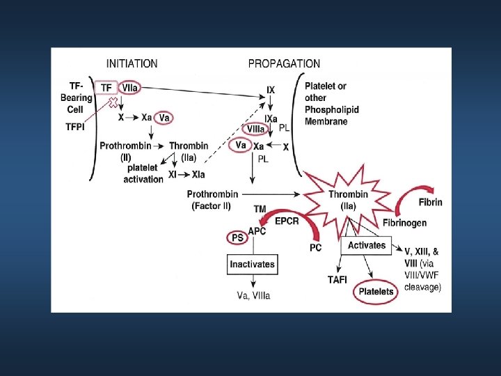
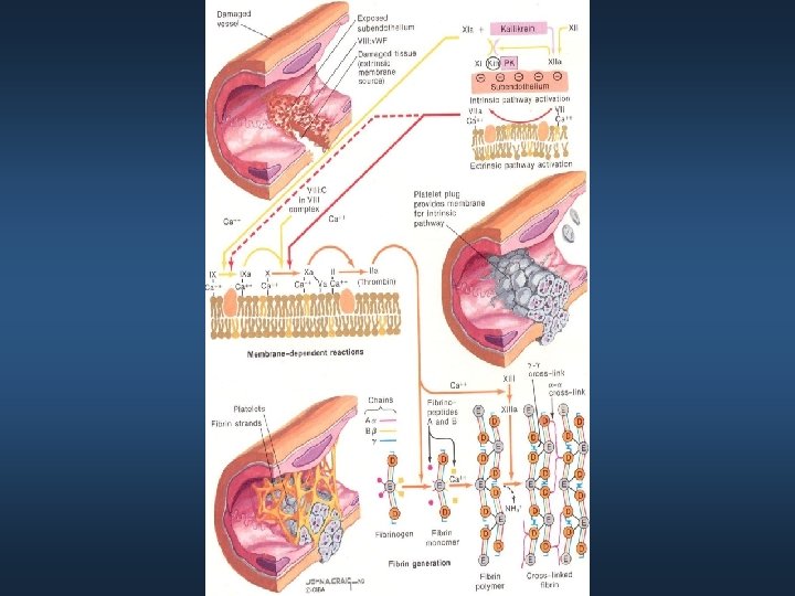
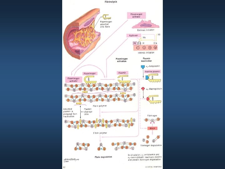
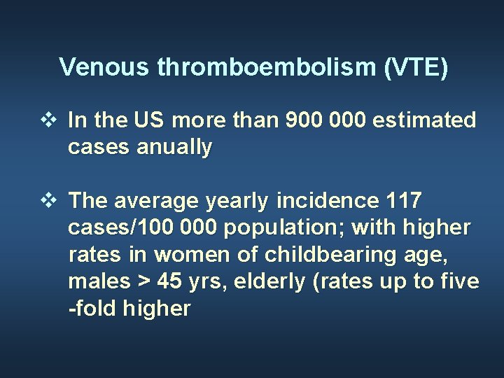
Venous thromboembolism (VTE) v In the US more than 900 000 estimated cases anually v The average yearly incidence 117 cases/100 000 population; with higher rates in women of childbearing age, males > 45 yrs, elderly (rates up to five -fold higher
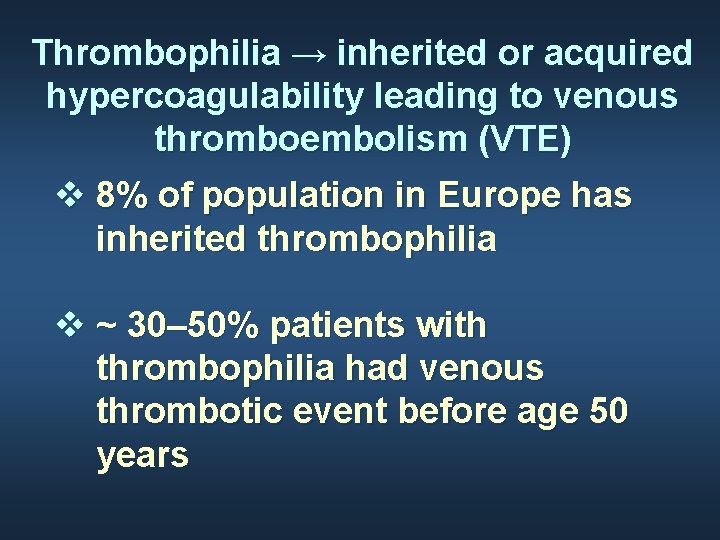
Thrombophilia → inherited or acquired hypercoagulability leading to venous thromboembolism (VTE) v 8% of population in Europe has inherited thrombophilia v ~ 30– 50% patients with thrombophilia had venous thrombotic event before age 50 years
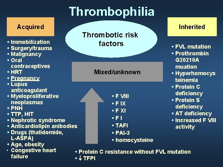
Thrombophilia Acquired Inherited Thrombotic risk factors w Immobilization w FVL mutation w Surgery/trauma w Prothrombin w Malignancy G 20210 A w Oral contraceptives muation w HRT Mixed/unknown w Hyperhomocys w Pregnancy teinemia w Lupus w Protein C anticoagulant deficiency w F VIII w Myeloproliferative w Protein S neoplasmas w F IX deficiency w PNH w F XI w AT deficiency w TTP, HIT w. F I w Increased F VIII w Nephrotic syndrome w TAFI w Anticardiolipin anibodies activity w Drugs (thalidomide, w PAI-3 L-ASPA) w homocysteine w Age, obesity • Congestive heart w Protein C resistance without FVL mutation failure w ¯ TFPI

Most common thrombophilias Inherited: v Factor V Leiden v prothrombin G 202 10 A mutation v 4 G/4 G mutation of the plasminogen activator inhibitor gene (PAI-1) v thermolabine variant of methylenotetrahydrofolate reductase, the most common cause of homocysteinamia v antithrombin (AT) deficiency v protein C deficiency v protein S deficiencyagulable state Acquired Antiphospholipid antibody
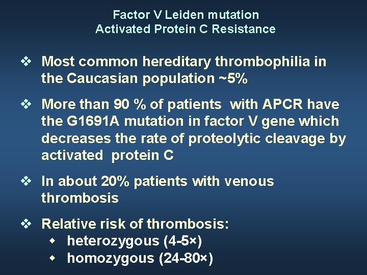
Factor V Leiden mutation Activated Protein C Resistance v Most common hereditary thrombophilia in the Caucasian population ~5% v More than 90 % of patients with APCR have the G 1691 A mutation in factor V gene which decreases the rate of proteolytic cleavage by activated protein C v In about 20% patients with venous thrombosis v Relative risk of thrombosis: w heterozygous (4 -5×) w homozygous (24 -80×)
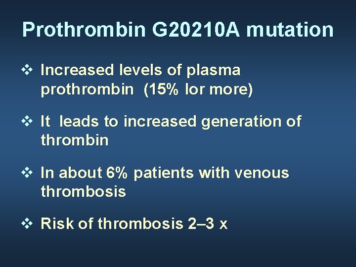
Prothrombin G 20210 A mutation v Increased levels of plasma prothrombin (15% lor more) v It leads to increased generation of thrombin v In about 6% patients with venous thrombosis v Risk of thrombosis 2– 3 x
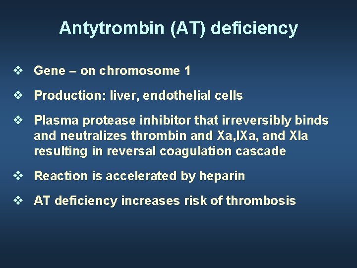
Antytrombin (AT) deficiency v Gene – on chromosome 1 v Production: liver, endothelial cells v Plasma protease inhibitor that irreversibly binds and neutralizes thrombin and Xa, IXa, and XIa resulting in reversal coagulation cascade v Reaction is accelerated by heparin v AT deficiency increases risk of thrombosis
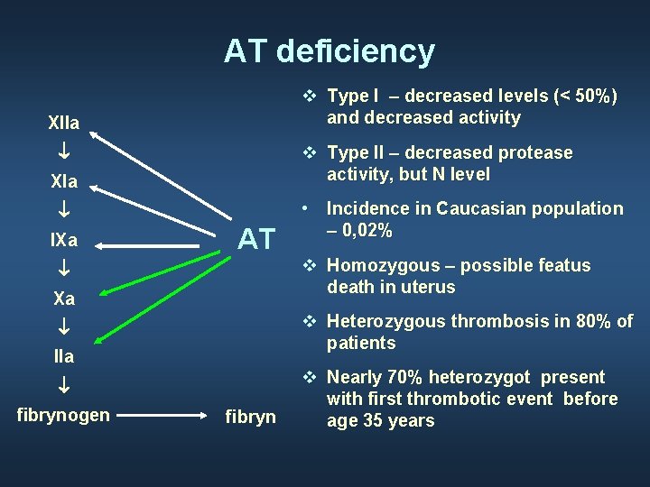
AT deficiency v Type I – decreased levels (< 50%) and decreased activity XIIa ¯ v Type II – decreased protease activity, but N level XIa ¯ IXa AT ¯ Xa v Homozygous – possible featus death in uterus v Heterozygous thrombosis in 80% of patients ¯ IIa ¯ fibrynogen • Incidence in Caucasian population – 0, 02% fibryn v Nearly 70% heterozygot present with first thrombotic event before age 35 years
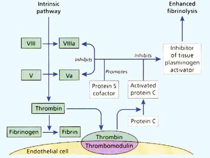
Protein C and protein S v Are vitamin K-dependent enogenous anticoagulants v Homozygous protein C deficiency can cause neonatal purpura fulminans v Patients with either protein C or protein S deficiency → warfin skin necrosis at the initiation of anticoagulation due to transient hypercoagulable state
Protein C and protein S deficiency v Protein C inhibits thrombin v Protein C and protein S are affected by liver disease, anticoagulation with warfin, nephrotic syndrome, DIC, vitamin K deficiency, oral contraceptives, pregnancy, and hormonal replacement therapy
Elevated F VIII levels v Relative risk of thrombosis=4. 8 v Are found with increased age, obesity, pregnancy, surgery, inflammation, liver disease, hyperthyreoidism, and diabetes v No gene alteration has been found, although familial clustering of increased FVIII levels is noted
Hereditary thrombotic dysfibrinogenemia v Qualitative defects in the fibrin molecule v VTE in 20 % of pts and bleeding tendency in 25 % of pts, but 55 % are asymptomatic v N or low levels of FBG v Prolonged thrombin time (TT)
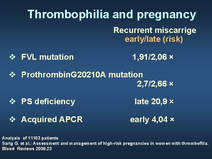
Thrombophilia and pregnancy Recurrent miscarrige early/late (risk) v FVL mutation 1, 91/2, 06 × v Prothrombin. G 20210 A mutation 2, 7/2, 66 × v PS deficiency v Acquired APCR late 20, 9 × early 4, 04 × Analysis of 11183 patients Sarig G. et al. : Assessment and management of high-risk pregnancies in women with thrombofilia. Blood Reviews 2009. 23
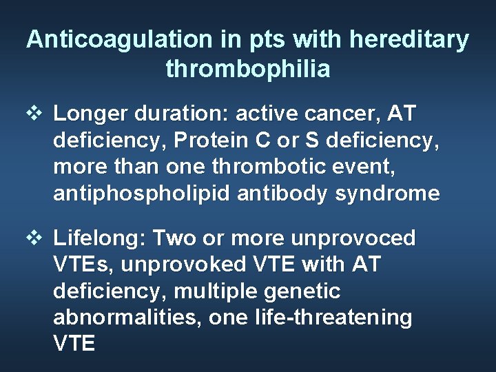
Anticoagulation in pts with hereditary thrombophilia v Longer duration: active cancer, AT deficiency, Protein C or S deficiency, more than one thrombotic event, antiphospholipid antibody syndrome v Lifelong: Two or more unprovoced VTEs, unprovoked VTE with AT deficiency, multiple genetic abnormalities, one life-threatening VTE
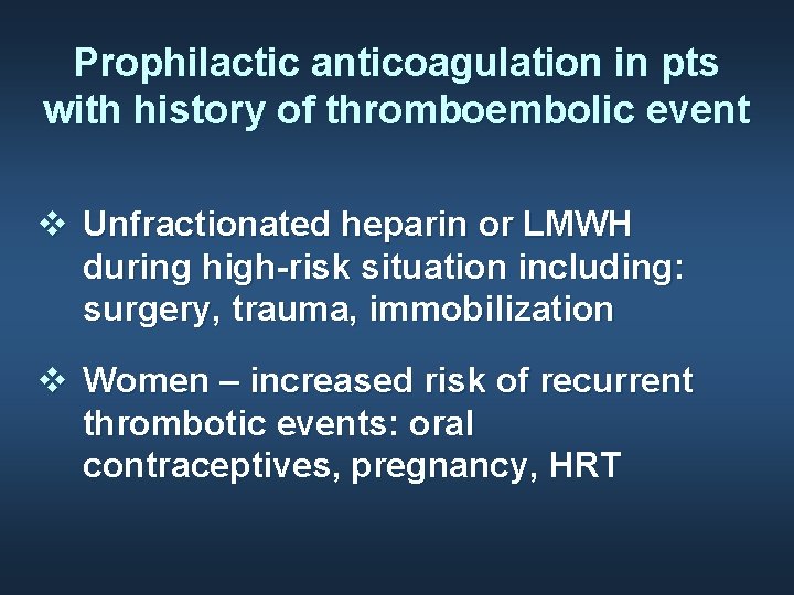
Prophilactic anticoagulation in pts with history of thromboembolic event v Unfractionated heparin or LMWH during high-risk situation including: surgery, trauma, immobilization v Women – increased risk of recurrent thrombotic events: oral contraceptives, pregnancy, HRT
Consideration of a hypercoagulable workup v Thrombosis at age < 50 years v Recurrent VTE v Thrombosis at unusual sites (cerebral sinus, mesenteric vein, portal vein, hepatic vein) v Recurrent second or third trimester fetal loss, placental abruption, or severe preeclampsia
Anticoagulation in the primary treatment and prophilaxis of VTE The purpuse of anticoagulation is to: v prevent additional clots from developing, v immediately stop further propagation of the existing clot v Permit endogenous fibrinolysis to begin to dissolve the clot physiologically
Anticoagulation in the primary treatment and prophilaxis of VTE v LMWH or fondaparinux or rivaroxaban v LMWH and fondaparinux are preferable to UFH v if warfin (VKA) is used, it should be started after initiation of UFH, LMWH or fondaparinus v anticoagulation – for at least 3 months
Patient No 1 v 28 years old, white man v Sudden symptoms: dyspnea, cough, chest pain, hemoptysis. BMI 19 kg/m 2 v No thrombotic history; one week prior to event he drived a car for 8 hours without any break v Laboratory tests: blood morphology – N, PT- N, APTT - N, dimer D 4054 ng/ml (N < 500 ng/ml) v CT: acute bilateral pulmonary embolism v Doppler USG: DVT in both legs!! (l. Shaheen K et al. : Factor V Leiden: How great is the risk of venous thromboembolism Cleveland Clin J Med. 2012; 79: 265 -272
Patient No 1 v FV Leiden mutation heterozygous type v Treatment: UFH, later warfin ► INR 2 -3 for 2 consecutive days v At home: oral anticoagulation for 6 months (INR 2 -3). Two weeks after end of treatment: AT, FVIII, PC i PS - normal
Patient No 2 v 17 years old girl with no thrombotic history; since 6 months on oral contraceptives v Sudden pain in left groin v DVT i PE v No family history of thrombosis v Diagnosis: FVL mutation heterozygous type, FII G 20210 A mutation and MTHFR C 677 T, 4 G/5 G PAI gen- type 1 mutation homozygous type Krieza L et al: Pathophysiol Haemost Thromb 2010; 37: 24 -29
Consideration of a hypercoagulable workup v Thrombosis at age < 50 years v Recurrent VTE v Thrombosis at unusual sites (cerebral sinus, mesenteric vein, portal vein, hepatic vein) v Recurrent second or third trimester fetal loss, placental abruption, or severe preeclampsia
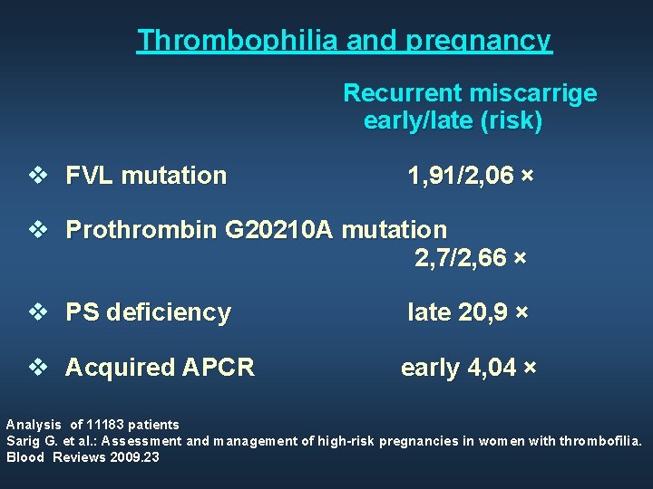
Thrombophilia and pregnancy Recurrent miscarrige early/late (risk) v FVL mutation 1, 91/2, 06 × v Prothrombin G 20210 A mutation 2, 7/2, 66 × v PS deficiency late 20, 9 × v Acquired APCR early 4, 04 × Analysis of 11183 patients Sarig G. et al. : Assessment and management of high-risk pregnancies in women with thrombofilia. Blood Reviews 2009. 23
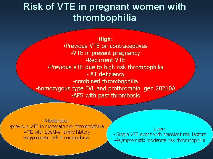
Risk of VTE in pregnant women with thrombophilia High: • Previous VTE on contraceptives • VTE in present pregnancy • Recurrent VTE • Previous VTE due to high risk thrombophilia - AT deficiency -combined thrombophilia -homozygous type FVL and prothrombin gen 20210 A • APS with past thrombosis Moderate: • previous VTE in moderate risk thrombophilia • VTE with positive family history • Asyptomatic risk thrombophilia Low: • Single VTE event with transient risk factors • Asymptomatic moderate risk thrombophilia
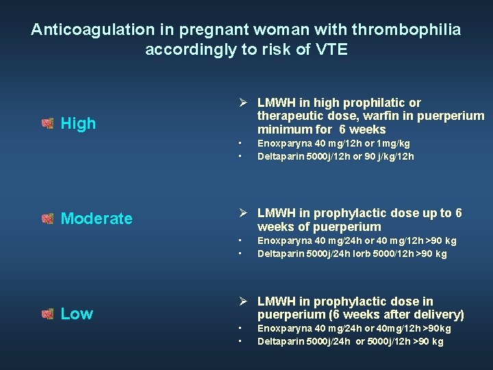
Anticoagulation in pregnant woman with thrombophilia accordingly to risk of VTE High LMWH in high prophilatic or therapeutic dose, warfin in puerperium minimum for 6 weeks • • Moderate LMWH in prophylactic dose up to 6 weeks of puerperium • • Low Enoxparyna 40 mg/12 h or 1 mg/kg Deltaparin 5000 j/12 h or 90 j/kg/12 h Enoxparyna 40 mg/24 h or 40 mg/12 h >90 kg Deltaparin 5000 j/24 h lorb 5000/12 h >90 kg LMWH in prophylactic dose in puerperium (6 weeks after delivery) • • Enoxparyna 40 mg/24 h or 40 mg/12 h >90 kg Deltaparin 5000 j/24 h or 5000 j/12 h >90 kg
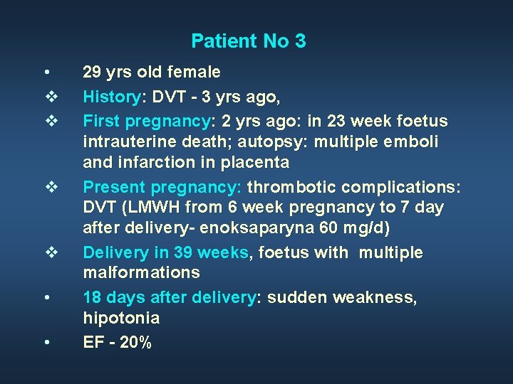
Patient No 3 • v v • • 29 yrs old female History: DVT - 3 yrs ago, First pregnancy: 2 yrs ago: in 23 week foetus intrauterine death; autopsy: multiple emboli and infarction in placenta Present pregnancy: thrombotic complications: DVT (LMWH from 6 week pregnancy to 7 day after delivery- enoksaparyna 60 mg/d) Delivery in 39 weeks, foetus with multiple malformations 18 days after delivery: sudden weakness, hipotonia EF - 20%
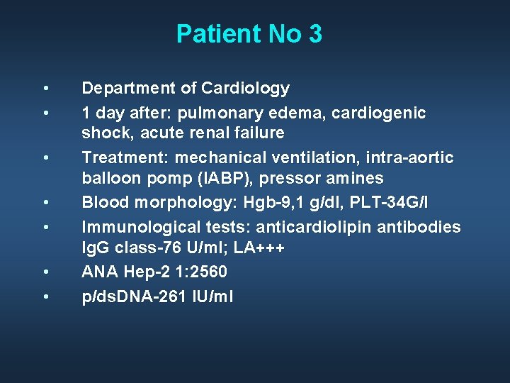
Patient No 3 • • Department of Cardiology 1 day after: pulmonary edema, cardiogenic shock, acute renal failure Treatment: mechanical ventilation, intra-aortic balloon pomp (IABP), pressor amines Blood morphology: Hgb-9, 1 g/dl, PLT-34 G/l Immunological tests: anticardiolipin antibodies Ig. G class-76 U/ml; LA+++ ANA Hep-2 1: 2560 p/ds. DNA-261 IU/ml
Diagnosis and therapy • • • Diagnosis: LED, antiphospholipid syndrome Differential: inherited thrombophilia, TTP Therapy: Metylprednison 1, 0 g for 5 days; IVIG- total dose 150 g LMWH in therapeutic dose EF-35%; PLT-143 G/l
• • • 15 days after: left side hemiplegia, unconcious MRI- acute multifocal cerebral ischemia USG Doppler- multiple thrombi in left jugulr athery Therapy: anticoagulation (ASA, warfin) immunosupression: CPD (5 pulses), metylprednisolon, plasmapheresis (5 times) A year after: clical improvement (no thrombotic or neurologic events), decrease in APS abs
- Slides: 36