March 31 2016 Orlando FL Vertigo Workshop Mike


 • (Vascular Loop Syndrome) • Multiple Sclerosis")


 by stimulating hair")


 Mercado 2013©")


, which includes caloric testing, is one of the most vital tests")


 33")


- Slides: 53
March 31, 2016 Orlando, FL Vertigo Workshop Mike Valdez, PA-C Updated 12/03/2015
Vertigo Workshop Clear Instruction Live Demonstration Hands-On Practice Learn by doing Vertigo examination Neurological examination Rhomberg Test Fukada Stepping Test Demonstration ENG/VNG Canalith Repositioning
Introduction There are multiple methods and techniques available to successfully complete all the topics presented in this workshop. Some are based on patient request, available equipment or supervising physician’s preference. The goal of this workshop is to correctly demonstrate the most common methods and give participants time for hands on training.
Vertigo Workshop Learning Objectives • Discuss and demonstrate vertigo examination; – Neurological examination – Rhomberg Test – Fukuda Stepping Test – Dix-Hallpike • Demonstrate ENG/VNG. • Demonstrate and practice canalith repositioning
Balance Mercado 2013©
Clinical Evaluation of Vertiginous Patient Central Peripheral Labrynthitis Vestibular Neuronitis BPPV Perilymphatic Fistula Meniere’s Disease Autoimmune Ataxia Vascular disorders (Vertibrobasilar Insufficiency) (Vascular Loop Syndrome) Multiple Sclerosis CNS Neoplasm (tumor) Cardio (orthostatic hypotension) Cerebrovascular (CVA/TIA) Migraine Syphilis Systemic Neurology/Cardiology Medication Endocrine Disequilibrium Mercado 2013© Otolaryngology
Algorithm History Central Physical Exam CN II-XII Romberg Fukuda Dix-Hallpike Peripheral Hearing Test Audio/Tymps ABR/OAE IMAGING CT/MRI/MRA Carotid U/S Diagnostic Tests LABS Mercado 2013© Balance Test ENG/VNG Ecog
Clinical Evaluation of Vertiginous Patient • History • Physical examination – Pneumatic Otoscopy • Middle ear disease • Hennebert’s Sign – CN II-XII – Romberg – Fukuda Stepping Test – Dix-Hallpike • Diagnostic tests • Goal is to Differentiate central vs. peripheral
Peripheral Vestibular Neuritis Dizziness/Vertigo Nausea/Vomiting Cochlear Neuritis Ear Pressure /Full Hearing loss Tinnitus Labyrinthitis Dizziness/Vertigo Nausea/Vomiting Ear Pressure /Full Hearing loss Tinnitus May be bacterial Very sick
Central • Vascular disorders • (Vertibrobasilar Insuficiency) • (Vascular Loop Syndrome) • Multiple Sclerosis • CNS Neoplasm (tumor) • Cardio (orthostatic hypotension) • Cerebrovascular (CVA/TIA)
Physical Examination Visualize tympanic membranes; • Infection • Perforation • Trauma • Cholesteatoma /otorrhea.
Physical Examination Develop routine and systematic approach to dizzy patient. Begin with brief neurological exam • CN II – confrontation testing and ophthalmoscopic exam. • CN III, IV, VI- extraoccular movement (EOM) • CN V – corneal reflex • CN VII – facial strength & symmetry • CN VIII – Webber/Renne audiogram • CN IX – gag reflex soft palate • CN XI – shoulder shrug • CN XII – tongue protrusion Not a neurologist – looking for gross abnormalities
Pneumatic Otoscopy • Hennebert’s Sign – nystagmus and vertigo with +/- pressure • Normally: No nystagmus • May be positive in: Perilymph fistula, Semicircular canal dehiscence syndrome, and Meniere’s disease Mercado 2011 ©
Romberg Test • Patient asked to stand with feet together and eyes closed • Increased sway with eyes closed suggests inner ear problem • Equal sway with eyes open and closed suggests CNS problem • Fall or step is positive test (usually towards side of lesion) • Positive usually posterior column disorder
Fukuda Stepping Test • Patients are asked to step with eyes closed and hands out in front • 100 steps. • Turn usually occurs to the side of the lesion • Forward motion is often normal poor sensitivity and specificity.
Tandem Gait Test • Patients are asked to walk heel to toe in a straight line or in a circle • Complex function evaluates many aspects of balance • Poor performance seen in cerebellar lesions, but can lesions be seen in many disorders • Poor sensitivity and specificity
Dix-Hallpike
Positional Vertigo • Dix-Hallpike Maneuver reproduces benign paroxysmal positional vertigo (BPPV) by stimulating hair cells. • Majority of BPPV is posterior canal. • Canalithiasis theory -Free floating debris (dislodged otoconia) in the endolymph of the posterior canal – inertial drag of endolymph causes displacement of the cupula resulting in latent vertigo which resolves when debris settles
• Dix-Hallpike – examiner stands to the side of the patient, who sits upright with head turned to examiner (LEFT). The patient is positioned so that when the body is supine, the head will extend BEYOND end of table. Consider a shoulder roll to provide adequate headhanging. • The examiner holds the head and moves the patient RAPIDLY from sitting to head hanging position. First with the head turned to one side, then the other. Once in the head hanging position, patients with BPPV will show a burst of nystagmus after a delay of 5 -10 seconds. Episodes last a few seconds (fatigueability).
Dix-Hallpike Mercado 2013© Demonstration Dix-Hallpike Maneuver Mercado 2013©
Canalith Repositioning If vertigo is reproducible with Dix-Hallpike Maneuver, patient requires canalith repositioning or Eply Maneuver.
Modified Eply Maneuver Patient’s head is systematically rotated so that the loose particles slide out of the semicircular canal and back into the utricle. 1. If vertigo affects RIGHT ear, the patient is brought to the head hanging position with right ear turned DOWNWARD. 2. Move the head to end of table, rotate head to the left with right ear turned UPWARD. 3. Hold for 30 seconds, then roll patient onto the left side while clinician rotates head LEFTWARD until the nose points down to floor. 4. Hold position for 30 seconds. 5. Then patients returns to sitting position with head facing left.
Canalith Repositioning Mercado 2013© Demonstration Canalith Repositioning (Eply Maneuver) Mercado 2013©
Diagnostic Tests • Audiology: assess Peripheral Vestibular System – – – Hearing: Audiogram, otoacoustic emissions Tympanogram Electrophysiologic: Ecog, ABR, VEMP ENG / VNG Rotary Chair Posturography • Imaging: assess CNS – CT / MRI / MRA – Carotid US • Blood Tests: assess Systemic
Elements of Videonystagmography Dr. Salvatore Gruttadauria, Au. D. Clinical Director-Balanceback 25
Electronystagmogram Electronystagmography (ENG), which includes caloric testing, is one of the most vital tests for evaluating the vertiginous patient. Helps differentiate central versus peripheral etiology and in addition in localizing the dysfunctioning ear. ENG requires approximately 45 to 90 minutes.
Electronystagmogram • Records eye movements during series of positional changes, oculomotor testing, calorics • Helpful in localizing lesion, confirming vestibular disorder present • Not always diagnostic
Electronystagmogram • An electrode is placed lateral to each eye with a ground electrode placed on the forehead. • Because of the voltage differences between the cornea and the retina, eye movements can be graphed on a strip chart recording
Frenzel Glasses Patient can often suppress nystagmus caused by a peripheral vestibulopathy by fixation. Many of the vestibulooculomotor tests in the office examination can be performed with +20 lenses (i. e. , cataract glasses), which prevent the patient from focusing on objects in the visual surround.
Oculomotor Testing Useful as cerebellar disorders and degenerative disorders of the central nervous system • Saccades • Smooth Pursuit • Optokinetics • Gaze 30
Saccades • Peak Velocity • Accuracy • Latency 31
Smooth Pursuit • The tracking test measures the ability of subjects to match eye movement to visual target movement 32
Smooth Pursuit (Abnormal) 33
Optokinetic Testing • The eye movement elicited by the tracking of a moving field. It differs from smooth pursuit which is the eye movement elicited by tracking of a single distinct target. 34
Optokinetics 35
Gaze Testing Central and peripheral implications based on pattern of nystagmus observed during eye positions with and without fixation(vision) • Gaze Right • Gaze Left • Gaze Up • Gaze Down Done with vision and in vision denied conditions 36
High Frequency Headshake Test • The head is shaken in the yaw plane for approximately 30 seconds. • In normal subjects or persons with symmetrical vestibular loss (such as bilateral vestibular loss), no nystagmus is expected. • In persons with a dynamic imbalance between the ears (such as due to unilateral vestibular neuritis or an acoustic neuroma), a nystagmus is often seen (usually beating towards the “better” ear. 37
Positioning Tests • Active movement • Typically Dix-Hallpike but there are many variations • Used to diagnosis BPPV 38
39
Positional Testing Static Test • Looking to see: Does a change in gravitational orientation cause abnormal eye movement? • Specifically looks at the Utricle and Saccule as our linear force detectors • Supine, Head Right, Head Left, Body Right, Body Left, Body Right Headshake, Body left Headshake • Done with vision and vision denied conditions 40
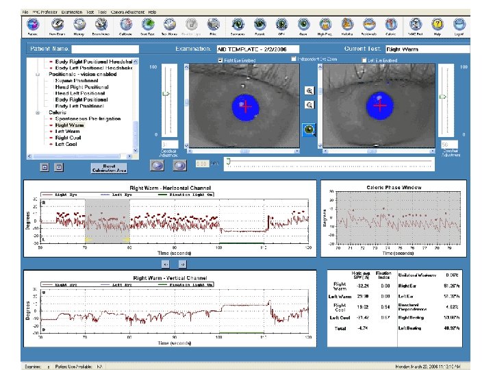
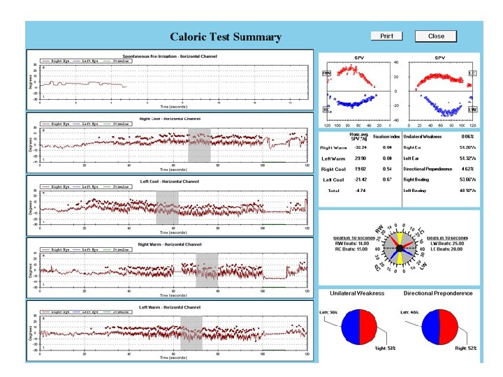
Calorics Air • Temperature Settings • 24 Celsius Cool • 50 Celsius Warm • Length of Stimulation • 50 -60 seconds 41
Calorics Water • Temperature Settings • 30 Celsius Cool • 44 Celsius Warm • Length of Stimulation • 30 Seconds 42
Calorics Limitations • Looks only at the VOR at very low frequencies • Tests the lateral/horizontal canals alone 43
Calorics • Allows us to compare two aspects • The strength of right ear versus left ear • The SPV of right beating nystagmus verses left beating nystagmus • Unilateral Weakness • Directional Preponderance • Bilateral Weakness 44
Demonstration VNG Fast, accurate and quantitative test that may be used to accurately diagnose balance disorders and differentiate between: Central Nervous System Disorders Vestibular Disorders Other Balance Disorders
Dizzy Pearls • THERE ARE NO ENT REASONS FOR SYNCOPE! • IF SYMPTOMS DON’T RESOLVE OR IMPROVE WITHIN A REASONABLE AMOUNT OF TIME (4 weeks), THINK NEOPLASM. • NYSTAGMUS WITHOUT VERTIGO, THINK CENTRAL LESION. • Don’t forget about syphilis as a cause of vertigo!
Otosyphilis Syphilis is presently uncommon in the United States although it is making a comeback in some populations (persons with HIV infection). Syphilis is caused by infection with Treponema Pallidum, a spirochete. It usually is spread through contact with infectious lesions or body fluids, and generally through sexual contact. Summary 1. Otosyphilis is a rare but very serious cause of hearing loss and dizziness 2. It is diagnosed with a lumbar puncture as it is a form of neurosyphilis 3. It is treated with daily penicillin 4. After treatment, follow-up lumbar punctures are needed every 3 months until spinal fluid returns to normal. http: //www. dizziness-and-balance. com/disorders/infections/otosyphilis. htm http: //www. acthiv. org/2013_presentations/ACTHIV 29. pdf
Vertigo Pearl Suspect syphilis as cause of vertigo “Otosyphilis mimics immune disorders of the inner ear. ” Acta. Oto. Laryngologica, 2006; 126: 679 -684. Dan, J et al. ACT HIV 2013 abstract. “Otosyphilis: A review of 85 cases. ” Otolaryngology-Head and Neck Surgery (2007)136, 67 -71. 5 CDC Sexually Transmitted Diseases Treatment Guidelines, 2010. “Diseases Characterized by Genital, Anal or Perianal Ulcers. ” “Neurosyphilis: A Historical Perspective and Review. ” CNS Neuroscience & Therapeutics. 16(2010) e 157 -e 168. 8 Little JP, et al. “Otosyphilis in a patient with human immunodeficiency virus: Internal auditory canal gumma. ” Otolaryngol Head Neck Surg (1995) 112(3): 5. 9 Mckenzie et al. “Otosyphilis and HIV: therapeutic dilemma and outcome of treatment. ” BMJ Case Rep. 2009; bcr 01. 1464.
Vertigo Workshop: Room Set Up Screen Station 3 VNG Demonstration Station 2 Neuro Exam Rhomberg Fukada Stepping Test Dix-Hallpike Canalith repositioning Station 2 Station 1 Neuro Exam Rhomberg Fukada Stepping Test Dix-Hallpike Canalith repositioning Station 1 Projector Speaker Proctors
Vertigo Workshop Evaluation Score cards will be used for admission to workshops and attendance. Credit will only be awarded for completed score cards. Name Session 1 2 3 4 5 On scale of 1 through 5 with 5 being most likely 1. Were learning objectives met? 2. Was instruction free of commercial bias? 3. Was there adequate instruction before practice? 4. Was there adequate supervision during practice? 5. Were training aids useful/realistic in learning skill? 6. How likely are you to perform these skills in future 7. Did this training improve your skills? Comments: Scale 1 -5
Vertigo Workshop Score Card Rotate and complete each station. “Go/No Go” for internal use only. Completion of workshop is NOT contingent on pass/fail. Name Session 1 2 3 4 5 Task Go Vertigo physical examination • Neurological examination • Rhomberg • Fukuda Stepping Test • Dix-Hallpike • Canalith repositioning Comments Proctor Name Proctor Signature No Go