
MANLI MIRAGE BRS Teguh Santoso Liew Houng Bang
MANLI MIRAGE BRS Teguh Santoso, Liew Houng Bang Erhan Tenekecioglu, Ricardo Costa, Daniel Chamié, J. Ribamar Costa J, Yoshinobu. Onuma, Alexander Abizaid Patrick W. Serruys Imperial College, London, UK Room: Congressional AB Feb 19 th 2017, Sunday-03: 58 -04: 05 pm 1

Disclosure Statement of Financial Interest Disclosure: Within the past 12 months, I or my spouse/partner have had a financial interest/arrangement or affiliation with the organization(s) listed below. Affiliation/Financial Relationship • • Grant/Research Support Consulting Fees/Honoraria Company • • • • Abbott Astra. Zeneca Biotronik Boston scientific Cardialysis GLG Research Medtronic Sinomedical Sciences Technology Société Europa Digital Publishing, Stentys France Svelte Medical Systems Volcano Qualimed St. Jude Medical Xeltis 2
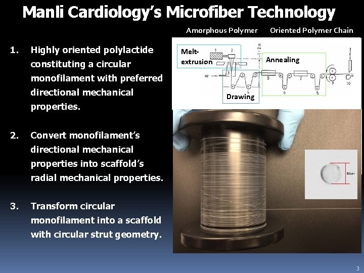
Manli Cardiology’s Microfiber Technology Amorphous Polymer 1. Highly oriented polylactide constituting a circular Meltextrusion Oriented Polymer Chain Annealing monofilament with preferred directional mechanical properties. 2. Convert monofilament’s directional mechanical properties into scaffold’s radial mechanical properties. 3. Drawing Annealing is heating of a polymer to around its glass transition temperature in order to relieve internal stress introduced during extrusion and drawing. Transform circular monofilament into a scaffold with circular strut geometry. 3
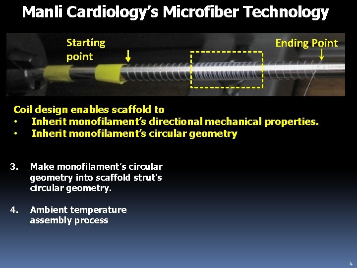
Manli Cardiology’s Microfiber Technology Starting point Ending Point Coil design enables scaffold to • Inherit monofilament’s directional mechanical properties. • Inherit monofilament’s circular geometry 3. Make monofilament’s circular geometry into scaffold strut’s circular geometry. 4. Ambient temperature assembly process 4
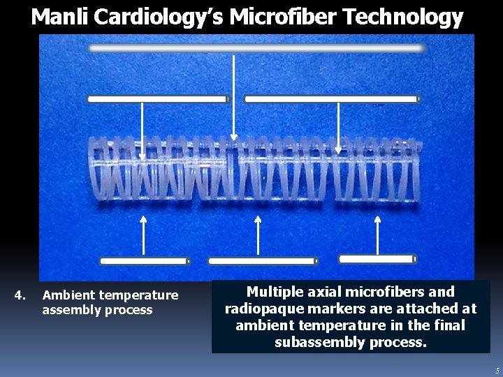
Manli Cardiology’s Microfiber Technology 4. Ambient temperature assembly process Multiple axial microfibers and radiopaque markers are attached at ambient temperature in the final subassembly process. 5
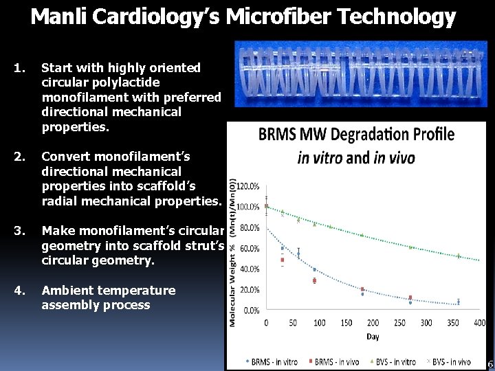
Manli Cardiology’s Microfiber Technology 2. 3. 4. Start with highly oriented circular polylactide monofilament with preferred directional mechanical properties. Convert monofilament’s directional mechanical properties into scaffold’s radial mechanical properties. Make monofilament’s circular geometry into scaffold strut’s circular geometry. Ambient temperature assembly process Radial Strength ( kilopascal) 1. 160 Example 140 PLLA 120 As. Polymerized Material Mirage Microfiber (125μm) 60 -70 MPa 300 MPa 2 -6% 35 % 100 Tensile 80 strength 60 40 Elongation to 20 break 0 Design BRMS Mirage Strut thickness 125 Modulus (mm) BVS Resorb 2 1 3. 1 -3. 7 156 150 GPa * for 3. 0 x 18 -mm devices Amaranth Xience V 3 > 3. 0 -3. 8 150 -200 GPa 81 1 6
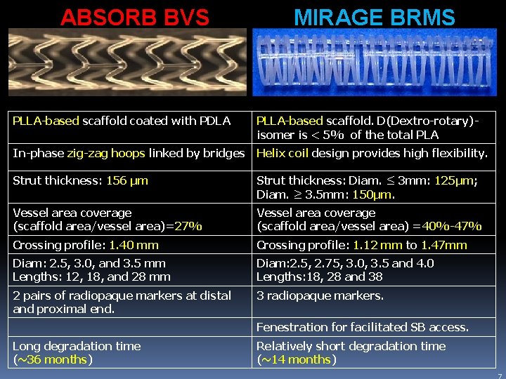
ABSORB BVS MIRAGE BRMS PLLA-based scaffold coated with PDLA PLLA-based scaffold. D(Dextro-rotary)isomer is < 5% of the total PLA In-phase zig-zag hoops linked by bridges Helix coil design provides high flexibility. Strut thickness: 156 μm Strut thickness: Diam. ≤ 3 mm: 125μm; Diam. ≥ 3. 5 mm: 150μm. Vessel area coverage (scaffold area/vessel area)=27% Vessel area coverage (scaffold area/vessel area) =40%-47% Crossing profile: 1. 40 mm Crossing profile: 1. 12 mm to 1. 47 mm Diam: 2. 5, 3. 0, and 3. 5 mm Lengths: 12, 18, and 28 mm Diam: 2. 5, 2. 75, 3. 0, 3. 5 and 4. 0 Lengths: 18, 28 and 38 2 pairs of radiopaque markers at distal and proximal end. 3 radiopaque markers. Fenestration for facilitated SB access. Long degradation time (~36 months) Relatively short degradation time (~14 months) 7
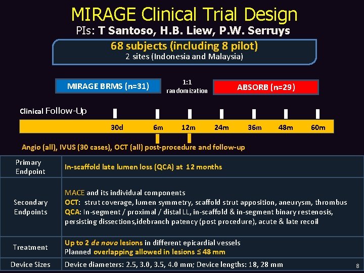
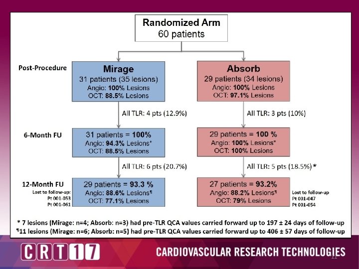
MIRAGE Clinical Trial Design PIs: T Santoso, H. B. Liew, P. W. Serruys 68 subjects (including 8 pilot) 2 sites (Indonesia and Malaysia) 1: 1 randomization MIRAGE BRMS (n=31) ABSORB (n=29) Clinical Follow-Up 30 d 6 m 12 m 24 m 36 m 48 m 60 m Angio (all), IVUS (30 cases), OCT (all) post-procedure and follow-up Primary Endpoint In-scaffold late lumen loss (QCA) at 12 months Secondary Endpoints MACE and its individual components OCT: strut coverage, lumen symmetry, scaffold strut apposition, aneurysm, thrombus QCA: In-segment / proximal / distal LL, in-scaffold & in-segment binary restenosis, persisting dissections, idebranch patency (post procedure), acute & late recoil Treatment Up to 2 de novo lesions in different epicardial vessels Planned overlapping allowed in lesions ≤ 48 mm Device Sizes Device diameters: 2. 5, 3. 0, 3. 5, 4. 0 mm; Device lengths: 18, 28 mm 8
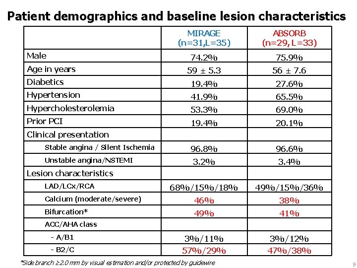
Patient demographics and baseline lesion characteristics MIRAGE (n=31, L=35) ABSORB (n=29, L=33) 74. 2% 75. 9% 59 ± 5. 3 56 ± 7. 6 Diabetics 19. 4% 27. 6% Hypertension 41. 9% 65. 5% Hypercholesterolemia 53. 3% 69. 0% Prior PCI 19. 4% 20. 1% 96. 8% 96. 6% 3. 2% 3. 4% 68%/15%/18% 49%/15%/36% Calcium (moderate/severe) 46% 38% Bifurcation* 49% 41% - A/B 1 3%/11% 3%/12% - B 2/C 57%/29% 47%/38% Male Age in years Clinical presentation Stable angina / Silent Ischemia Unstable angina/NSTEMI Lesion characteristics LAD/LCx/RCA ACC/AHA class *Side branch ≥ 2. 0 mm by visual estimation and/or protected by guidewire 9
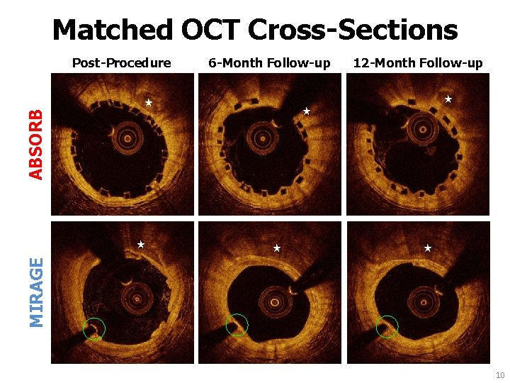
Matched OCT Cross-Sections Post-Procedure 6 -Month Follow-up 12 -Month Follow-up ★ ★ ABSORB ★ ★ ★ MIRAGE ★ 10
11
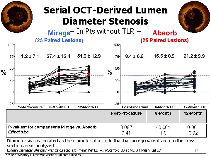
Serial OCT-Derived Lumen Diameter Stenosis Mirage− In Pts without TLR − (25 Paired Lesions) 11. 2 ± 7. 1 27. 4 ± 12. 4 31. 8 ± 12. 9 P-values* for comparisons Mirage vs. Absorb Effect size Absorb (26 Paired Lesions) 16. 6 ± 8. 9 21. 2 ± 9. 9 Post-Procedure 6 -Month 12 -Month 0. 097 0. 41 <0. 001 1. 0 0. 001 0. 92 8. 4 ± 6. 6 Diameter was calculated as the diameter of a circle that has an equivalent area to the crosssection areas analyzed Lumen Diameter Stenosis was calculated as: (Mean Ref LD – In-Scaffold LD at MLA) / Mean Ref LD *Mann-Whitney U test was used for all comparisons 12
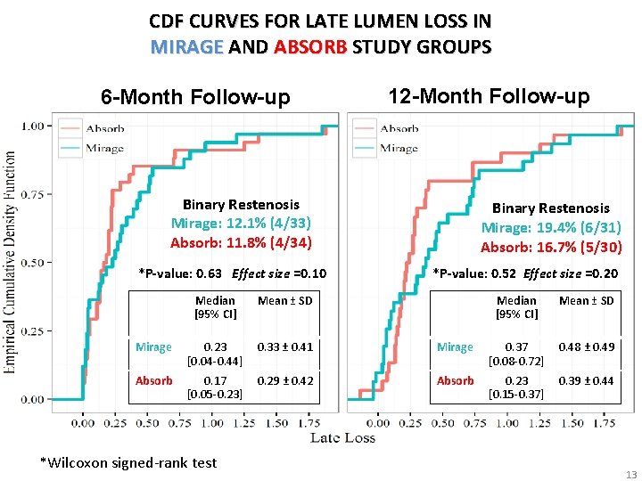
CDF CURVES FOR LATE LUMEN LOSS IN MIRAGE AND ABSORB STUDY GROUPS 6 -Month Follow-up 12 -Month Follow-up Binary Restenosis Mirage: 12. 1% (4/33) Absorb: 11. 8% (4/34) *P-value: 0. 63 Effect size =0. 10 Median [95% CI] Mean ± SD Mirage 0. 23 [0. 04 -0. 44] 0. 33 ± 0. 41 Absorb 0. 17 [0. 05 -0. 23] 0. 29 ± 0. 42 *Wilcoxon signed-rank test Binary Restenosis Mirage: 19. 4% (6/31) Absorb: 16. 7% (5/30) *P-value: 0. 52 Effect size =0. 20 Median [95% CI] Mean ± SD Mirage 0. 37 [0. 08 -0. 72] 0. 48 ± 0. 49 Absorb 0. 23 [0. 15 -0. 37] 0. 39 ± 0. 44 13
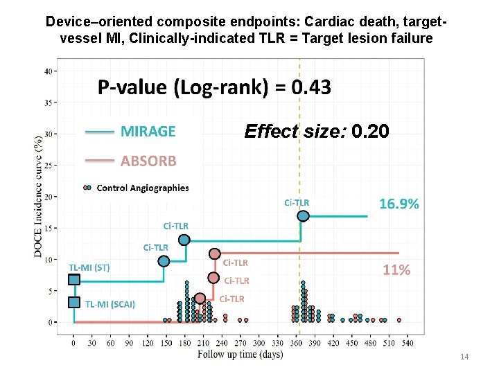
Device–oriented composite endpoints: Cardiac death, targetvessel MI, Clinically-indicated TLR = Target lesion failure Effect size: 0. 20 14
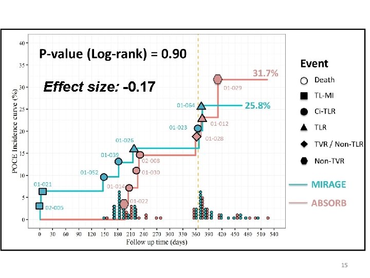
Effect size: -0. 17 15
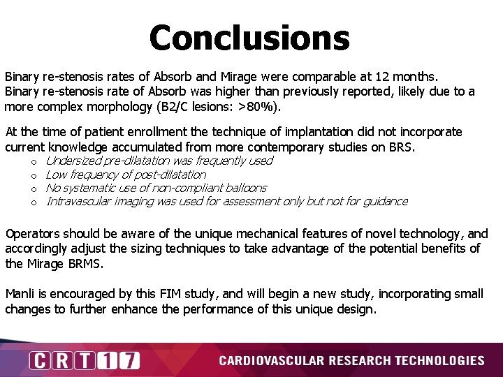
Conclusions Binary re-stenosis rates of Absorb and Mirage were comparable at 12 months. Binary re-stenosis rate of Absorb was higher than previously reported, likely due to a more complex morphology (B 2/C lesions: >80%). At the time of patient enrollment the technique of implantation did not incorporate current knowledge accumulated from more contemporary studies on BRS. o o Undersized pre-dilatation was frequently used Low frequency of post-dilatation No systematic use of non-compliant balloons Intravascular imaging was used for assessment only but not for guidance Operators should be aware of the unique mechanical features of novel technology, and accordingly adjust the sizing techniques to take advantage of the potential benefits of the Mirage BRMS. Manli is encouraged by this FIM study, and will begin a new study, incorporating small changes to further enhance the performance of this unique design.
- Slides: 16