
Manifestasi dan Tatalaksana TB pada Sistem Saraf Rivan
Manifestasi dan Tatalaksana TB pada Sistem Saraf Rivan Danuaji Staff Bagian Neurologi RSUD Dr Moewardi / FK UNS
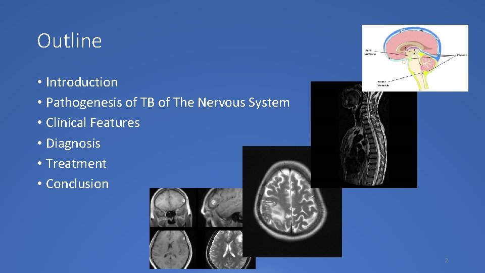
Outline • Introduction • Pathogenesis of TB of The Nervous System • Clinical Features • Diagnosis • Treatment • Conclusion 2

Introduction • Tuberculosis remains a major global problem and a public health issue of considerable magnitude, not mention in Indonesia • Several risk factors have been observed for this serious phenomenon. • The increasing prevalence of HIV infection, • over-crowding in the urban population and in abnormal communities (such as prisons, concentration camps, refugee colonies), • poor nutritional status, • appearance of drug-resistant strains of tuberculosis, • ineffective tuberculosis control programmes, and • an increase in migration from countries where tuberculosis is prevalent to the developed world. 3
Introduction • The incidence of tuberculosis varies from 9 cases per 100 000 population per year in the US; to 110– 165 cases per 100 000 population in the developing countries of Asia and Africa. • In Indonesia there were 156. 733 new cases (Ministry of Health, 2017) • Tuberculous involvement of the central nervous system (CNS) is an important and serious type of extra-pulmonary involvement. • It has been estimated that approximately 10% of all patients with tuberculosis have CNS involvement (Wood M, Anderson M, 1998). 4

Statistic of TB in Indonesia 5
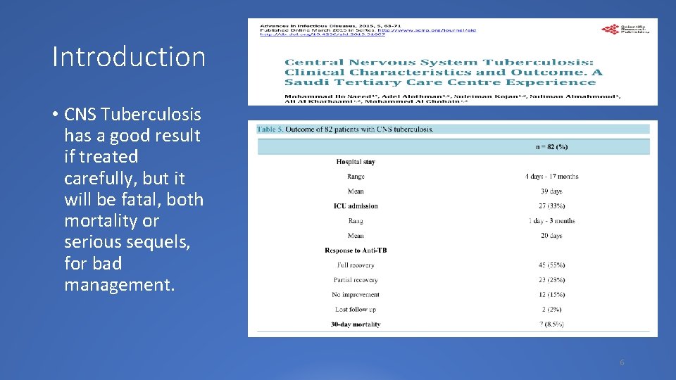
Introduction • CNS Tuberculosis has a good result if treated carefully, but it will be fatal, both mortality or serious sequels, for bad management. 6

Pathogenesis of CNS Tuberculosis 7

Pathogenesis of CNS Tuberculosis • Most tuberculous infections of the CNS are caused by Mycobacterium tuberculosis. Less frequently, other mycobacteria may be involved. • It is believed that the bacilli reach the CNS by the haematogenous route secondary to disease elsewhere in the body 8

Pathogenesis of CNS Tuberculosis • CNS tuberculosis develops in two stages: • Initially small tuberculous lesions (Rich’s foci) develop in the CNS, either during the stage of bacteremia of the primary tuberculous infection or shortly afterwards. • These initial tuberculous lesions may be in the meninges, the sub-pial or subependymal surface of the brain or the spinal cord, and may remain dormant for years after initial infection • Later, rupture or growth of one or more of these small tuberculous lesions produces development of various types of CNS tuberculosis. • The specific stimulus for rupture or growth of Rich’s foci is not known, although immunological mechanisms are believed to play an important role. • Rupture into the subarachnoid space or into the ventricular system results in meningitis. 9
Pathogenesis of CNS Tuberculosis • The type and extent of lesions that result from the discharge of tuberculous bacilli into the cerebrospinal fluid (CSF), depend upon the number and virulence of the bacilli, and the immune response of the host • The pathogenesis of localized brain lesions is also thought to involve haematogenous spread from a primary focus in the lung (which is visible on the chest radiograph in only 30% of cases) Classification of CNS tuberculosis: • Intracranial • Tuberculous meningitis (TBM) • TBM with miliary tuberculosis • Tuberculous encephalopathy • Tuberculous vasculopathy • Space-occupying lesions: • tuberculoma (single or multiple); • multiple small tuberculoma with miliary tuberculosis; • tuberculous abscess • Spinal • Pott’s spine and Pott’s paraplegia • Tuberculous arachnoiditis (myeloradiculopathy) • Non-osseous spinal tuberculoma • Spinal meningitis 10
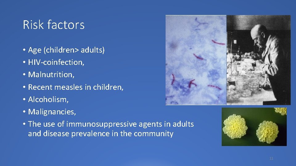
Risk factors • Age (children> adults) • HIV-coinfection, • Malnutrition, • Recent measles in children, • Alcoholism, • Malignancies, • The use of immunosuppressive agents in adults and disease prevalence in the community 11

Clinical Features 12
Clinical features • These nonspecific symptoms include • • • malaise, anorexia, fatigue, fever, myalgias, and headache. • Adults with tuberculous meningitis (TBM) can often present with the classic meningitis symptoms: • fever, • headache and stiff neck along with focal neurological deficits, • behavioral changes, and • alterations in consciousness • Cerebrovascular complications of tuberculous meningitis that occur typically as multiple or bilateral lesions in the territories of the middle cerebral artery perforating vessels are termed as tuberculous vasculopathy 13
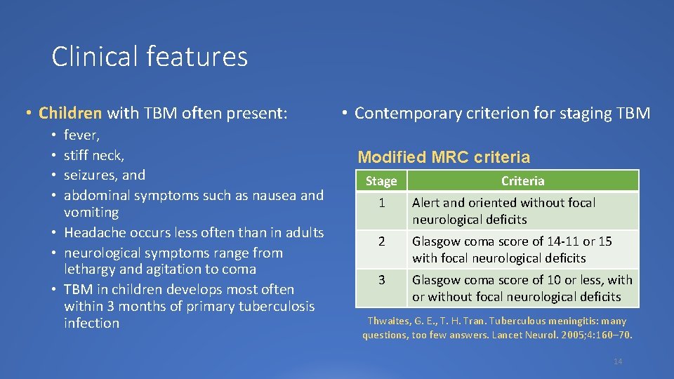
Clinical features • Children with TBM often present: fever, stiff neck, seizures, and abdominal symptoms such as nausea and vomiting • Headache occurs less often than in adults • neurological symptoms range from lethargy and agitation to coma • TBM in children develops most often within 3 months of primary tuberculosis infection • • • Contemporary criterion for staging TBM Modified MRC criteria Stage Criteria 1 Alert and oriented without focal neurological deficits 2 Glasgow coma score of 14 -11 or 15 with focal neurological deficits 3 Glasgow coma score of 10 or less, with or without focal neurological deficits Thwaites, G. E. , T. H. Tran. Tuberculous meningitis: many questions, too few answers. Lancet Neurol. 2005; 4: 160– 70. 14

Staging of TBM is classified into 3 stages according to the British Medical Research Council (MRC) criteria Stage I: Prodromal phase with no definite neurologic symptoms. Stage II: Signs of meningeal irritation with slight or no clouding of sensorium and minor (cranial nerve palsy) or no neurological deficit. Stage III: Severe clouding of sensorium, convulsions, focal neurological deficit and involuntary movements.
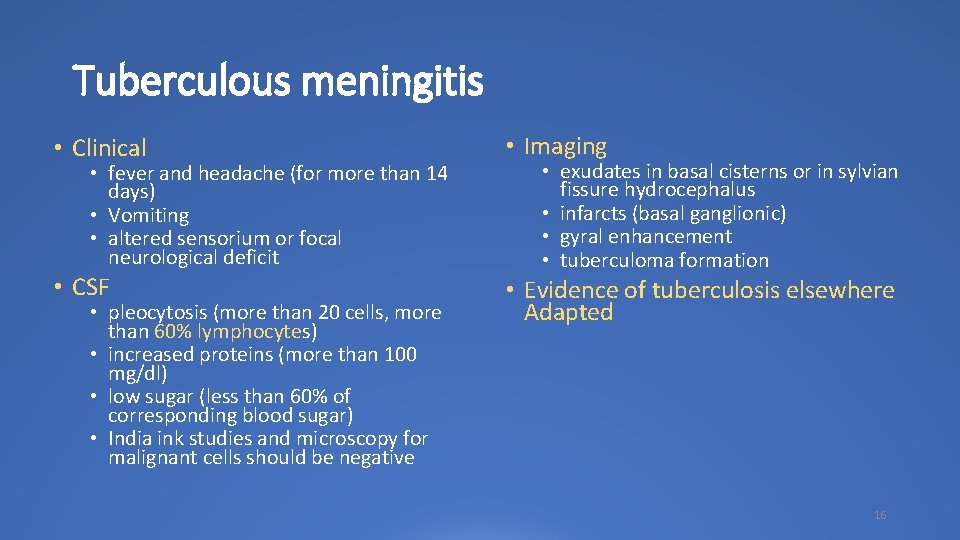
Tuberculous meningitis • Clinical • Imaging • CSF • Evidence of tuberculosis elsewhere Adapted • fever and headache (for more than 14 days) • Vomiting • altered sensorium or focal neurological deficit • pleocytosis (more than 20 cells, more than 60% lymphocytes) • increased proteins (more than 100 mg/dl) • low sugar (less than 60% of corresponding blood sugar) • India ink studies and microscopy for malignant cells should be negative • exudates in basal cisterns or in sylvian fissure hydrocephalus • infarcts (basal ganglionic) • gyral enhancement • tuberculoma formation 16
Tuberculous meningitis Clinical manifestations of tuberculoma or tuberculous brain abscess depend largely on their location, and patients often present with headache, seizures, papilledema, or other signs of increased intracranial pressure. The presentation of brain abscess is more sub acute (1 week to 3 months) than tuberculoma but slower in onset than pyogenic brain abscesses 17
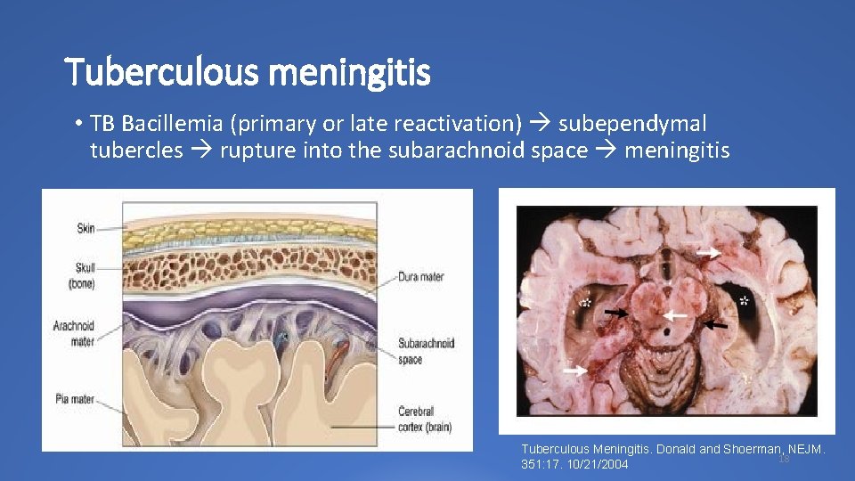
Tuberculous meningitis • TB Bacillemia (primary or late reactivation) subependymal tubercles rupture into the subarachnoid space meningitis Tuberculous Meningitis. Donald and Shoerman, NEJM. 18 351: 17. 10/21/2004
Tuberculous Encephalopathy (TBE) • TBE is a rare outcome usually more common in younger population and is characterized by diffuse brain edema and demyelination, which usually is extensive • Clinical features: Impaired consciousness, seizures, disseminated intravascular coagulation, signs and symptoms of meningitis with or without spinal fluid changes characterize this syndrome. • This syndrome may be one of the leading causes of neurologic devastation and death in CNS TB patients 19
Spinal tuberculosis • Involvement of the spine occurs in less than 1% of TB patients and it can be secondary to Pott’s spine or as non-osseous spinal cord tuberculosis or spinal tuberculous meningitis. • It is a leading cause of paraplegia • Spinal cord compression in Pott’s spine is mainly caused by pressure from a paraspinal abscess. • Neurological deficits may also result from dural invasion by granulation tissue and compression from the debris of sequestrated bone, a destroyed intervertebral disc, or a dislocated vertebra • Neurological involvement can occur at any stage of Pott’s spine and even years later, when there has been apparent healing, because of stretching of the cord in the deformed spinal canal. 20
Spinal tuberculosis • Typically, there is a history of local pain, tenderness over the affected spine or even overlying bony deformity in the form of gibbus. • Paravertebral abscess may be palpated on the back of a number of patients. • These patients usually have acute or subacute, progressive, spastic type of sensorimotor paraparesis. • The incidence of paraparesis in patients with Pott’s spine varies from 27% to 47%. • Non-osseous spinal cord tuberculosis canoccur in the form of tuberculomas: • • extradural tuberculomas arachnoid lesions without dural involvement subdural/extramedullary lesions Intramedullary tuberculomas 21
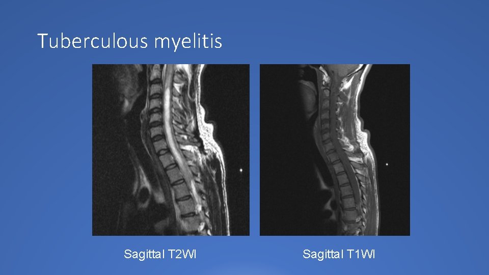
Tuberculous myelitis Sagittal T 2 WI Sagittal T 1 WI
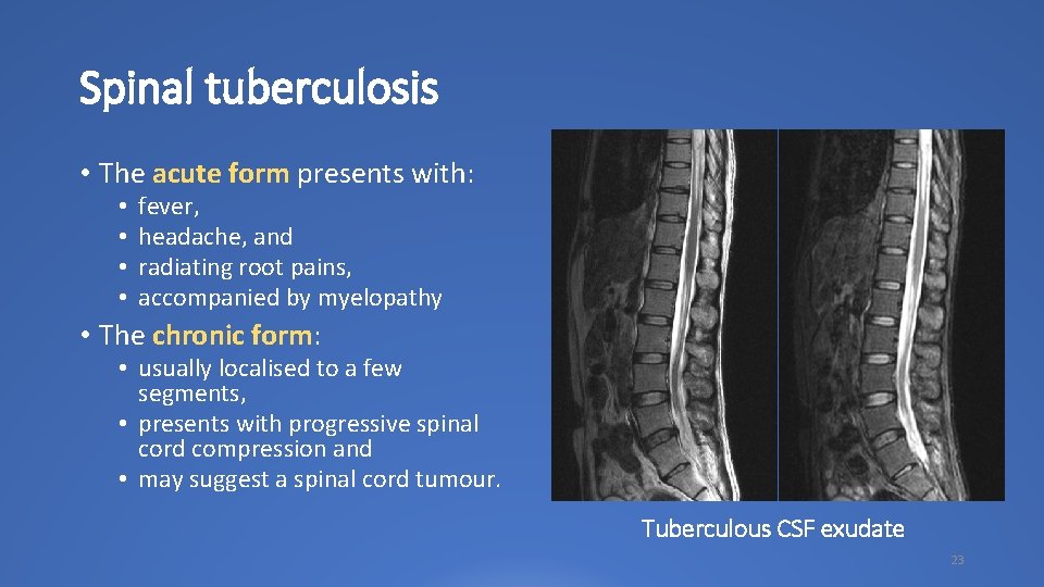
Spinal tuberculosis • The acute form presents with: • • fever, headache, and radiating root pains, accompanied by myelopathy • The chronic form: • usually localised to a few segments, • presents with progressive spinal cord compression and • may suggest a spinal cord tumour. Tuberculous CSF exudate 23
Diagnosis 24
Investigation CSF Analysis • Definitive diagnosis of tuberculous meningitis depends upon the detection of the tubercle bacilli in the CSF, either by smear examination or by bacterial culture. • Rates of CSF culture positivity for clinically diagnosed cases range from 25% to 70% • Once anti-tuberculosis medication is commenced, the sensitivity of smear and culture falls rapidly Molecular and Biochemical Analysis: • nucleic acid amplification (NAA) • polymerase chain reaction (PCR) • antibody detection, • antigen detection, or • chemical assays such as • adenosine deaminase (ADA) and • tuberculostearic acid measurements 25

Tuberculin skin test (TST) • The diagnostic utility of skin testing being positive for CNS tuberculosis varies from 10 -20% to 50%. • The performance of the tuberculin skin test for the diagnosis of tuberculosis varies according to: • • • TST provide indication of previous tuberculosis infection; neither is sufficiently sensitive nor specific to diagnose active disease age, vaccination with BCG, nutritional status, HIV infection, and technique of administration 26
Radiological Evaluation • Every patient with TBM should • All patients should have a chest-Xpreferably be evaluated with ray as part of the diagnostic contrast enhanced CT imaging assessment. before the start or within the first • Serial transcranial doppler 48 hours of treatment ultrasonography (TCD) with blood • MRI shows diffuse, thick, flow velocity (Vm) and pulsatility meningeal enhancement index (PI) measurments, can be efficiently utilized to prognosticate • Contrast enhanced MRI is outcome in tuberculous meningitisgenerally considered as the related vasculopathy modality of choice. It is useful for assessment of the location of lesions and their margins, as well as ventriculitis, meningitis and spinal involvement (sensitivity 86%, specificity 90%) 27
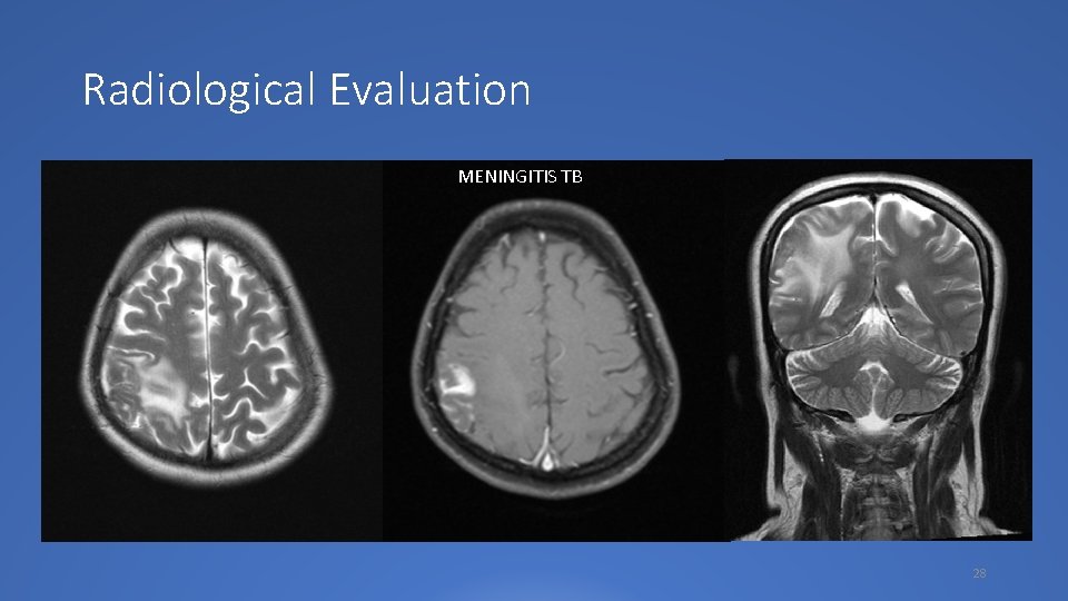
Radiological Evaluation MENINGITIS TB 28
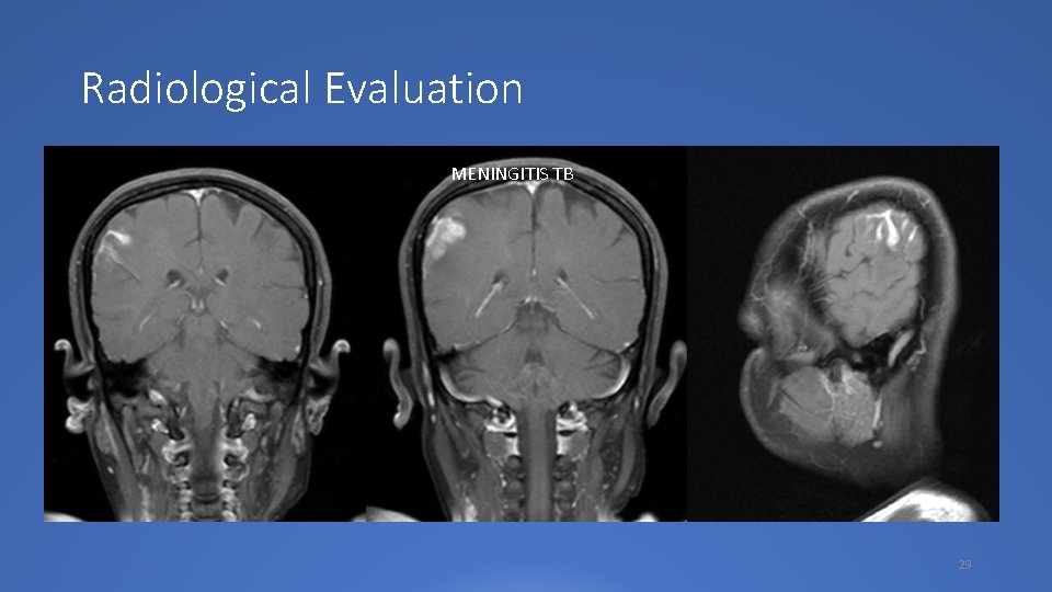
Radiological Evaluation MENINGITIS TB 29
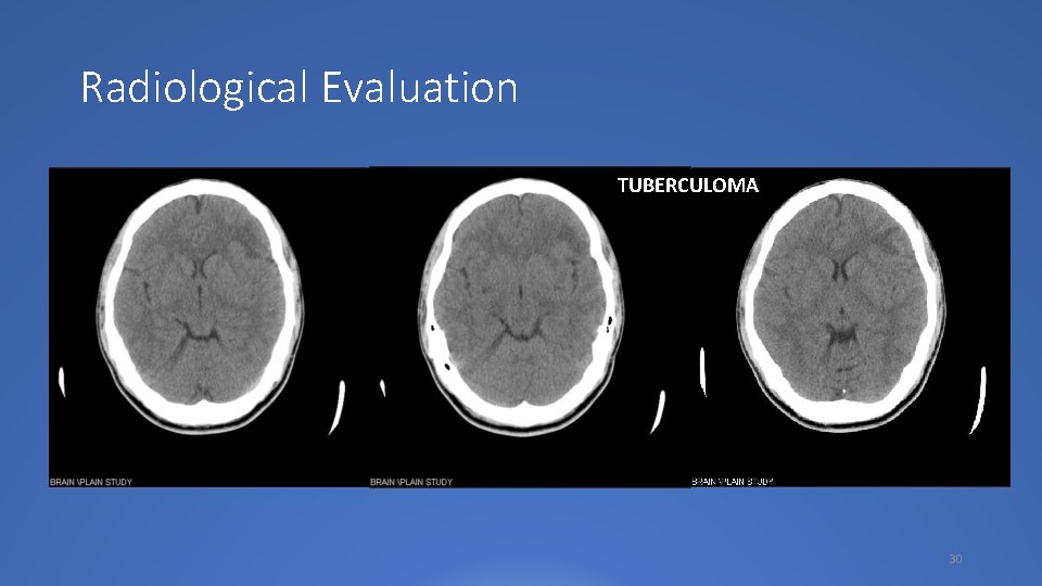
Radiological Evaluation TUBERCULOMA 30
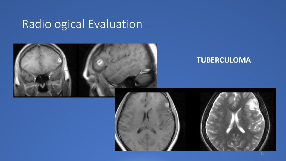
Radiological Evaluation TUBERCULOMA 31
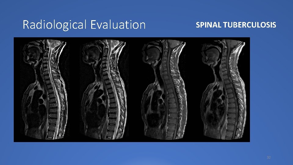
Radiological Evaluation SPINAL TUBERCULOSIS 32

Treatment of CNS TB 33

Treatment of CNS TB • Rationale use of steroids in CNS TB: • All patients with TBM may receive adjunctive corticosteroids regardless of disease severity at presentation • Adults (>14 years) should start treatment with dexamethasone 0. 4 mg/kg/24 hours with a tapering course over 6 to 8 weeks • Children (d” 14 years) should be given prednisolone 4 mg/ kg/24 hrs (or equivalent dose dexamethasone: 0. 6 mg/kg/24 hrs) for 4 weeks, followed by a tapering course over 4 weeks 34
Prognosis and sequelae • The single most important determinant of outcome, for both survival and sequelae, is the stage of tuberculous meningitis at which treatment has been started. • If treatment is started in stage I, mortality and morbidity is very low, • while in stage III almost 50% of patients die, and those who recover may have some form of neurological deficit 36
Conclusion • Early recognition and timely treatment of CNS TB is critical if the considerable mortality and morbidity associated with the condition is to be prevented • A minimum of 10 month-treatment is warranted, and the single most important determinant of outcome is the stage of tuberculous meningitis at which treatment has been started 37
“AKU TELAH MEMBERIKAN OBAT YANG AKU KENAL TERHADAP PENYAKIT YANG AKU PAHAMI KEPADA PASIEN YANG SEBAGIAN BESAR TIDAK TAHU APA-APA” (Lucas Meliala, 2009) 38

Come and Joint us at SUNSHINE 2018 Visite our website: www. soloneuro. org 39

THANK YOU 40
- Slides: 39