Mandible The mandible Skeleton of lower jaw Rami


Mandible

The mandible Skeleton of lower jaw Rami Body Horse-shoe shaped body

External surface of body of mandible Has 2 borders and 2 surfaces Oblique line Horse-shoe shaped body Symphysis menti mental protuberance alveolar border Mental foramen base of mandible

Internal surface of body of mandible Subligual fossa Superior genial tubercle Inferior genial tubercle Mylohyoid line Submandibular fossa digastric fossa

Has 2 borders, 2 surfaces and 2 processes Coronoid process Anterior border Mandibular notch Pterygoid fovea Condylar process Head Neck of mandible Outer surface of Ramus Posterior border Oblique line Angle of mandible External surface of ramus of mandible

Coronoid process Lingula Mylohyoid line Mandibular notch Condylar process Mandibular foramen Mylohyoid groove Rough area Internal surface of ramus of mandible
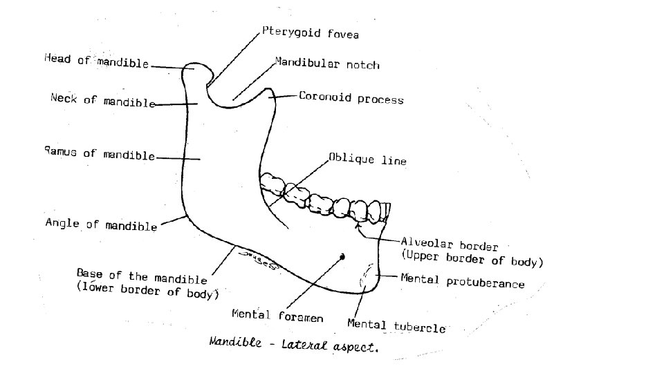
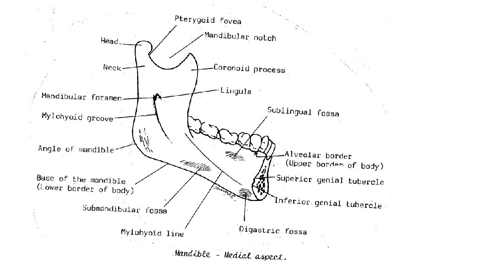

• Body of the mandible - It is horse-shoe shaped havening; - 2 borders (upper and lower) - 2 surfaces (inner and outer) A- Upper border (alveolar margin), carries the sockets for the teeth. B- Lower border shows digastric fossa close to the symphysis menti on each side. C- Outer surface; shows the following features. 1 - Symphysis ﺍﻻﻟﺘﺼﺎﻕ menti; a faint median ridge. 2 - Mental ﺫﻗﻨﻲ protuberance; a median elevation in front of body close to lower border. 3 - Mental tubercle, a projection on each side of the mental protuberance. 4 - Mental ﺫﻗﻨﻲ foramen; on the outer surface transmits the mental nerve and vessels. 5 - Oblique line; from the anterior border of the ramus to the mental foramen. D- Inner surface. 1 - Superior and inferior genial ﺫﻗﻨﻲ tubercles; close to the middle line. 2 - Mylohyoid line, an oblique Line on the inner surface of the body. 3 - Submandibular fossa, below the mylohyoid line to submandibular gland. 4 - Sublingual fossa, above the mylobyoid line to sublingual gland.

Ramus of the mandible, 1. Upper concave border, Mandibular notch. 2. Lower border, it continuous with the base of the mandible. 3. Angle of the mandible is the meeting of the posterior and inferior borders. 4. Inner surface, shows the following features, a. Mandibular foramen in the center of the ramus → mandibular canal and transmits the inferior alveolar nerve and vessels. b. Lingula, a small tongue like process medial to the mandibular foramen. c. Mylohyoid groove; starts below the mandibular foramen and passes downward and forwards to end below the posterior end of the mylohoid line. - It lodges the mylohyoid nerve and vessels. - 2 processes, a- Coronoid process, a sharp projection in front of the mandibular notch. b- Condylar process, the projection behind the mandibular notch. It constitutes of 1 - Head of the mandible to form temporo-mandibular joint. 2 - Neck of the mandible, a constriction below the head. Pterygoid fovea, a small depression on front of the neck.

** Muscles attached to the mandible A- The ramus, receives the insertion of the 4 muscles of mastication; 1. Masseter muscle, into the outer surface of the ramus. 2. Temporalis, into the tip and anterior border and medial surface of the coronoid process. 3. Lateral pterygoid muscle, into the pterygoid fovea. 4. Medial pterygoid muscle, into the inner surface of the angle. B- The body (1 insertion and 6 origins); 1 - Platysma muscle, inserted into the base of the mandible. 2 - Buccinator muscle from the oblique line. 3 - Anterior belly of digastric muscle from the digastric fossa. 4 - Mylohoid muscle from the mylohoid line. 5 - Geniohoid muscle from the inferior genial tubercle. 6 - Genioglossus muscle from the superior genial tubercle. 7 - Superior constrictor muscle of the pharynx from the posterior end of the mylohoid line. ** Ligaments attached to the mandible 1. Temporomandibular ligament extends from articular eminence of e skull to lateral aspect of neck. 2. Stylomandibular ligament; extends from the styloid process to the angle. 3. Sphenomandibular ligament; from the spine of the sphenoid to the lingula. 4. Pterygomandibular ligament; from pterygoid Hamulus to posterior end of mylohyoid line.

** Nerves related to the mandible A- 2 Nerves related to the foramina; 1. Inferior alveolar nerve enters the mandibular foramen. 2. Mental nerve emerges from the mental foramen. B- 2 Nerves related to the grooves, 1 - Nerve to mylohyoid, in the mylohyoid groove. 2 - Lingual nerve runs forwards along groove on the medial aspect of the last molar tooth. ** Arteries related to the mandible 1 - Inferior alveolar artery: passes through the manndibular foramen and canal 2 - Mental artery: comes out of the mental foremen. 3 - Mylohyoid artery runs in the mylohoid groove. 4 - Facial artery, curves around the lower border of the mandible at the antero-inferior angle of masseter muscle. ** Glands related to mandible, 1. Submandibular salivary gland, related to the submandibular fossa. 2. Sublingual salivary gland, related to the sublingual fossa. 3. Parotid gland related to the posterior border of the ramus.

Cervical Vertebrae
 Transverse process Anterior tubercle Posterior tubercle Articular")
Typical vertebrae (C 3 to C 6) Transverse process Anterior tubercle Posterior tubercle Articular processes Carotid tubercle=anterior Articular tubercle of transverse facet process of C 6 Foramen Transverserium Vertebrae Body Uncinate process Vertebral foramen Bifid Spine

Cervical Vertebrae - All are characterized by the presence of foramen transversarium in the transverse process. - They are classified into a- Typical vertebrae, these are 3, 4, 5, 6. b- Non-typical vertebrae, these are 1 (atlas), 2 (axis) and 7. Typical Cervical Vertebra The spine is short and bifid. ** Particular features, 1 - The upper surface of the body is concave from side to side with bilateral lips (uncinate process), 2 - The bodies give attachment anteriorly to the anterior longitudinal ligament and posteriorly to the posterior longitudinal ligament. 3 - The laminae give attachment to the ligamenta flava. 4 - The spines → Iigamentum nuchae. 5 - The tubercles (anterior and posterior) of the transverse processes. 6 - The foramina transversarium from the 6 th up to the 1 st transmit the 1) Second part of the vertebral artery. 2) Sympathetic plexus around the artery. 3) Vertebral vein.

lateral mass Anterior tubercle Anterior arch dental fovea Kidney-shaped superior Articular facet Groove for vertebral artery Atlas posterior arch Posterior tubercle Superior surface C 1 Atlas

Kidney-shaped Articular facet Superior surface C 1 Circular Articular facet Inferior surface C 1
 ** General features; 1 - Absence of a body.")
First Cervical Vertebra (Atlas) ** General features; 1 - Absence of a body. - Short anterior arch and a longer posterior arch. 2 - Superior articular facet → kidney-shaped (atlanto-occipital joint) and inferior articular facet→ circular (lateral atlanto-axial joint) 4 - The anterior arch carries an anterior tubercle. Posteriorly, the anterior arch carries an articular Facet for the dens of the axis (median atlantoaxial joint). 5 - The posterior arch presents a groove on its upper surface to vertebral artery 6 - The medial side of each lateral mass presents a tubercle for attachment of the transverse ligament of the atlas.
 Posterior articular facet for transverse ligament Posterio- superior surface C 2")
Dens (odontoid process) Posterior articular facet for transverse ligament Posterio- superior surface C 2 Axis Second Cervical Vertebra (Axis) • It is characterized by the presence of dens or odontoid process which projects from the upper part of the body. • The dens is grooved posteriorly by transverse ligament. Spinous process

Foramen Transversarium Body Transmits only vertebral vein Spine Has long and non-bifid Spinous processes C 7 Vertebra
 Above: the occipital condyles of")
• ATLANTO- OCCIPITAL JOINTS - Articular surfaces: a) Above: the occipital condyles of the skull. b) Below: the superior articular kidney-shaped facets of the atlas. Flexion and extension (nodding), We move the head to say “YES’ • ATLANTO-AXIAL JOINTS 1) 2 lateral atlanto-axial joints : - Between the inferior articular facet of the atlas and a superior articular facet of the axis. 2) Median atlanto – axial joint: a synovial joint of pivot variety between Odontoid process of the axis and Facet on the posterior surface of the anterior arch of atlas and the transverse ligament of atlas. • Movements side to side or rotatory movements of the head. we move the head to say “NO” • Applied anatomy; - Hanging leads to backward dislocation of the axis from the atlas and tear of the transverse ligament. - The dens of the axis is thrust backwards leading to damage of medulla oblongata and spinal cord. Atlas Dens of axis Transverse ligament of atlas

Thoracic Vertebrae

Thoracic vertebrae Twelve Typical 2 -8 th. v. Atypical 1, 9, 10, 11, 12 th. v.

Body Superior demifacet Vertebral foramen T. process Pedicle Articular facet of T. process Lamina Typical thoracic vertebra Sup. Articular facet Spinus process

Superior demifacet sup. Articular process and facet Articular facets transverse process pedicle Inferior demifacet Articular facets Spinous process inf. Articular processes
 transverse process 12 th th.")
Complete costal facet sup. Articular process (thoacic in type) transverse process 12 th th. v. inf. Articular process (lumber in type)

11 th thoracic vertebra 1 - Transverse process has no facet. 2 - The body kidney shaped. 3 - Complete circular facet close to the upper border of the body 4 - Inferior articular directed forward. 12 th thoracic vertebra 1 - Transverse process has no facet. 2 - The body kidney shaped. 3 - Complete circular facet away from the upper border of the body. It encroaches on the middle of the pedicle. 4 - Inferior articular process directed process laterally.

Lumbar Vertebrae
 1 - A")
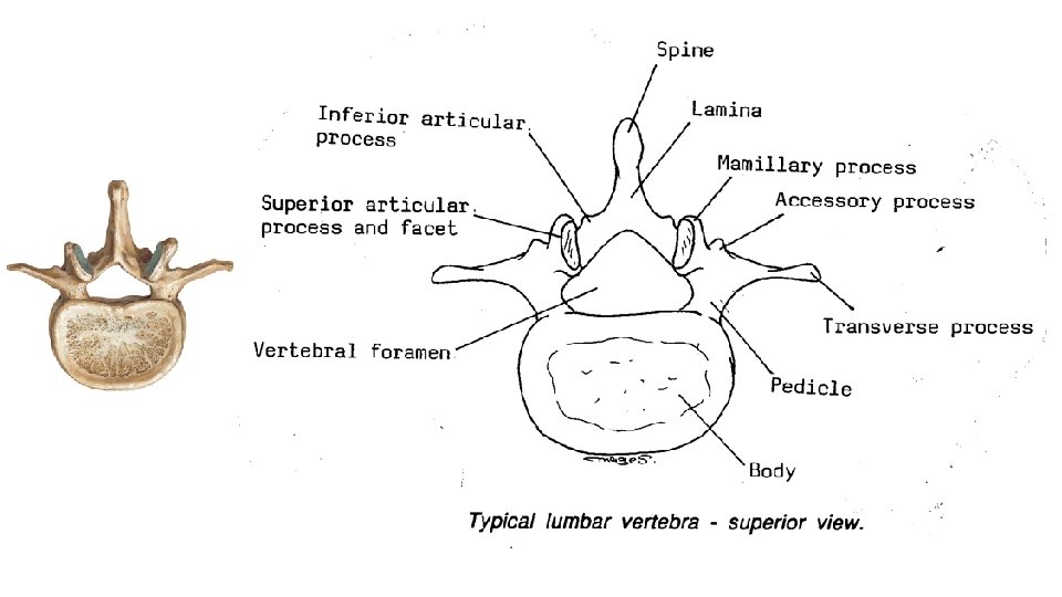
General features of the typical lumbar vertebrae (L 1 -L 4) 1 - A large body which increases in size gradually from the 1 st to the 4 th. 2 - A wide triangular vertebral foramen. 3 - A thin (long and tapering) transverse process. 4 An accessory process behind the root of the transverse process. 5 - The superior articular process is curved with the superior articular facet concave medially. 6 - The inferior articular process is curved with the inferior articular facet convex laterally. - The distance between two superior articular processes (facets) is wider than the inferior. 7 - Mammillary process on posterior edge of superior articular process. 8 - Spine is broad and quadrilateral,
 The transverse processes are thick, strong and are attached to whole sides of")
1) The transverse processes are thick, strong and are attached to whole sides of the pedicles. 2) The distance between two superior articular processes is nearly equal to inferior articular processes

- Slides: 33