Mandible Fractures Jacques Peltier MD Matthew Ryan MD

Mandible Fractures Jacques Peltier MD Matthew Ryan MD UTMB – Dept of Otolaryngology May 2004

History • Edwin Smith Papyrus 1650 described Hx, Phy, Diagnosis. Often fatal disease • Hippocrates – Described monomaxillary dental fixation and binding • Sulicetti – 1492 Described “tie teeth of jaw to teeth of uninjured jaw”

History • Schede 1888 – Bone plate of steel secured with 4 screws • Luhr 1960 – Developed mandibular compression plates • Michelet and Champy 1970’s – Placement of small bendable non-compression plates

Epidemiology • Mandible most common after nasal fractures • Mandible : Zygoma : Maxilla 6: 2: 1 • Ellis 4711 facial fractures, 45% with mandible fractures • Assault>MVA>Fall>Sports
 – Socket of canine")
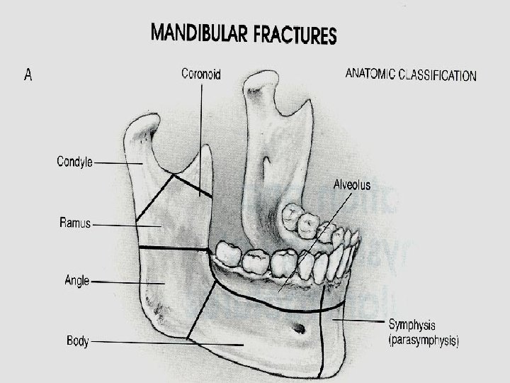
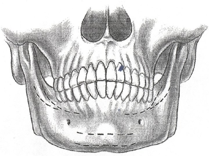
Epidemiology • Sites of weakness – Third molar (esp. impacted) – Socket of canine tooth – Condylar neck
 5196 fractures – Young military men – Angle")
Epidemiology • Boole et al (laryngoscope) 5196 fractures – Young military men – Angle 35%, Symphysis 20%, Body 12%, Condylar 9%, Subcondylar 4%, Ramus 4%, Alveolar 3%, Coronoid 1% – 70% 1 fracture, 30% 2 fractures, . 2% more than 2 – Facial lacs 30%, other facial fx. 16%, C-spine 0. 8%

Haug et al

Fischer et al

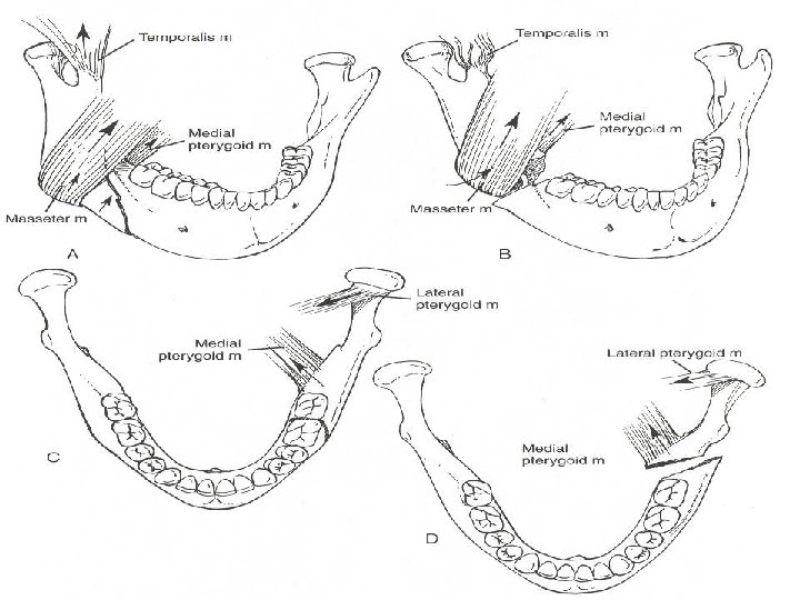
Favorable vs. Unfavorable • Masseter, Medial and Lateral Pterygoid, and Temporalis tend to draw fractures medial and superior • Almost all fractures of angle unfavorable

Evaluation • Stabilization via ATLS protocol • Part of secondary survey – Pain, malocclusion, trismus, V 3 sensory deficit – History of TMJ (earlier mobilization) – Blow to face favors parasymphyseal fracture and contralateral angle fracture – Fall to chin (bilateral condylar fractures)
 • Psychiatric, nutritional, gastrointestinal, seizure disorders • Previous")
Evaluation • Previous occlusion (Class I-III) • Psychiatric, nutritional, gastrointestinal, seizure disorders • Previous facial trauma • Other injuries (c-spine, intra-abdominal, likely prolonged intubation)

Physical Exam • Complete Head and Neck exam – Palpable step off – Tenderness to palpation – Malocclusion – Trismus (35 mm or less) – FOM hematoma – Altered sensation of V 3 – Crepitus

Physical Exam • Dental Exam – Lost, fractured, or unstable teeth – Dental Health – Relation to fracture – Quantity

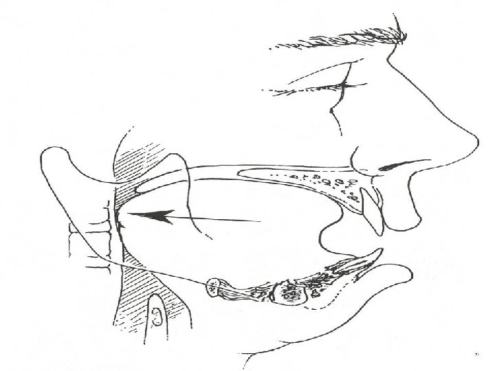
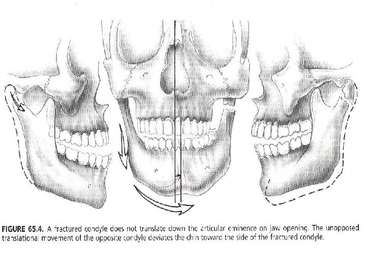
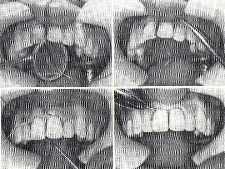
Physical Exam • Unilateral fractures of Condyle – Decreased translational movement, functional height of condyle – Deviation of chin away from fracture, open bite opposite side of fracture Bilateral fractures of condyle - Anterior open bite

Picture of open bites

Evaluation • Panorex, mandible series • CT scan – Not as diagnostic as plain films for nondisplaced fractures of mandible. – Most useful for coronoid and condylar fractures, associated midface fractures

Physiology • Primary Healing – In rigid fixation techniques – Lag screws, compression plates, Recon plate, external fixation, Wire fixation, Miniplate fixation – No callus formation – Question of bone resorption

Physiology • Secondary bone healing – Callus formation – Remodeling and strengthening – MMF, Wire fixation, Miniplate fixation

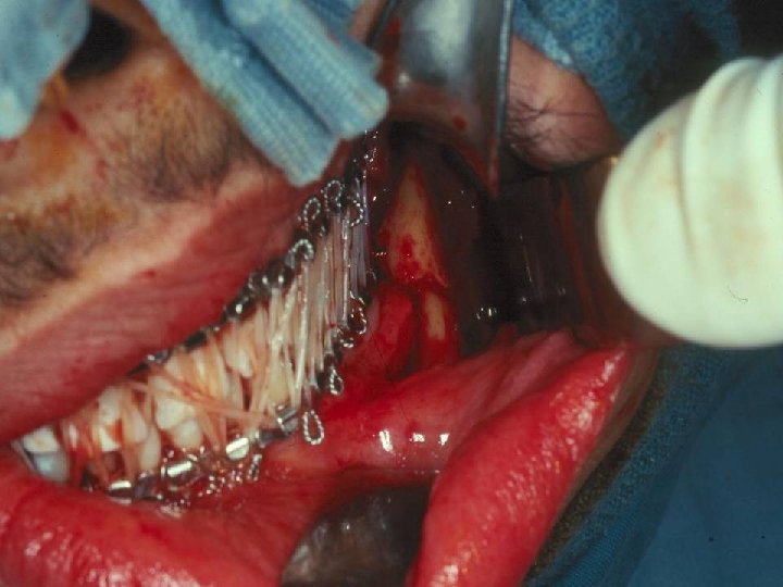
Closed Reduction • Favorable, non-displaced fractures • Grossly comminuted fractures when adequate stabilization unlikely • Severely atrophic edentulous mandible • Children with developing dentition

Closed Reduction • Length of MMF – De Amaratuga – 75% of children under 15 healed by 2 weeks, 75% young adults 4 wks – Juniper and Awty – 82% had healed at 4 wks – Longer period for edentulous fractures 610 wks

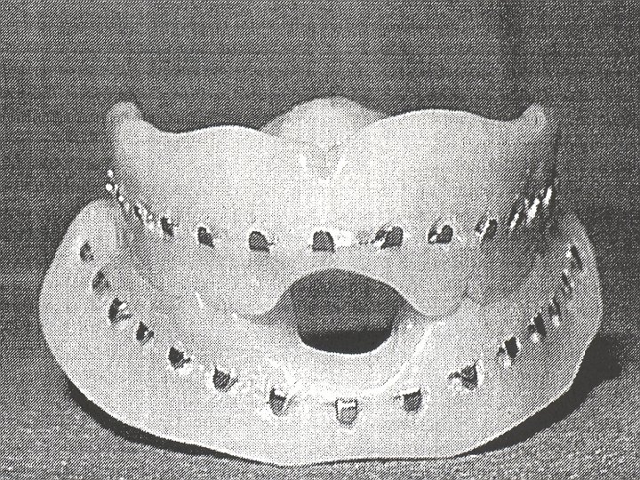
Closed Reduction • Edentulous fractures – Bradley found absent inferior alveolar artery in 40% 60 -80 yo’s – Periosteal blood supply disturbed by stripping – Up to 20% non-union despite type of treatment – May consider Gunning Splints

Open Reduction • Displaced unfavorable fractures • Mandible fractures with associated midface fractures • When MMF contraindicated or not possible • Patient comfort • Facilitate return to work

Open Reduction • Contraindications – General Anesthetic risk too high – Severe comminution and stabilization not possible – No soft tissue to cover fracture site – Bone at fracture site diffusely infected (controversial)

Open Reduction • Associated condylar fracture • Associated Midface fractures • Psychiatric illness • GI disorders involving severe N/V • Severe malnutrition • To avoid tracheostomy in patients who need postoperative intubation

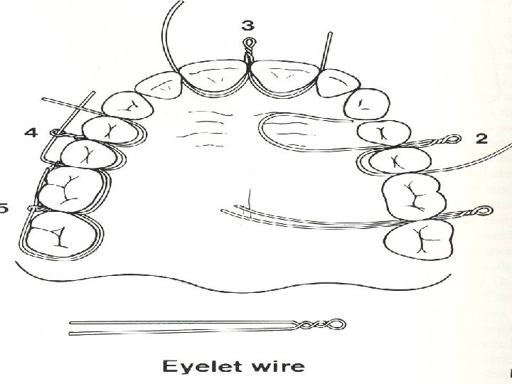
Open Reduction • Intraosseous wiring – Semirigid fixation – Cheap – Technically difficult – Primary and Secondary bone healing
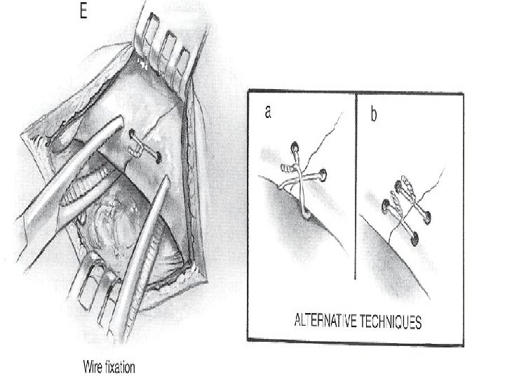
 – Good for anterior mandible")
Open Reduction • Lag Screws – Rigid fixation (Compression) – Good for anterior mandible fractures, Oblique body fractures, mandible angle fractures – Cheap – Technically difficult – Injury to inferior alveolar neurovascular bundle

Open reduction • Ellis 41 patients with anterior lag screw technique • 4. 9% infection rate • No malocclusion • No Non-union

Lag Screw Technique

Lag Screw Technique

Lag Screw Technique

Rigid Fixation • Compression plates – Rigid fixation – Allow primary bone healing – Difficult to bend – Operator dependent – No need for MMF

Rigid Fixation • Miniplates – Semi-rigid fixation – Allows primary and secondary bone healing – Easily bendable – More forgiving – Short period MMF Recommended

Rigid Fixation • Schierle et al studied experimental model, then applied in patients. – Model suggested two plates more stable – Patients divided into two groups with equal complication rates, equal functional results

Miniplates, Champy technique

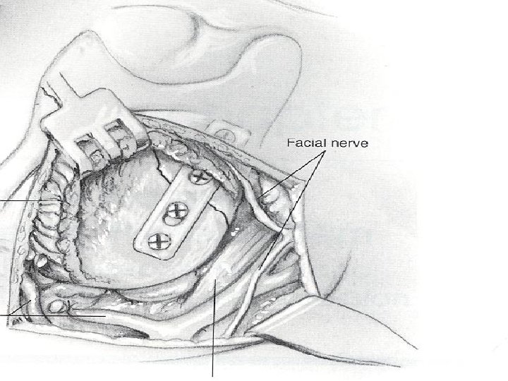
Rigid Fixation • Reconstruction Plates – Good for comminuted fractures – Bulky, palpable – Difficult to bend – Locking plates more forgiving

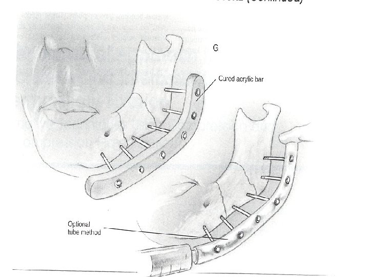
External Fixation • Alternative form of rigid fixation • Grossly comminuted fractures, contaminated fractures, non-union • Often used when all else fails

Edentulous Fractures • Chalmers and Lyons 1976 – Recommended closed reduction to preserve periosteal blood supply • Chalmers and Lyons 1995 – 167 fractures in edentulous mandibles – ORIF 82% – 15% complications – 12% Fibrous union

Edentulous Fractures • ORIF – Inferior alveolar canal more superior in location – Vertical height 20 mm compatible with standard plating systems – Vertical height 10 mm or less, likely need rib graft – Plate removal after fracture healing if interferes with denture placement

Teeth in line of fracture • Keep teeth if – Previously healthy – Peridontal plexus intact – No major structural injury – Tooth does not interfere with reduction of fracture

Teeth in line of fracture • Neal and associates – 32% incidence of morbidity with teeth in line of fracture – No statistical difference if tooth was removed

Teeth in line of fracture • Amaratunga – 16% complication rate in retained teeth – 13% in removed teeth – Retain teeth for 4 -6 weeks if important for MMF

Condylar and Subcondylar • Lindhal and Hollender – Closed reduction in children, teens, adults – Intracapsular fractures – Higher incidence of postoperative sequelae in adults – Children and Teens with less sequelae, more remodeling

Condylar and Subcondylar • Norholt – Children 5 -20 with intracapsular condylar fractures – Increased dysfunction with increasing age

Condylar and Subcondylar • Closed reduction with arch bars MMF 2 -3 weeks mainstay for youths – Ankylosis of TMJ and facial asymmetry most feared complication – Less effective for • increasing age • decreased ramus height • more displaced

Condylar and Subcondylar • ORIF, Absolute indications – Displacement into middle cranial fossa – Inability to achieve occlusion with closed reduction – Foreign body in joint space

Condylar and Subcondylar • Relative indications – Bilateral condylar fractures to preserve vertical height – Associated injuries that dictate earlier function • Soft tissue swelling causing airway compromise with MMF • Intracapsular fracture on opposite side where early mobilization important

Immediate Mobilization • Kaplan et al. – Studied ORIF in two groups, one with MMF for 2 weeks, one with immediate mobilization – No statistical difference in rates of complications, postoperative pain, dental health, nutritional status

Bioabsorbable Plates • Plating can relieve stress, no bone remodeling • Bulky plates, thermal sensitivity, palpable • Absorbable plates expensive • Better in children? • Use of poly-L-lactide in 69 fractures by Kim et al – – – 12% complication 8% infection No malunion

References Kim et al “Treatment of Mandible Fractures using Bioabsorbable plates”, Plastic and Reconstructive Surgery, vol 110, july 2002, 25 -31 Bailey, Byron J. Head and Neck Surgery - Otolaryngology. Third Edition. Lippincott Williams and Wilkins, 2001. Ellis, E. “Treatment Methods for Fractures of the Mandibular Angle. " Journal of Craniomaxillofacial Trauma, vol. 28. 1999: 243 -252. Ellis, E. , et. al. “Lag Screw Fixation of Mandibular Angle Fractures. ” Journal of Oral Maxillofacial Surgery , vol. 49. 1991: 234 -243. Kim et. al. "Treatment of Mandible Fractures Using Bioabsorable Plates. " Journal of Plastic and Reconstructive Surgery , vol. 110. 2002: 25 -31. Boole et. al. "5196 Mandible Fractures Among 4381 Active Duty Army Soldiers, 1980 to 1998. " Laryngoscope, 111(10). Oct. 2001: 1691 -6, Kaplan et al. "Immediate Mobilization Following Fixation of Mandible Fractures, A Prospective Randomized Study. " Laryngoscope, vol. 111(9). Sept 2001: 1520 -1524 Spina and Marciani. Mandibular Fractures, pages 85 - 105 Schierle et. al. "One or Two Plate Fixation of Mandible Fractures? " Journal of Cranio-Maxillofacial Surgery. Vol. 25, 1997: 162 -168.
- Slides: 62