
Management of obstetric hemorrhage APH PPH Hala Nsour
Management of obstetric hemorrhage APH & PPH Hala Nsour
obstetric hemorrhage �Hemorrhage is one of the most common cause of maternal death
APH �DIFINTION : vaginal bleeding from second half of pregnancy( 20 week) to delivery of the baby �Critical for fetus �Emergent the well being of both mother and
Causes � Common Placenta previa Abruptio placentae bloody show � Uncommon Uterine rupture Vasa praevia Morbidly adherent placenta (MAP) marginal separation � Local Cervical, vaginal, or uterine pathology � Systemic cause
Initial evaluation �If a patient is bleeding profusely, profusely a team approach to the assessment and management should be instituted to maintain hemodynamic stability. �This team should include an obstetrician, obstetrician an anesthesiologist, anesthesiologist and nurses who are knowledgeable about the management of the critically ill patient. �two large bore intravenous line should be placed.
Then …. . �Complete medical history …. . Past medical hx , known bleeding disorder or liver disease �The vital signs and amount of bleeding should be checked immediately, as should the patient's mental status �Physical examination Start with general look to pt (pale , anxious , jaundice) Then go to abdominal exam (SFH/Leoppold’s/tenderness) A pelvic examination should not be performed until placenta previa has been excluded by ultrasonography ( so start with abdominal US )
Cont…. . � Once placenta previa has been excluded, a sterile speculum examination can be safely done to rule out genital tears or lesions (e. g. , cervical cancer) that may be responsible for the bleeding. � If none are identified, a digital examination or pelvic ultrasound may be performed to determine whether cervical dilatation is present
� A complete blood count should be obtained and compared with previous evaluations to help assess the amount of blood loss. � An assessment of the patient's coagulation profile should be done and cross matched for at least 6 units of blood (packed cells). � The patient should be typed
Cont…. �Indirect coombs test (ICT) and antibody titer should be done (if –ve mother blood group and +ve partner ) �In case of –ve ICT and low AB titer …… anti D should be order �Regular monitoring of fetus and mother by US and NST.
�The most important and accurate method of determining the cause is US � The ultrasonographic evaluation should include not only the location and character of the placenta but also an assessment of gestational age, an estimate of fetal weight, a determination of the fetal presentation, and a screening for fetal anomalies. Other than liqour volume Uterine activity and the fetal heart rate should be assessed with a monitored strip(CTG) to rule out labor and establish fetal well being �
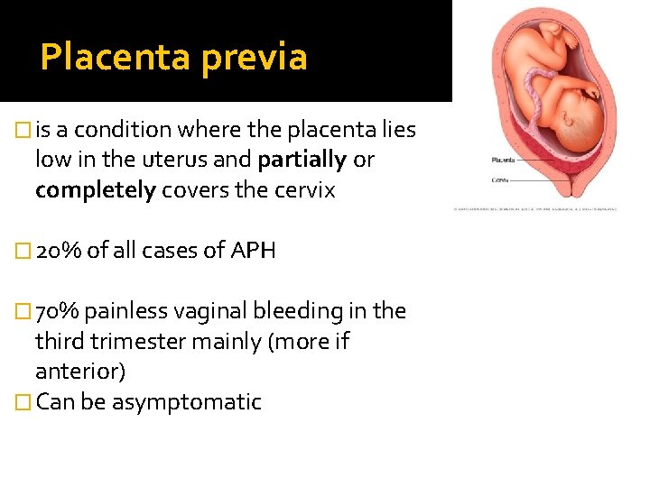
Placenta previa � is a condition where the placenta lies low in the uterus and partially or completely covers the cervix � 20% of all cases of APH � 70% painless vaginal bleeding in the third trimester mainly (more if anterior) � Can be asymptomatic
Risk factors �Multiparty �Increase maternal age �Smoking �Malpresentation � multiple gestation �Cesarean delivery �Prior placenta previa �Uterine abnormality
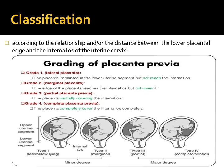
Classification � according to the relationship and/or the distance between the lower placental edge and the internal os of the uterine cervix.
Diagnosis �US : transabdominal US has an accuracy of 95% transvaginal US has an accuracy of 100% Note : When P. P diagnosed in second trimester a repeat US is indicated at 30 to 32 wks for follow up evaluation.
Managements Depend on GA and extent of bleeding P. P antenatal care : � Hospitalization Depend on distance between home and hospital , availability of transportation , previous bleeding episodes, hematology laboratory results , and acceptance of receiving donor blood products � corticosteroids Consider it up to 34 week � Excessive bleeding : CS regaedless of GA � Mild not repetitive : expectant management in the hospital � After 36 weeks : cs if fetal lungs are mature � PV and intercourse are contraindicated
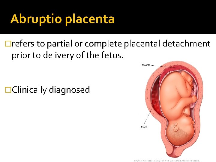
Abruptio placenta �refers to partial or complete placental detachment prior to delivery of the fetus. �Clinically diagnosed
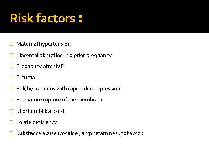
Risk factors : � Maternal hypertension � Placental abruption in a prior pregnancy � Pregnancy after IVF � Trauma � Polyhydramnios with rapid decompression � Premature rupture of the membrane � Short umbilical cord � Folate deficiency � Substance abuse (cocaine , amphetamines , tobacco )
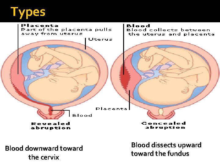
Types Blood downward toward the cervix Blood dissects upward toward the fundus
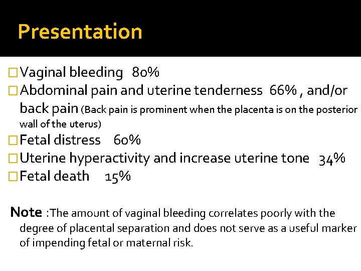
Presentation �Vaginal bleeding 80% �Abdominal pain and uterine tenderness 66% , and/or back pain (Back pain is prominent when the placenta is on the posterior wall of the uterus) �Fetal distress 60% �Uterine hyperactivity and increase uterine tone �Fetal death 15% 34% Note : The amount of vaginal bleeding correlates poorly with the degree of placental separation and does not serve as a useful marker of impending fetal or maternal risk.
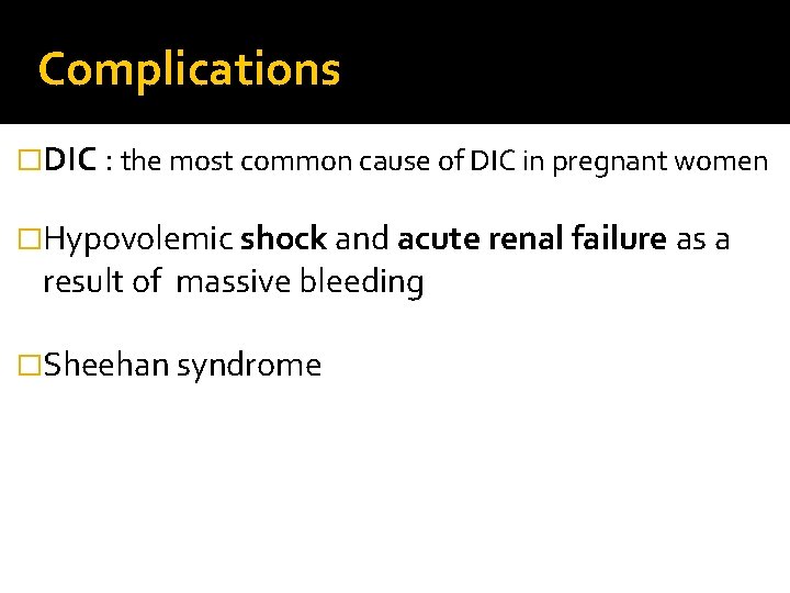
Complications �DIC : the most common cause of DIC in pregnant women �Hypovolemic shock and acute renal failure as a result of massive bleeding �Sheehan syndrome
Focus management �Intensive monitoring for both mother and fetus � 2 large IV line and blood prepair �The use of tocolytics or uterine relaxants is not advisable �Uterine tone must be maintained to control bleeding after delivery �If > 36 week deliver , <36 expectant if mild
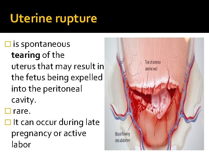
Uterine rupture � is spontaneous tearing of the uterus that may result in the fetus being expelled into the peritoneal cavity. � rare. � It can occur during late pregnancy or active labor
Risk factor �Prior uterine scar (40% of cases) , high vertical (classic ) scar �Obstructed labor in multiparous pt �Induction of labor using prostaglandins �Obesity
Sign and symptoms �sudden onset fetal heart rate (FHR) abnormalities �vaginal bleeding �sudden onset intense constant abdominal pain � cessation of uterine contractions � recession of the presenting part �maternal hypotension and tachycardia.
Focus management � Total abdominal hysterectomy is the treatment of choice , although debridement of rupture site and primary closure maybe consider in women of low parity who desire more children �Repair vs hysterectomy : v pt plans for future pregnancy v extent of damage v hemodynamic stability v surgeon’s skills
Morbidly adherent placenta (MAP) �Abnormal attachment of the placenta through the uterine myometrium �Risk factor : - hx of uterine surgery - prior cs delivery (abnormal implantation )
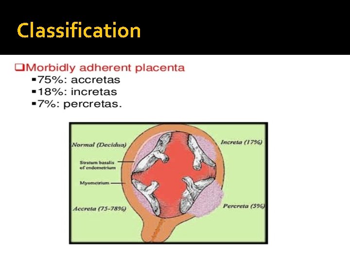
Classification
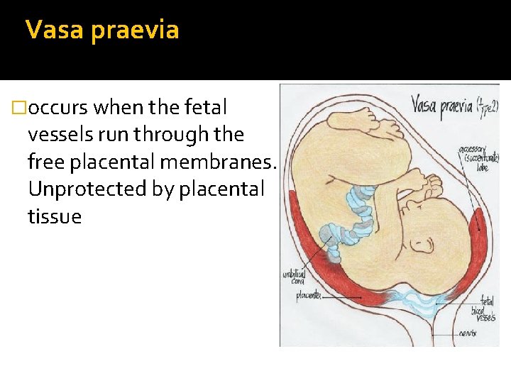
Vasa praevia �occurs when the fetal vessels run through the free placental membranes. Unprotected by placental tissue
Diagnosis �Pervaginal examination during early labour detecting the pulsating fetal vessels inside the internal os, or by the presence of dark‐red vaginal bleeding and acute fetal compromise after spontaneous or artificial rupture of the placental membranes. �Apt test If fetus blood the fluid of color will remain red (not denatured by KOH ) � A combination of both transabdominal and transvaginal colour Doppler imaging (CDI) ultrasonography provides the best diagnostic accuracy for vasa praevia.
Management emergency caesarean delivery and neonatal resuscitation, including the use of blood transfusion if required, are essential in the management of ruptured vasa praevia diagnosed during labor � Because of the speed at which fetal exsanguination can occur and the high perinatal mortality rate associated with ruptured vasa praevia, delivery should not be delayed while trying to confirm the diagnosis, particularly if there is evidence that fetal wellbeing is compromised. � In the presence of confirmed vasa praevia in the third trimester, elective caesarean section should ideally be carried out prior to the onset of labour. � A decision for prophylactic hospitalisation from 30– 32 weeks of gestation in women with confirmed vasa praevia should be individualised and based on a combination of factors, including multiple pregnancy, antenatal bleeding and threatened premature labour. �
�In cases of vasa praevia that develop premature rupture of membranes and/or labour at viable gestational ages, a caesarean section should be performed without delay. �� To avoid unnecessary anxiety, admissions, prematurity and caesarean section, it is essential to confirm persistence of vasa praevia by ultrasound in the third trimester.

- Slides: 32