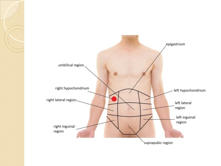
Management of Mass in Right Hypochondrium Dr Naeem


- Slides: 36
Management of Mass in Right Hypochondrium Dr Naeem Zia FCPS, FACS, FRCS Professor of Surgery
Learning Objectives �At the end of the lecture the student should be able to �Describe anatomy of right hypochondrium �Construct differential diagnosis of masses in RHC �Describe the clinical features of different masses �Outline different investigations �Make a management plan
Clinical features �General Physical Examination �Anaemia �Jaundice �Supraclavicular lymph nodes �Emaciation
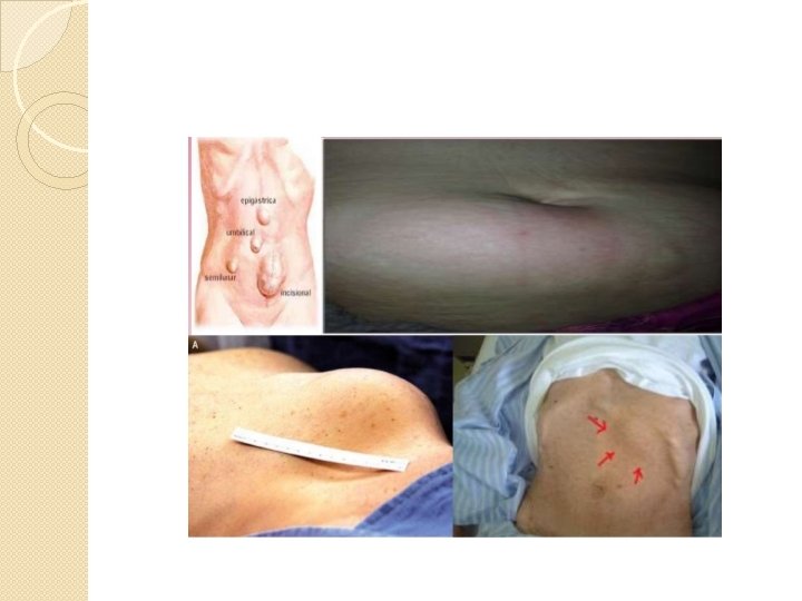
Inspection
Palpation � Local temperature � Tender? � Muscular rigidity � Confirming positive findings of inspection � Note margins on palpation. Can you get around the margin? Well or ill-defined. � Consistency: Soft, firm or hard. Hard swellings are usually malignant, soft swelling may be cystic. � Mobility: � Is swelling parietal or intra-abdominal? � Hernial sites for expansile impulse on coughing � Pulsations if present: expansile (aortic) or transmitted
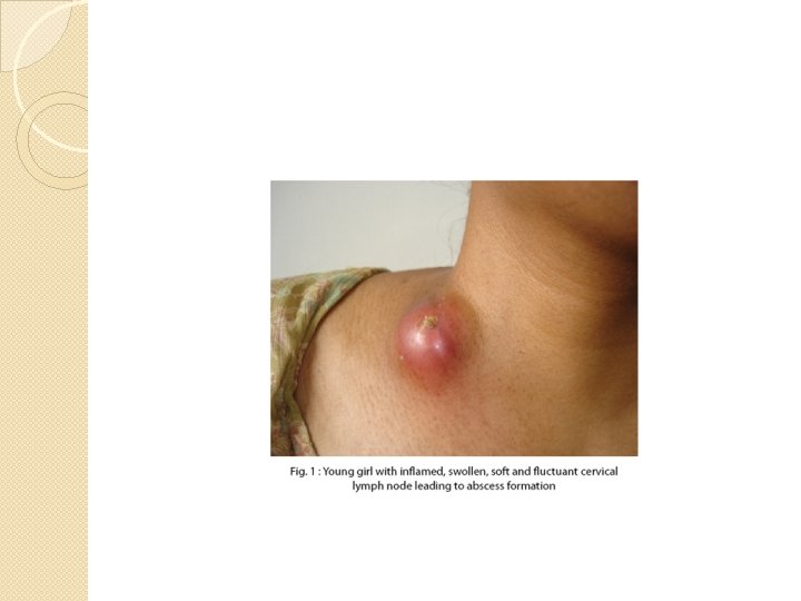
Swellings in abdominal wall �Cold Abscess � 1. Fluctuant swelling with no signs of inflammation 2. Swelling becomes prominent when the abdominal muscles contract 3. Irregularity in the affected rib or deformity of the spine
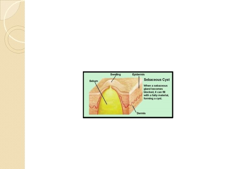
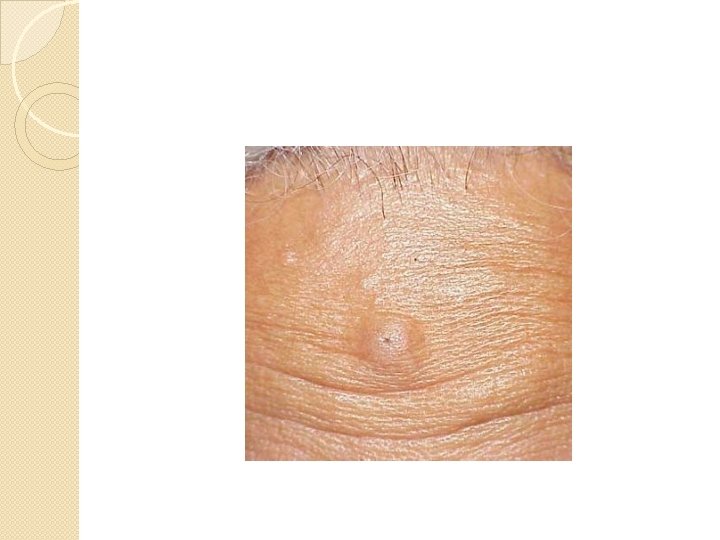
�Sebacous cyst
Intra-abdominal Swellings �Hepatic � 1. It moves with respiration but is not mobile sideways 2. The swelling is continuous with the liver dullness without a band of colonic resonance
�Gall Bladder � 1. Oval smooth swelling, the size of an egg 2. Moves with respiration, can be moved sideways but cannot be pushed down into the loin (like kidney swelling)
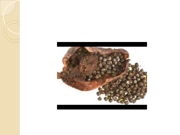
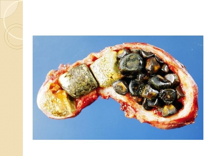
�Chronic Cholecystitis and Cholelithiasis Gall bladder may be palpable. Murphy’s sign is positive: ◦ Tenderness under the right costal margin at the lateral border of the rectus muscle when the patient takes a deep breath. ◦ If a stone is present in the common bile duct there is a triad of intermittent colic, intermittent jaundice and fever with chills and rigors. ◦ By Courvoisier’s law, gall bladder is not palpable.
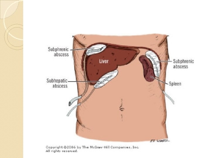
�Sub-Phrenic Abscess � 1. Pain in the right hypochondrial region referred to the shoulders 2. Diffuse tender swelling in the right hypochondrial region 3. Signs of septicemia: High fever with rigors, sweating and marked tachycardia 4. Screening: Raised and fixed diaphragm with gas under it 5. Features of the causative condition e. g. perforated peptic ulcer, liver abscess
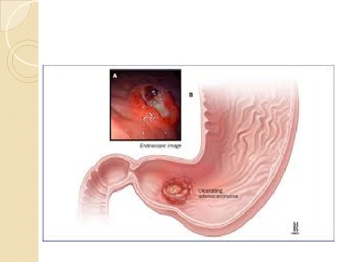
Stomach and Duodenum �Carcinoma of Pylorus: � 1. There is irregular firm lump which moves on respiration 2. Patient is usually elderly and has anorexia and weight loss 3. Barium meal would show filling defect
�Sub-Acute Perforation of a Peptic Ulcer � 1. Localized, tender, inflammatory mass may be present with a central abscess 2. History of peptic ulcer 3. Barium meal would reveal the ulcer
�Hepatic Flexure of Colon �Hypertrophic Tuberculosis �This usually causes a lump in the right iliac fossa which may be drawn towards the right hypochondriac region by fibrosis.
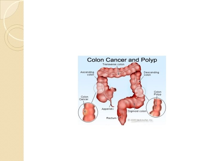
�Carcinoma of Colon ◦ 1. This commonly occurs in men above the age 40 years 2. There is alternate diarrhea and constipation. 3. The lump is irregular, firm and moves poorly on respiration 4. Occult blood may be present in stools 5. Filling defect may be seen on barium enema.
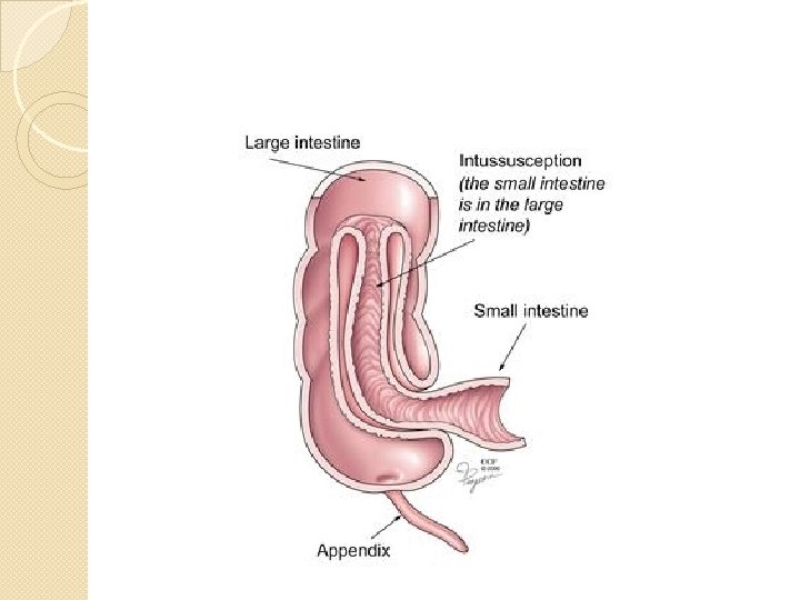
�Intussusception � 1. There is sudden intermittent abdominal pain with vomiting. 2. Absolute constipation may be replaced later by passage of blood and mucus (red current jelly) per anum without fecal odour. 3. There may be curved, sausage shaped lump in the line of the colon with its concavity towards the umbilicus. The lump may harden under examining fingers synchronously with an attack of screaming. 4. Barium enema would show typical pincer shaped ending of the radio-opaque material.
�Renal
�Supra renal
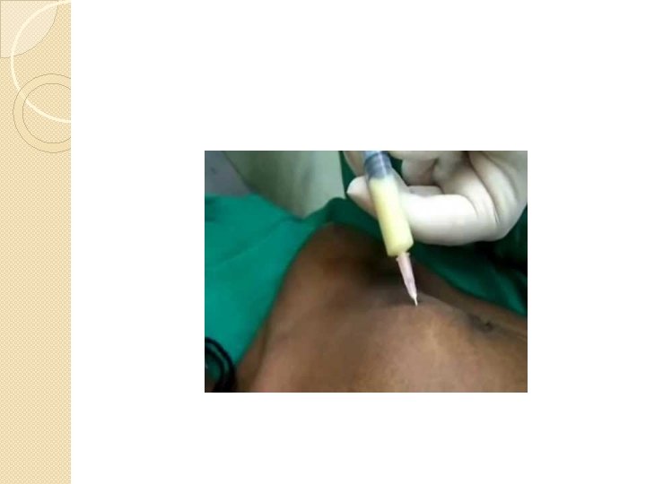
Investigations �Laboratory studies ◦ Routine ◦ special �Imaging Tissue FNAC Histopathology
Imaging �X-ray �Ultrasound �CT scan �MRI �ERCP �Nuclear scans
Endoscopy �EUS �ERCP