
Management of Headache and Facial Pain Haley Burke
Management of Headache and Facial Pain Haley Burke, MD, DABPN
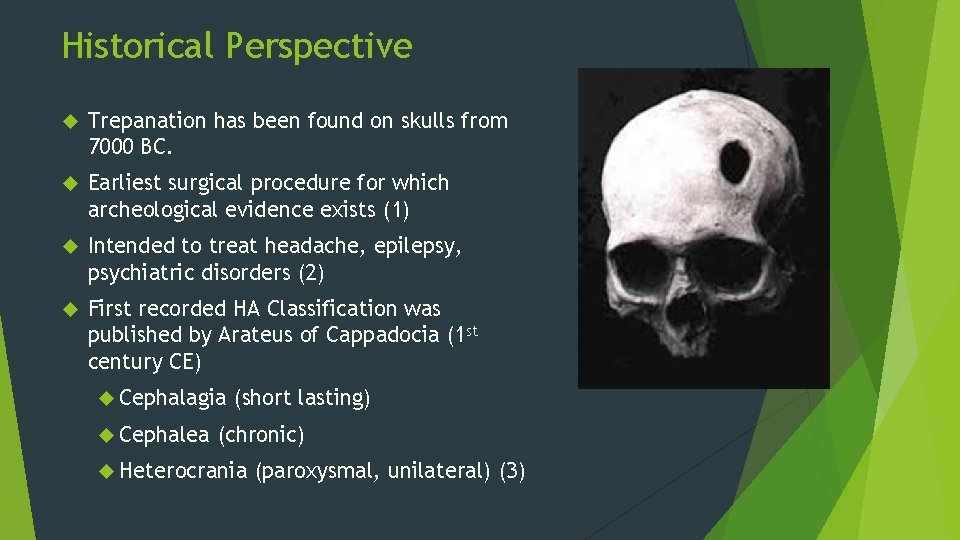
Historical Perspective Trepanation has been found on skulls from 7000 BC. Earliest surgical procedure for which archeological evidence exists (1) Intended to treat headache, epilepsy, psychiatric disorders (2) First recorded HA Classification was published by Arateus of Cappadocia (1 st century CE) Cephalagia Cephalea (short lasting) (chronic) Heterocrania (paroxysmal, unilateral) (3)
Epidemiology 50% of the population will experience a headache during any given year Affect > 28 million Americans (migraine alone) > 90% report a lifetime history of headache Average lifetime prevalence of Migraine: 18% 3% of the population with have a chronic headache (>15 days per month) Sex ratio for migraine: 3: 1 Sex ratio for TTH: 1: 1 (4) Prevalence of migraine: peaks age 25 -55 yo (5)
Migraine Impact on Lost Productivity Total costs of disability attributed to migraine: > $13 Billion annually (1998) $19. 6 Billion attributed to any headache type (2002) Impact of migraine on the labor force likely to increase as more women continue to enter the workforce.
Current Headache Classification International Classification of Headache Disorders – ICHD 3 -Beta www. ICHD-3. org I. Primary headaches II. Secondary headaches No associated underlying etiology Due to other pathology III. Painful cranial neuropathies, other facial pains and other headaches
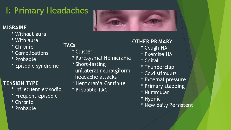
I: Primary Headaches MIGRAINE * Without aura * With aura OTHER PRIMARY TACs * Chronic * Cough HA * Cluster * Complications * Exercise HA * Paroxysmal Hemicrania * Probable * Coital * Short-lasting * Episodic syndrome * Thunderclap unilateral neuralgiform * Cold stimulus headache attacks * External pressure TENSION TYPE * Hemicrania Continue * Primary stabbing * Infrequent episodic * Probable TAC * Nummular * Frequent episodic * Hypnic * Chronic * New daily Persistent * Probable
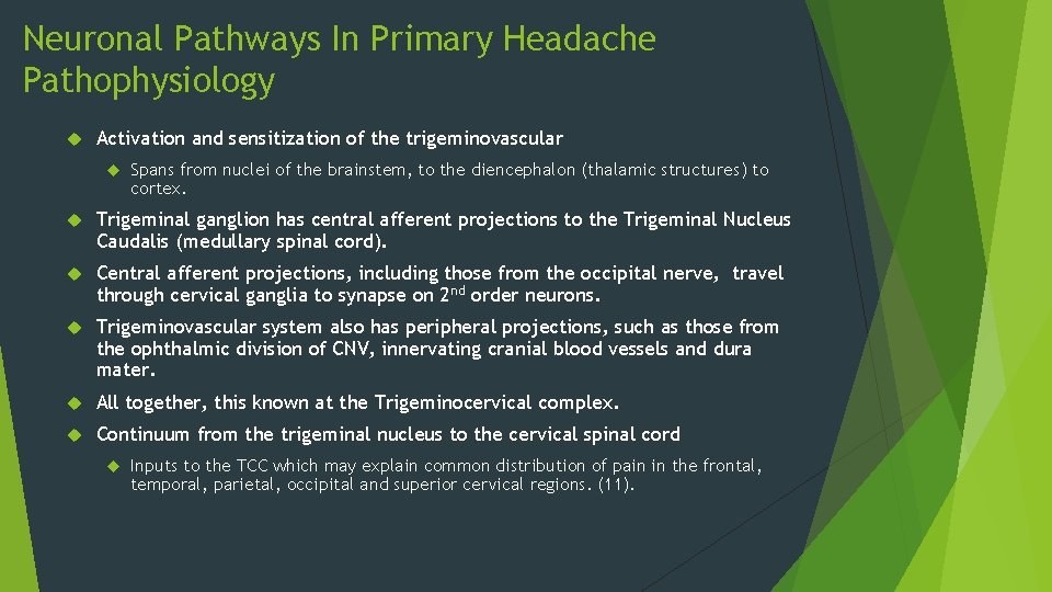
Neuronal Pathways In Primary Headache Pathophysiology Activation and sensitization of the trigeminovascular Spans from nuclei of the brainstem, to the diencephalon (thalamic structures) to cortex. Trigeminal ganglion has central afferent projections to the Trigeminal Nucleus Caudalis (medullary spinal cord). Central afferent projections, including those from the occipital nerve, travel through cervical ganglia to synapse on 2 nd order neurons. Trigeminovascular system also has peripheral projections, such as those from the ophthalmic division of CNV, innervating cranial blood vessels and dura mater. All together, this known at the Trigeminocervical complex. Continuum from the trigeminal nucleus to the cervical spinal cord Inputs to the TCC which may explain common distribution of pain in the frontal, temporal, parietal, occipital and superior cervical regions. (11).
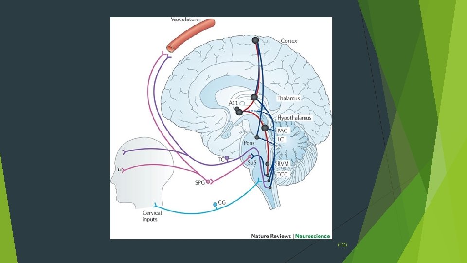
(12)
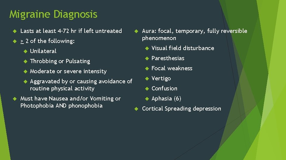
Migraine Diagnosis Lasts at least 4 -72 hr if left untreated > 2 of the following: Aura: focal, temporary, fully reversible phenomenon Unilateral Visual field disturbance Throbbing or Pulsating Paresthesias Moderate or severe intensity Focal weakness Aggravated by or causing avoidance of routine physical activity Vertigo Confusion Aphasia (6) Must have Nausea and/or Vomiting or Photophobia AND phonophobia Cortical Spreading depression
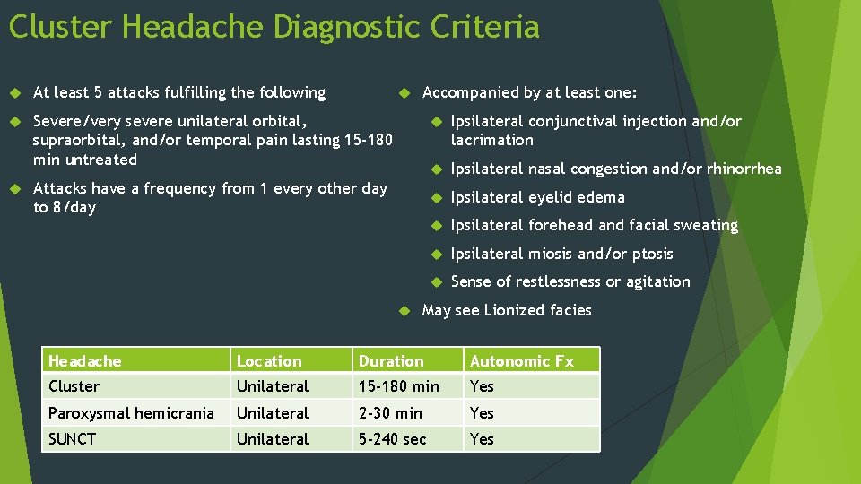
Cluster Headache Diagnostic Criteria At least 5 attacks fulfilling the following Severe/very severe unilateral orbital, supraorbital, and/or temporal pain lasting 15 -180 min untreated Accompanied by at least one: Attacks have a frequency from 1 every other day to 8/day Ipsilateral conjunctival injection and/or lacrimation Ipsilateral nasal congestion and/or rhinorrhea Ipsilateral eyelid edema Ipsilateral forehead and facial sweating Ipsilateral miosis and/or ptosis Sense of restlessness or agitation May see Lionized facies Headache Location Duration Autonomic Fx Cluster Unilateral 15 -180 min Yes Paroxysmal hemicrania Unilateral 2 -30 min Yes SUNCT Unilateral 5 -240 sec Yes
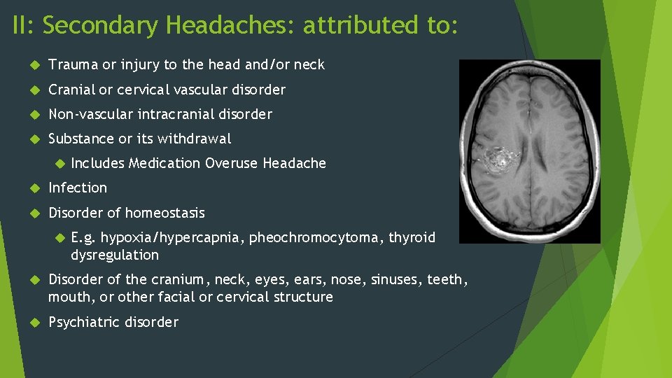
II: Secondary Headaches: attributed to: Trauma or injury to the head and/or neck Cranial or cervical vascular disorder Non-vascular intracranial disorder Substance or its withdrawal Includes Medication Overuse Headache Infection Disorder of homeostasis E. g. hypoxia/hypercapnia, pheochromocytoma, thyroid dysregulation Disorder of the cranium, neck, eyes, ears, nose, sinuses, teeth, mouth, or other facial or cervical structure Psychiatric disorder
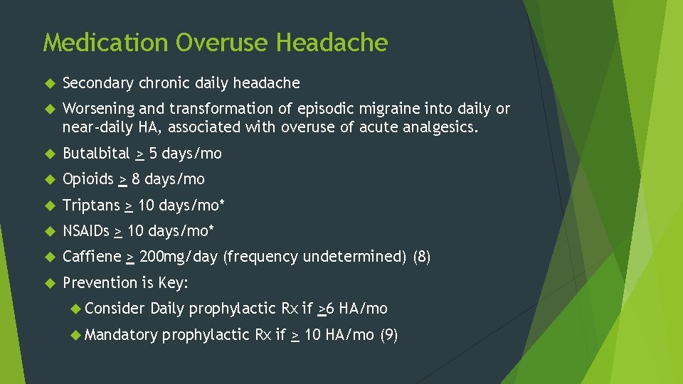
Medication Overuse Headache Secondary chronic daily headache Worsening and transformation of episodic migraine into daily or near-daily HA, associated with overuse of acute analgesics. Butalbital > 5 days/mo Opioids > 8 days/mo Triptans > 10 days/mo* NSAIDs > 10 days/mo* Caffiene > 200 mg/day (frequency undetermined) (8) Prevention is Key: Consider Daily prophylactic Rx if >6 HA/mo Mandatory prophylactic Rx if > 10 HA/mo (9)
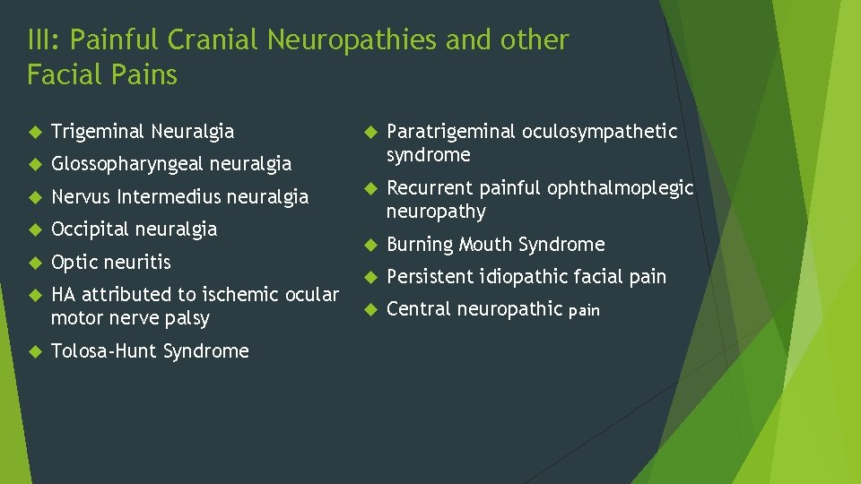
III: Painful Cranial Neuropathies and other Facial Pains Trigeminal Neuralgia Glossopharyngeal neuralgia Nervus Intermedius neuralgia Occipital neuralgia Optic neuritis HA attributed to ischemic ocular motor nerve palsy Tolosa-Hunt Syndrome Paratrigeminal oculosympathetic syndrome Recurrent painful ophthalmoplegic neuropathy Burning Mouth Syndrome Persistent idiopathic facial pain Central neuropathic pain
Treatment Options Lifestyle changes Prophylactic Abortive medications Procedural Surgical Medications Intervention Psychologic/Psychiatric treatment
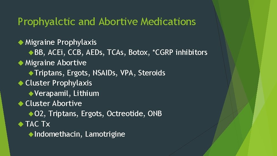
Prophyalctic and Abortive Medications Migraine Prophylaxis BB, ACEi, CCB, AEDs, TCAs, Botox, *CGRP inhibitors Migraine Abortive Triptans, Ergots, NSAIDs, VPA, Steroids Cluster Prophylaxis Verapamil, Lithium Cluster Abortive O 2, Triptans, Ergots, Octreotide, ONB TAC Tx Indomethacin, Lamotrigine
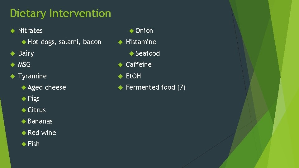
Dietary Intervention Onion Nitrates Hot dogs, salami, bacon Histamine Seafood Dairy MSG Caffeine Tyramine Et. OH Aged Fermented food (7) cheese Figs Citrus Bananas Red Fish wine
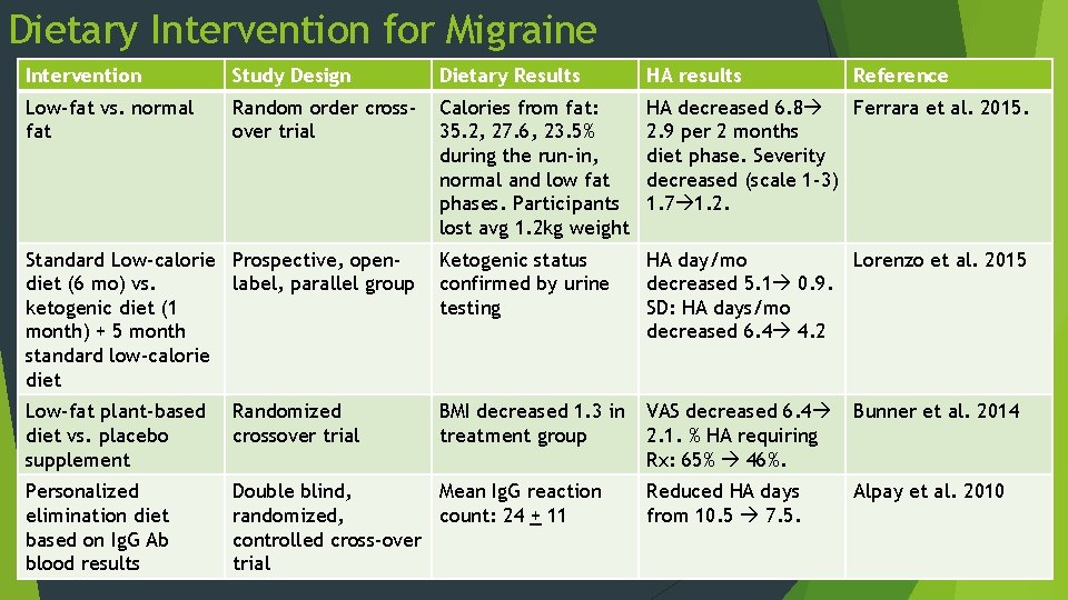
Dietary Intervention for Migraine Intervention Study Design Dietary Results HA results Reference Low-fat vs. normal fat Random order crossover trial Calories from fat: 35. 2, 27. 6, 23. 5% during the run-in, normal and low fat phases. Participants lost avg 1. 2 kg weight HA decreased 6. 8 Ferrara et al. 2015. 2. 9 per 2 months diet phase. Severity decreased (scale 1 -3) 1. 7 1. 2. Standard Low-calorie Prospective, opendiet (6 mo) vs. label, parallel group ketogenic diet (1 month) + 5 month standard low-calorie diet Ketogenic status confirmed by urine testing HA day/mo Lorenzo et al. 2015 decreased 5. 1 0. 9. SD: HA days/mo decreased 6. 4 4. 2 Low-fat plant-based diet vs. placebo supplement Randomized crossover trial BMI decreased 1. 3 in treatment group VAS decreased 6. 4 2. 1. % HA requiring Rx: 65% 46%. Bunner et al. 2014 Personalized elimination diet based on Ig. G Ab blood results Double blind, Mean Ig. G reaction randomized, count: 24 + 11 controlled cross-over trial Reduced HA days from 10. 5 7. 5. Alpay et al. 2010
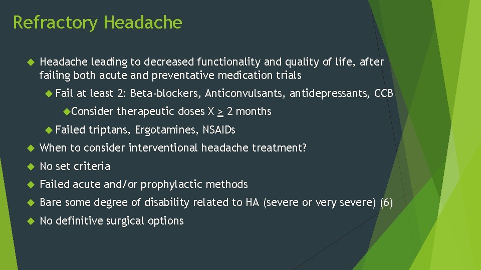
Refractory Headache leading to decreased functionality and quality of life, after failing both acute and preventative medication trials Fail at least 2: Beta-blockers, Anticonvulsants, antidepressants, CCB Consider Failed therapeutic doses X > 2 months triptans, Ergotamines, NSAIDs When to consider interventional headache treatment? No set criteria Failed acute and/or prophylactic methods Bare some degree of disability related to HA (severe or very severe) (6) No definitive surgical options
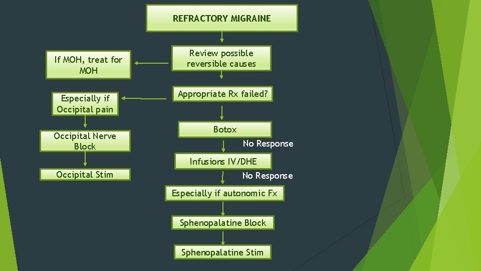
REFRACTORY MIGRAINE If MOH, treat for MOH Especially if Occipital pain Occipital Nerve Block Review possible reversible causes Appropriate Rx failed? Botox No Response Infusions IV/DHE Occipital Stim No Response Especially if autonomic Fx Sphenopalatine Block Sphenopalatine Stim
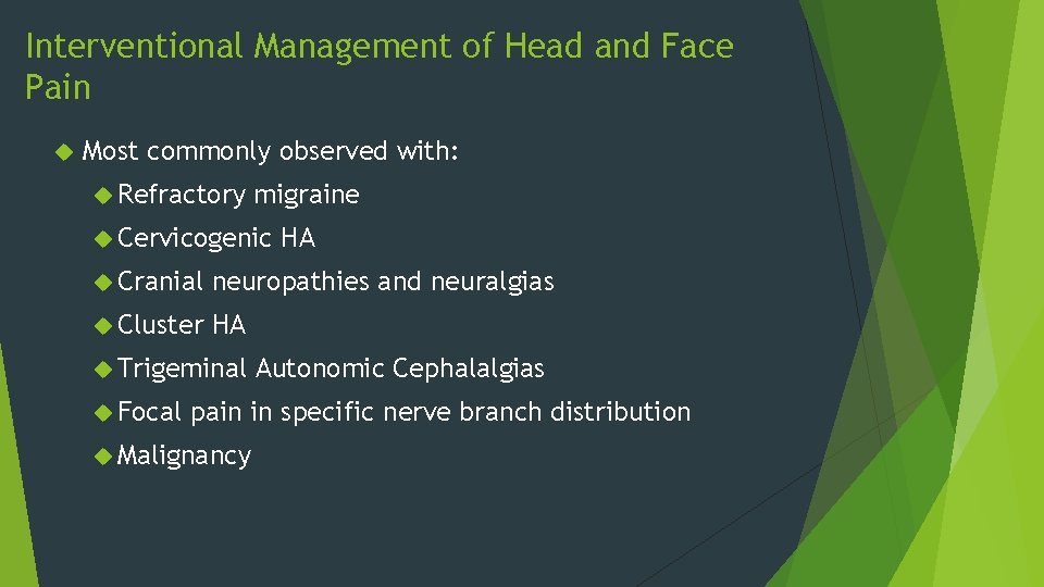
Interventional Management of Head and Face Pain Most commonly observed with: Refractory migraine Cervicogenic HA Cranial neuropathies and neuralgias Cluster HA Trigeminal Focal Autonomic Cephalalgias pain in specific nerve branch distribution Malignancy
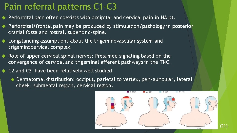
Pain referral patterns C 1 -C 3 Periorbital pain often coexists with occipital and cervical pain in HA pt. Periorbital/frontal pain may be produced by stimulation/pathology in posterior cranial fossa and rostral, superior c-spine. Longstanding assumptions about the trigeminovascular system and trigeminocervical complex. Role of upper cervical spinal nerves: Presumed signaling based on the convergence of cervical and trigeminal afferent pathways in the TNC. C 2 and C 3 have been relatively well studied Dermatomal distribution: occiput, parietal to vertex, peri-auricular, lateral cheek, submental region, cervical region. (21)
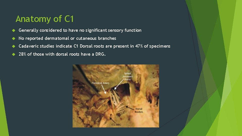
Anatomy of C 1 Generally considered to have no significant sensory function No reported dermatomal or cutaneous branches Cadaveric studies indicate C 1 Dorsal roots are present in 47% of specimens 28% of those with dorsal roots have a DRG.
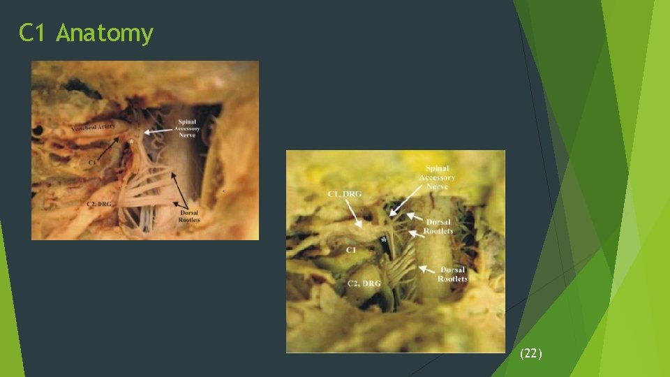
C 1 Anatomy (22)
Referral Patterns of C 1 -C 3 Johnston et al. 2013 N = 10 Patients with known occipital pain underwent C 1, C 2, C 3 stimulation to evaluate therapeutic procedures for chronic occipital pain. All patients failed conservative treatments Anticonvulsants, NSAIDs including indomethacin, physical therapy. All patients had pain reproduced by pressure over GON. C 1: RF needle was placed at posterior superior edge of the arch of the atlas by the C 1 spinal nerve, inferior to vertebral artery. C 2: needle placed next to C 2 DRG in the intervertebral foramen. C 3: Targeted transforaminally
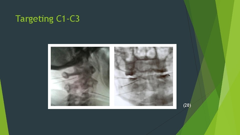
Targeting C 1 -C 3 (20)
Targeting C 1 -C 3 Motor responses (2 Hz, 1. 5 -2 m. A) confirmed needle placement. C 1 motor response: contraction of rectus capitis lateralis, rectus capitis anterior and longus capitis. Sensory stim (50 Hz, 0. 5 -1. 0 m. A) was then recorded Sensation from mechanical pressure due to needle tap and pressure through injectate administration were also recorded. Pain referral patterns were reported by the patient. Patients then underwent perineural injection of 2% chloroprocaine or 0. 5% bupivacaine with 10 mg Dexamethasone.
Results of C 1 -C 3 Stimulation All 6/6 patients with concomitant Dx of Migraine experienced periorbital and frontal pain with C 1 stim. Remaining patients (4) had pain in the parietal or occipital pattern with C 1 stim. In all patients: C 2 C 3 pain in occipital and parietal distribution pain in occipital, periauricular, submental, and/or lateral cervical distributions
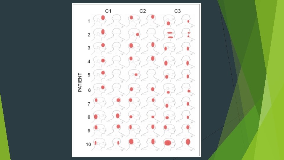
C 1 -C 3 Pain Referral Demonstrated periorbital and frontal distribution of pain elicited by direct stimulation of C 1 spinal nerve in subjects with migraine. Findings suggest sensory fibers innervating periorbital and frontal regions, which has not previously been described. Alternative explanation: sensory input from C 1 referred pain C 1 has been reported to innervate the dura mater of posterior fossa and upper c-spine. Stimulation of these regions has been reported to periorbital pain. Again – answer may reside in the TNC. Implications: C 1 may be an important therapeutic target.
Headaches and the Lower C-spine ICHD-3: addresses “upper cervical radiculopathy-induced headache” Specified as presence of clinically or radiologically clear evidence of radiculopathy associated with the 2 nd to 3 rd cervical levels Case reports of HA in context of: Tumor infiltration of C 2 Schwannoma at craniocervical junction Trigeminal Neuralgia due to compression at the spinal nucleus of CNV Pathophysiology of headaches from middle-lower cervical levels?
Headache and the Lower C-spine Mechanism likely related to the spinal nucleus of CNV Most pain stimuli at lower levels pass through the dorsal horn of the cord via central gray matter Signals ascend anterior spinothalamic tract on opposite side. Some stimuli ascend through the spinocervicothalamic tract. Descends to C 3 and synapses with C 3 nerve. Communicates with the TNC through some type of anastomosis. Spinal roots of CNXI with fibers from anterior branches of C 2 -C 4 innervate SCM and trapezius. Contain sensory nerves: Proprioceptive and Noxious signals (26)
Headache and the lower C-spine: Alternative Mechanism Overlap of dermatome and myotome as underlying explanation Dykes and Terzis (1981) cutaneous region served by one spinal nerve is wider and more variable in location than generally recognized. Myotome territory is larger than the corresponding dermatome. Schirmer (2011) significant # of roots innervated a broader range of muscles than previously known during intraoperative nerve root stimulation Ex: C 5 and C 6 contributed to all muscle of the upper extremity including trapezius. 129 pts – evaled c-spine.
Headache and the Lower C-Spine Persson et al 2006: evaluated effect of cervical SNRB on HA 275 pts with cervical radiculopathy. 161 of these pts reported associated daily or recurrent HA in addition to neck pain 59% of pts had > 50% reduction in HA 30 min post-procedure. 69% of these had total relief Pathophysiology unclear Authors suggest HA secondary to signals from disc capsule, cervical ligaments or muscles. Caution that HA as a singular symptom in not an indication for surgical decompression treatments.
Peripheral Nerve Blocks for HA and Facial Pain Few controlled studies regarding effect of LA procedures for HA and facial pain exist Placebo effects often common Complete or near complete pain relief after placebo 32. 4% (10) Goal: Block C-fibers Mechanism: Reduce afferent input to decrease activity at the trigeminal Nucleus caudalis, cervical dorsal horn, and converging circuits.
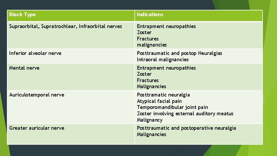
Block Type Indications Supraorbital, Supratrochlear, Infraorbital nerves Entrapment neuropathies Zoster Fractures malignancies Inferior alveolar nerve Posttraumatic and postop Neuralgias Intraoral malignancies Mental nerve Entrapment neuropathies Zoster Fractures Malignancies Auriculotemporal nerve Posttramatic neuralgia Atypical facial pain Temporomandibular joint pain Zoster involving esternal auditory meatus Malignancy Greater auricular nerve Posttraumatic and postoperative neuralgia Malignancies
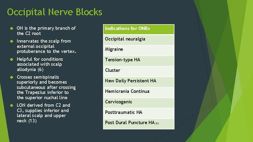
Occipital Nerve Blocks ON is the primary branch of the C 2 root Innervates the scalp from external occipital protuberance to the vertex. Occipital neuralgia Helpful for conditions associated with scalp allodynia (6) Tension-type HA Crosses semispinalis superiorly and becomes subcutaneous after crossing the Trapezius inferior to the superior nuchal line LON derived from C 2 and C 3, supplies inferior and lateral scalp and upper neck (13) Indications for ONBs Migraine Cluster New Daily Persistent HA Hemicrania Continua Cervicogenic Posttraumatic HA Post Dural Puncture HA (6)
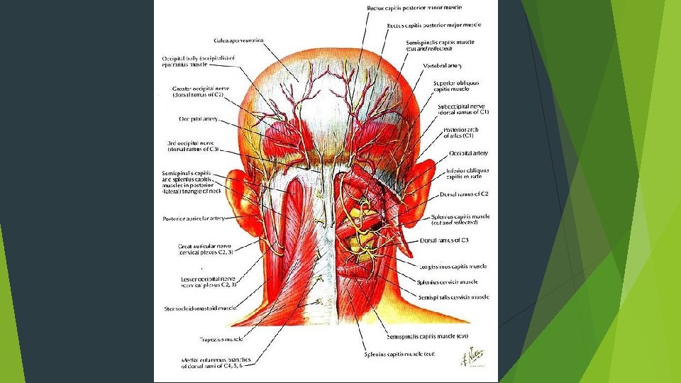
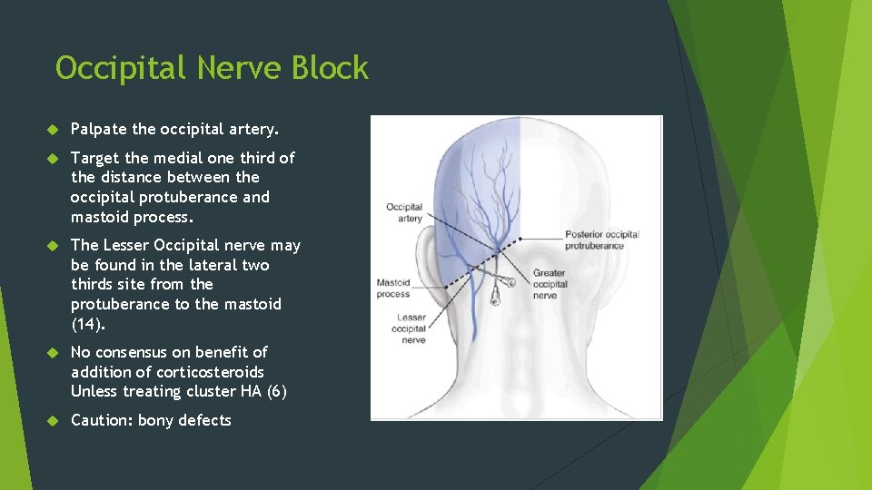
Occipital Nerve Block Palpate the occipital artery. Target the medial one third of the distance between the occipital protuberance and mastoid process. The Lesser Occipital nerve may be found in the lateral two thirds site from the protuberance to the mastoid (14). No consensus on benefit of addition of corticosteroids Unless treating cluster HA (6) Caution: bony defects
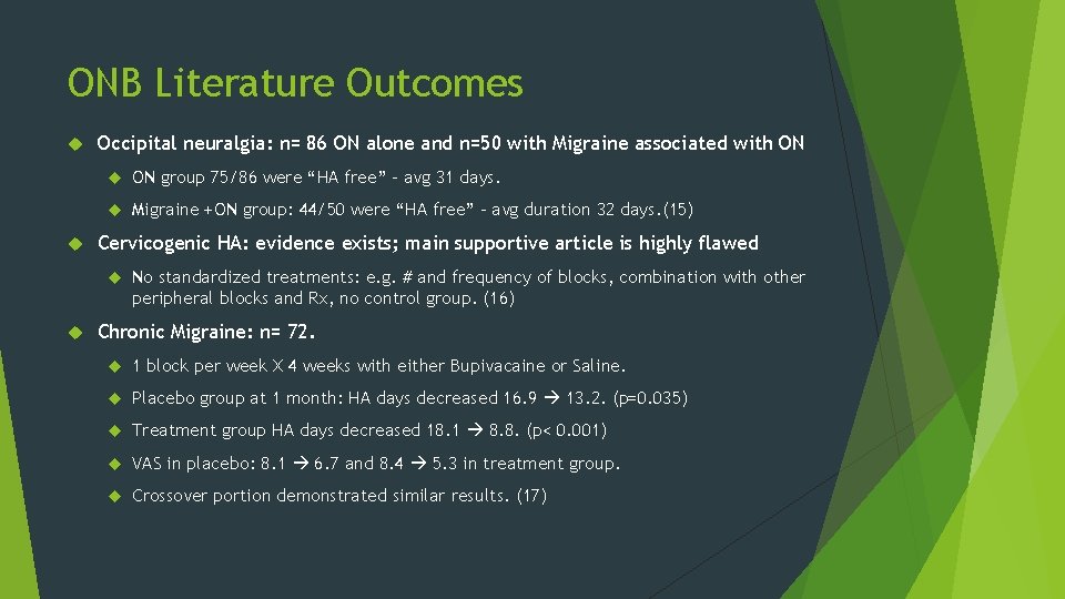
ONB Literature Outcomes Occipital neuralgia: n= 86 ON alone and n=50 with Migraine associated with ON group 75/86 were “HA free” – avg 31 days. Migraine +ON group: 44/50 were “HA free” – avg duration 32 days. (15) Cervicogenic HA: evidence exists; main supportive article is highly flawed No standardized treatments: e. g. # and frequency of blocks, combination with other peripheral blocks and Rx, no control group. (16) Chronic Migraine: n= 72. 1 block per week X 4 weeks with either Bupivacaine or Saline. Placebo group at 1 month: HA days decreased 16. 9 13. 2. (p=0. 035) Treatment group HA days decreased 18. 1 8. 8. (p< 0. 001) VAS in placebo: 8. 1 6. 7 and 8. 4 5. 3 in treatment group. Crossover portion demonstrated similar results. (17)
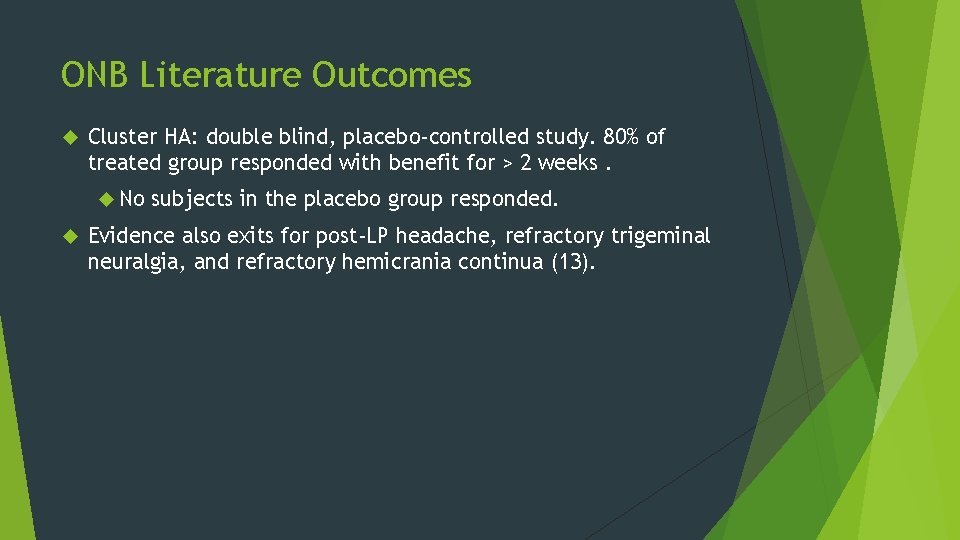
ONB Literature Outcomes Cluster HA: double blind, placebo-controlled study. 80% of treated group responded with benefit for > 2 weeks. No subjects in the placebo group responded. Evidence also exits for post-LP headache, refractory trigeminal neuralgia, and refractory hemicrania continua (13).
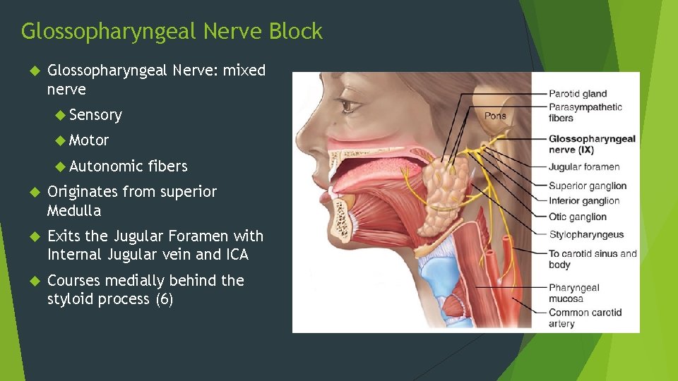
Glossopharyngeal Nerve Block Glossopharyngeal Nerve: mixed nerve Sensory Motor Autonomic fibers Originates from superior Medulla Exits the Jugular Foramen with Internal Jugular vein and ICA Courses medially behind the styloid process (6)
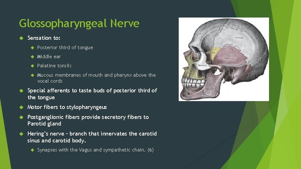
Glossopharyngeal Nerve Sensation to: Posterior third of tongue Middle ear Palatine tonsils Mucous membranes of mouth and pharynx above the vocal cords Special afferents to taste buds of posterior third of the tongue Motor fibers to stylopharyngeus Postganglionic fibers provide secretory fibers to Parotid gland Hering’s nerve – branch that innervates the carotid sinus and carotid body. Synapses with the Vagus and sympathetic chain. (6)
Glossopharyngeal Neuralgia Uncommon, unilateral. Neuralgic pain in ear, base of tongue, tonsillar fossa, or beneath angle of the jaw. Attacks last seconds -2 min Sx may be precipitated by swallowing, talking, coughing, chewing, yawning. Usually begins after 6 th decade. May see bradycardia and asystole with glossopharyngeal neuralgia paroxysms. Up to 2% of pt may experience LOC (6) Possibly secondary to microvascular compression by posterior cerebellar artery. Eagle syndrome Elongated styloid and ossified stylohyoid ligament. Consider block if: Diagnosis in question Refractory to conservative management
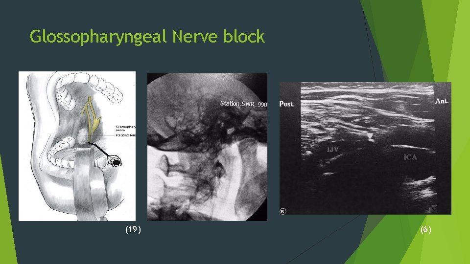
Glossopharyngeal Nerve block (19) (6)
The Sphenopalatine Ganglion Largest collection of neurons outside of the brain Composed of parasympathetic ganglia from the greater petrosal nerve Resides in the pterygopalatine fossa (PPF) bilaterally Axons Lacrimal gland nasal mucosa. Controls local blood flow Implicated in numerous headache and facial pain conditions Initially blocked with cocaine, followed by silver nitrate, 0. 4% gaseous formaldehyde, and 5% phenol in 1908 Studied for Cluster HA in the 1980 s 2006: delivery devices on the market 2009: Stim Implants (23)
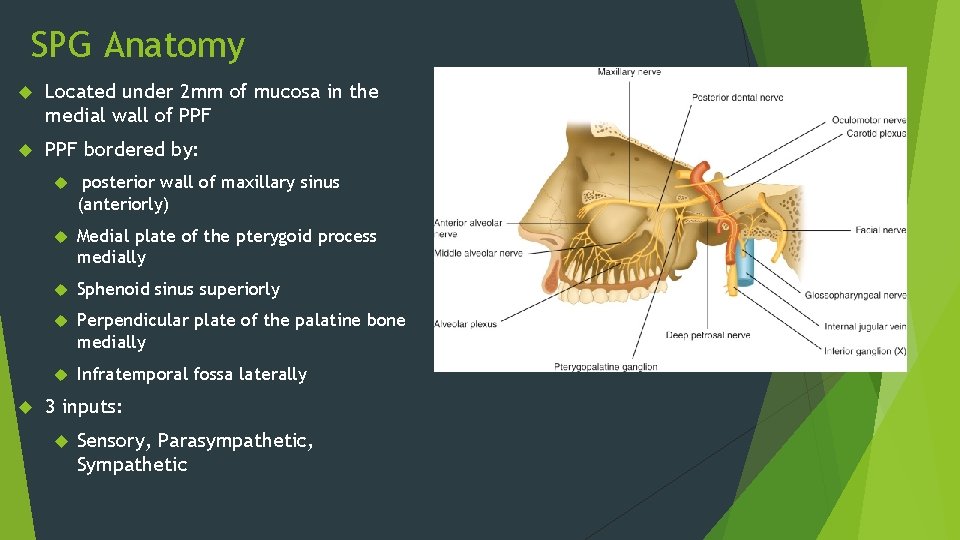
SPG Anatomy Located under 2 mm of mucosa in the medial wall of PPF bordered by: posterior wall of maxillary sinus (anteriorly) Medial plate of the pterygoid process medially Sphenoid sinus superiorly Perpendicular plate of the palatine bone medially Infratemporal fossa laterally 3 inputs: Sensory, Parasympathetic, Sympathetic
SPG Anatomy Sensory branches supplying: Bony palate Gingiva Mucosa of buccal cavity Uvula Tonsils Soft palate Orbit Connections to CN V blur sensory connections of SPG alone
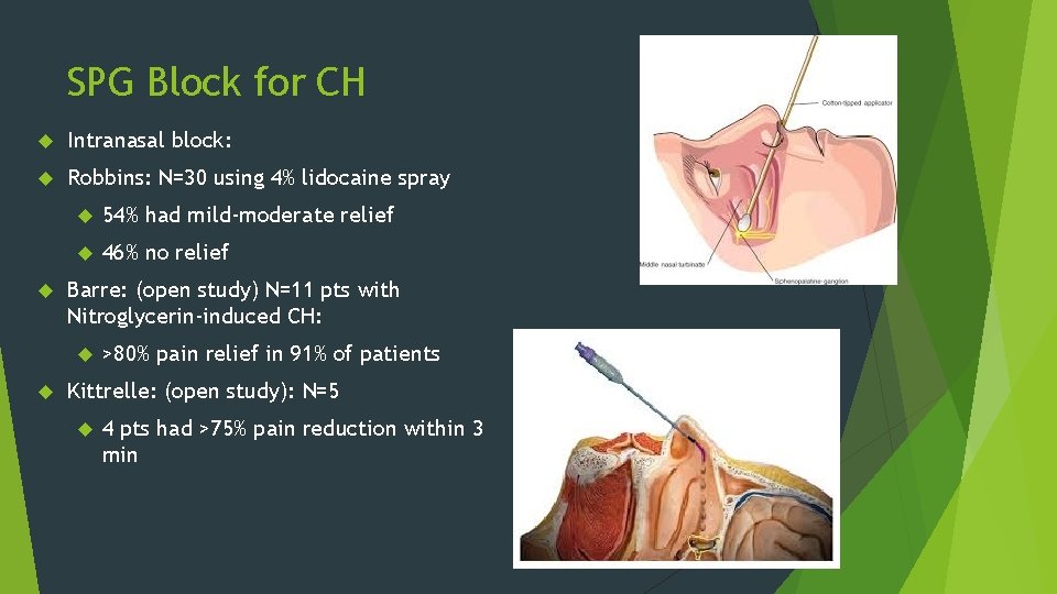
SPG Block for CH Intranasal block: Robbins: N=30 using 4% lidocaine spray 54% had mild-moderate relief 46% no relief Barre: (open study) N=11 pts with Nitroglycerin-induced CH: >80% pain relief in 91% of patients Kittrelle: (open study): N=5 4 pts had >75% pain reduction within 3 min
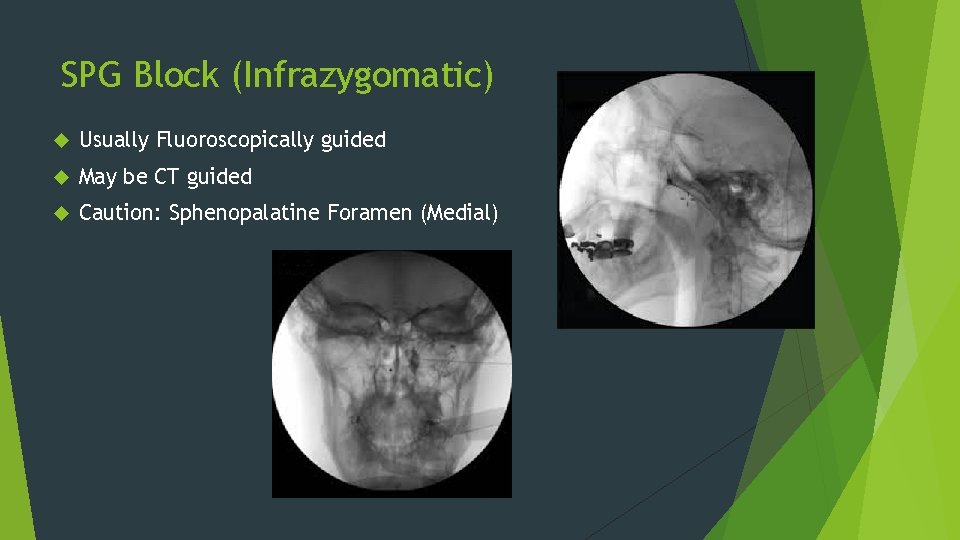
SPG Block (Infrazygomatic) Usually Fluoroscopically guided May be CT guided Caution: Sphenopalatine Foramen (Medial)
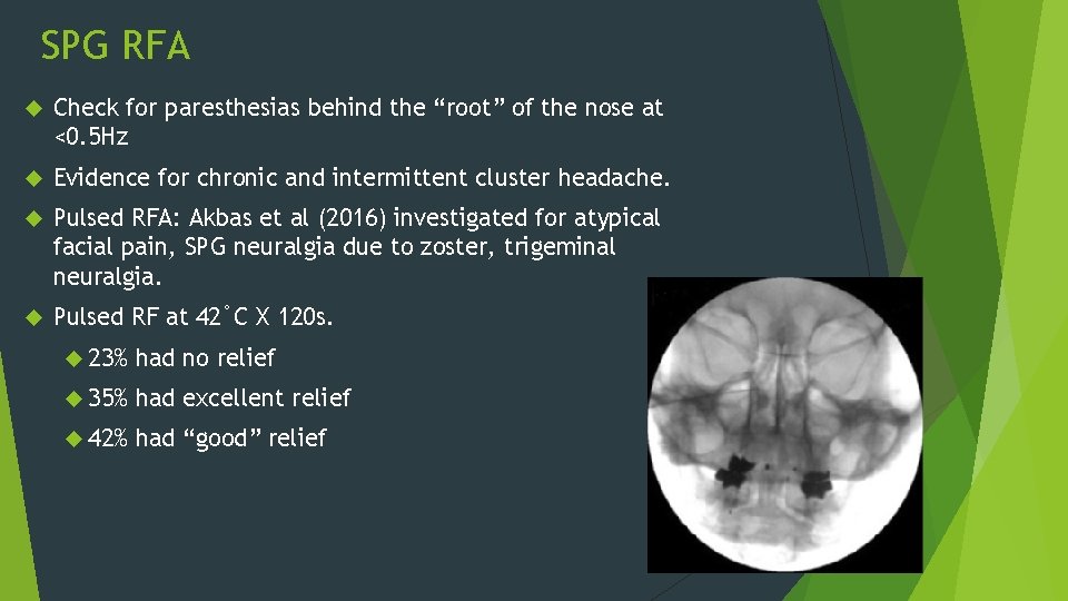
SPG RFA Check for paresthesias behind the “root” of the nose at <0. 5 Hz Evidence for chronic and intermittent cluster headache. Pulsed RFA: Akbas et al (2016) investigated for atypical facial pain, SPG neuralgia due to zoster, trigeminal neuralgia. Pulsed RF at 42˚C X 120 s. 23% had no relief 35% had excellent relief 42% had “good” relief
SPG Block and RFA Complications Epistaxis Intravascular injection Hematoma formation Infection Reflex bradycardia Hypoesthesia/dysesthesia: palate, maxilla, posterior pharynx. Dry eye Diplopia – injectate spread from PPF to inferior orbital fissure (limit injectate volume)
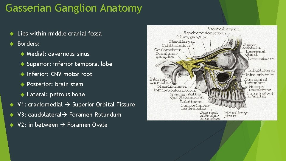
Gasserian Ganglion Anatomy Lies within middle cranial fossa Borders: Medial: cavernous sinus Superior: inferior temporal lobe Inferior: CNV motor root Posterior: brain stem Lateral: petrous bone V 1: craniomedial Superior Orbital Fissure V 3: caudolateral Foramen Rotundum V 2: in between Foramen Ovale
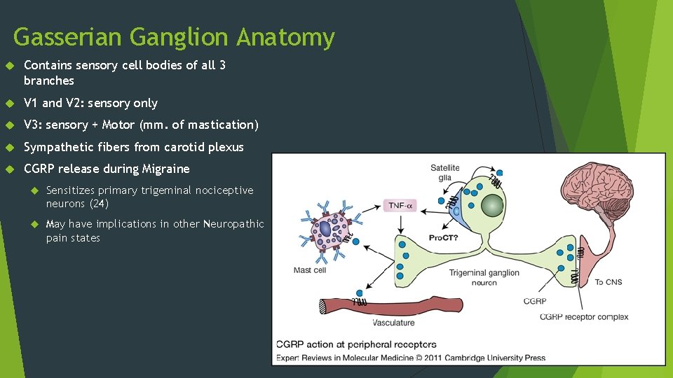
Gasserian Ganglion Anatomy Contains sensory cell bodies of all 3 branches V 1 and V 2: sensory only V 3: sensory + Motor (mm. of mastication) Sympathetic fibers from carotid plexus CGRP release during Migraine Sensitizes primary trigeminal nociceptive neurons (24) May have implications in other Neuropathic pain states
Gasserian Ganglion Block: Indications: Diagnostic or therapeutic treatment for Trigeminal Neuralgia symptoms Trigeminal Neuropathy ? To predict prognosis prior to neuroablative or surgical procedure of the Trigeminal ganglion Malignancy Orofacial pain syndromes NOS
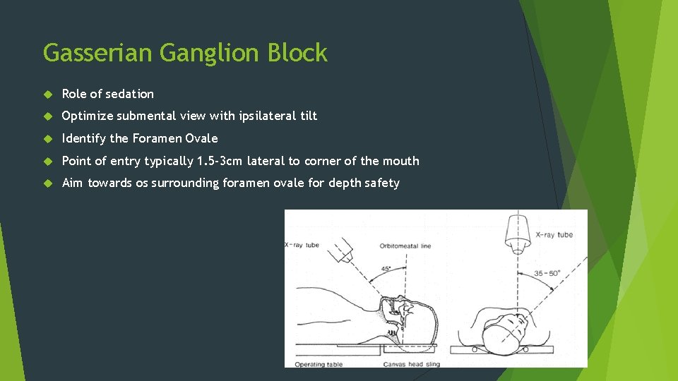
Gasserian Ganglion Block Role of sedation Optimize submental view with ipsilateral tilt Identify the Foramen Ovale Point of entry typically 1. 5 -3 cm lateral to corner of the mouth Aim towards os surrounding foramen ovale for depth safety
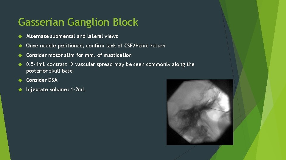
Gasserian Ganglion Block Alternate submental and lateral views Once needle positioned, confirm lack of CSF/heme return Consider motor stim for mm. of mastication 0. 5 -1 m. L contrast vascular spread may be seen commonly along the posterior skull base Consider DSA Injectate volume: 1 -2 m. L
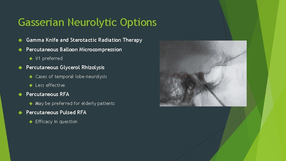
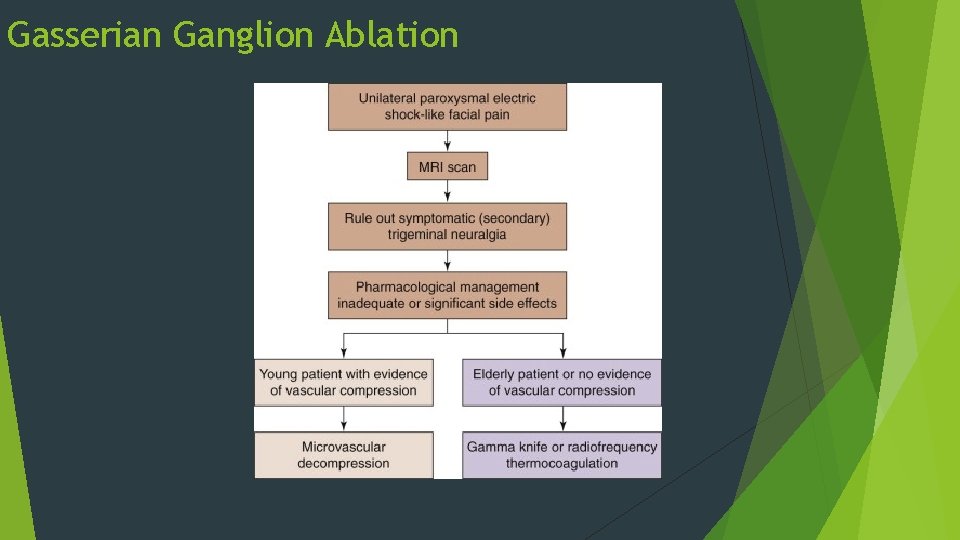
Gasserian Neurolytic Options Gamma Knife and Sterotactic Radiation Therapy Percutaneous Balloon Microcompression Percutaneous Glycerol Rhizolysis Cases of temporal lobe neurolysis Less effective Percutaneous RFA V 1 preferred May be preferred for elderly patients Percutaneous Pulsed RFA Efficacy in question
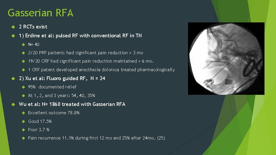
Gasserian RFA 2 RCTs exist 1) Erdine et al: pulsed RF with conventional RF in TN N= 40 2/20 PRF patients had significant pain reduction > 3 mo 19/20 CRF had significant pain reduction maintained > 6 mo. 1 CRF patent developed anesthesia dolorosa treated pharmacologically 2) Xu et al: Fluoro guided RF, N = 24 95% documented relief At 1, 2, and 3 years: 54, 40, 35% Wu et al: N= 1860 treated with Gasserian RFA Excellent outcome 78. 8% Good 17. 5% Poor 3. 7 % Pain recurrence 11. 1% during first 12 mo and 25% after 24 mo. (25)
Complications of Gasserian Block and RFA Anesthetic deposited to CSF Intravascular injection Anesthesia dolorosa Corneal Hypoesthesia Masseter weakness/paralysis Dysesthesia CN III and VI palsy (possibly permanent) CSF leakage Carotid-Cavernous fistula formation Infection/Meningitis Bleeding/hematoma
References: 1) Capasso, Luigi (2002). Principi di storia della patologia umana: corso di storia della medicina per gli studenti della Facoltà di medicina e chirurgia e della Facoltà di scienze infermieristiche (in Italian). 2) Brothwell, Don R. (1963). Digging up Bones; the Excavation, Treatment and Study of Human Skeletal Remains. London: British Museum (Natural History). p. 126. 3) Levin, Morris; Baskin, Steven M. ; Bigal, Marcelo E. (2008). Comprehensive Review of Headache Medicine. Oxford University Press. ISBN 0 -19 -536673 -5. 4) IASP-pain. org 5) Burton et al. The impact of migraine and the effect of migraine treatment on workplace productivity in the united states and suggestons for future research. Mayo Clin Proc. 2009 May; 84(5). 6) Narouze S. Interventional Management of Head and Face Pain. 2014. 7) Slavin M & Ailani J. A clinical approach to addressing diet with migraine patients. Current neurology and Neuroscience reports. 2017. 8) Americanmigrainefoundation. org 9) Tepper S & Tepper D. The Cleveland Clinic Manual of Headache Therapy. 2011. 10) de Craen et al. Placebo effect in the acute treatment of migraine: subcutaneous placebos are better than oral placebos. J Neurol. 2000. 11) Bartsch T, Goadsby PJ. Anatomy and physiology of pain referral in primary and cervicogenic headache disorders. Headache Curr. 2, 42– 48 (2005). 12) Akerman et al. Diencephalic and brainstem mechanisms in migraine. Nature Reviews: Neuroscience. 2011. 13) Kleen & Levin. Injection therapy for headache and facial pain. Oral and Maxillofacial surgery clinics. 2016 (28). 14) Kashipazha et al. Preventive effect of greater occipital nerve block on severity and frequency of migraine headache. Glob J Health Sci. 2014 15) Anthony M. : Headache and the greater occipital nerve. Clin Neurol Neurosurg 1992; 94: pp. 297 -301 16) Rothbart P. , Fiedler K. , Gale G. , et al: A descriptive study of 100 patients undergoing palliative nerve blocks for chronic intractable headache and neck ache. Pain Res Manag 2000; 5: pp. 243 -248 17) Inan L. E. , Inan N. , Karadas O. , et al: Greater occipital nerve blockade for the treatment of chronic migraine: a randomized, multicenter, double-blind, and placebo-controlled study. Acta Neurol Scand 2015 18) Ambrosini et al. Suboccipital injection with a mixture of rapid- and long-acting steroids in cluster headache: a double-blind placebo-controlled study. Pain. 2005. 19) Brown D. Atlas of Interventional Pain Management. 2017. 20) Johnston M et al. Pain referral patterns of the C 1 to C 3 nerves: implications for headache disorders. Annals of Neurology 2013. 21) Bogduk N & Govind J. Cervicogenic headache: an assessment of the evidence on clinical diagnosis, invasive tests, and treatment. Lancet Neurol. 2009. 22) Tubbs et al. Clinical anatomy of C 1 Dorsal Root, Ganglion, and Ramus: A review and anatomical study. Clinical Anatomy. 2007. 23) Robbins et al. The sphenopalatine ganglion: anatomy, pathophysiology, and therapeutic targeting in headache. Headache. 2015. 24) Cornelison et al. Elevated levels of calcitonin gen-related peptide in upper spinal cord promotes sensitization of primary trigeminal nociceptive neurons. Neuroscience. 2016. 25) Wu CY, Meng FG, Xu SJ, et al: Selective percutaneous radiofrequency thermocoagulation in the treatment of trigeminal neuralgia: report on 1860 cases. Chin Med J 2004; 117: pp. 467 -470 26) Gondo et al. A case of cervicogenic headache caused by C 5 nerve root derived schwannoma. Cephalagia. 22016.

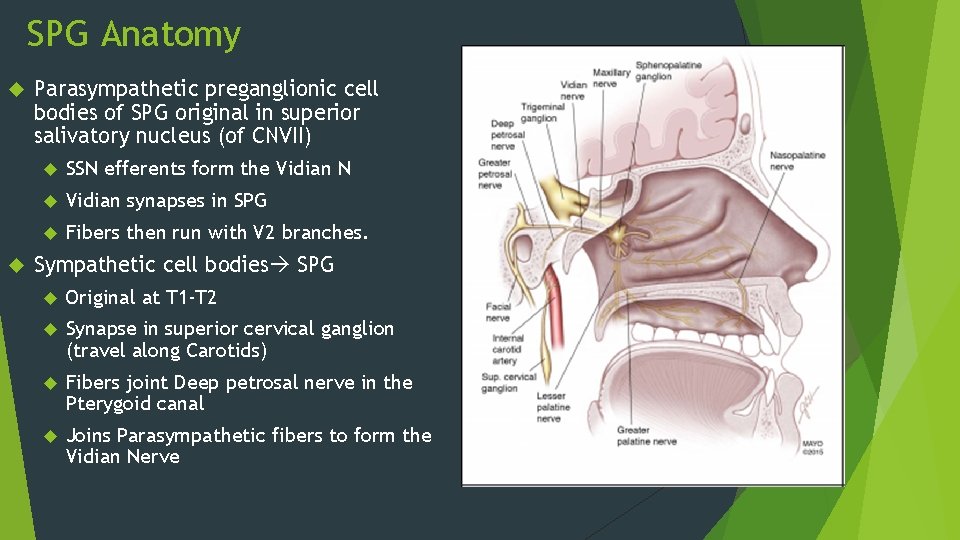
SPG Anatomy Parasympathetic preganglionic cell bodies of SPG original in superior salivatory nucleus (of CNVII) SSN efferents form the Vidian N Vidian synapses in SPG Fibers then run with V 2 branches. Sympathetic cell bodies SPG Original at T 1 -T 2 Synapse in superior cervical ganglion (travel along Carotids) Fibers joint Deep petrosal nerve in the Pterygoid canal Joins Parasympathetic fibers to form the Vidian Nerve
Gasserian Ganglion Ablation
- Slides: 63