
Management of Diarrheal disease in children Dr KURE




- Slides: 74
Management of Diarrheal disease in children Dr KURE HAJARA ISMAILA © NITMED TUTORIALS
OUTLINE �INTRODUCTION �DEFINITION �AETIOLOGY �PATHOPHYSIOLOGY �CLINICAL FEATURES �TREATMENT �COMPLICATIONS �PROGNOSIS
Introduction �DIARRHEAL DISEASE Are among the leading causes of morbidity and mortality in under fives in Nigeria. �Inadequate safe water, sanitation and hygiene account for the disease burden. �In 2016 UNICEF the diarrhea prevalence rate was 18. 8% and in the worst in sub-Saharan africa and above the average of 16%.
Introduction � 1. 2 -1. 9 EPISODES/ person annually in the general population � 2. 4 episodes per child < 3 years old annually � 5 episodes/year for children <3 years old and in day care �Worldwide it plays a central role in the vicious cycle of nutrient malabsorbtion, ensuing malnutrition and susceptibility to systemic infections such as measles.
Introduction �It is particularly problematic in infants because of their propensity to undergo acute volume and nutrient depletion during a phase of rapid somatic and brain growth. �Diarrhea is a reversal of the normal net absorptive status of water and electrolyte absorption.
Definition �Diarrhea is a disease of the gastro intestinal track characterized by abnormal fluidity and frequency of fecal evacuation, three or more or at least one bloody stool in a 24 hr period. �Diarrhea represents an excessive loss of fluid and electrolytes in stool and is defined quantitatively as a total daily volume exceeding 20 g/kg or > 10 ml/ stool/kg body weight /day)
�Frequency- normal for young infants(breast fed) to have 3 -10 stools per day. �Older infants have 1 -2/day �Consistency and colour- this changes with age and differs from person to person depending on your diet
High risk groups �Young age groups �Immune deficiency �Measles �Malnutrition �Travel to endemic region �Lack of breast feeding �Exposure to unsanitary conditions �Attendance of child care centre �Poor maternal education
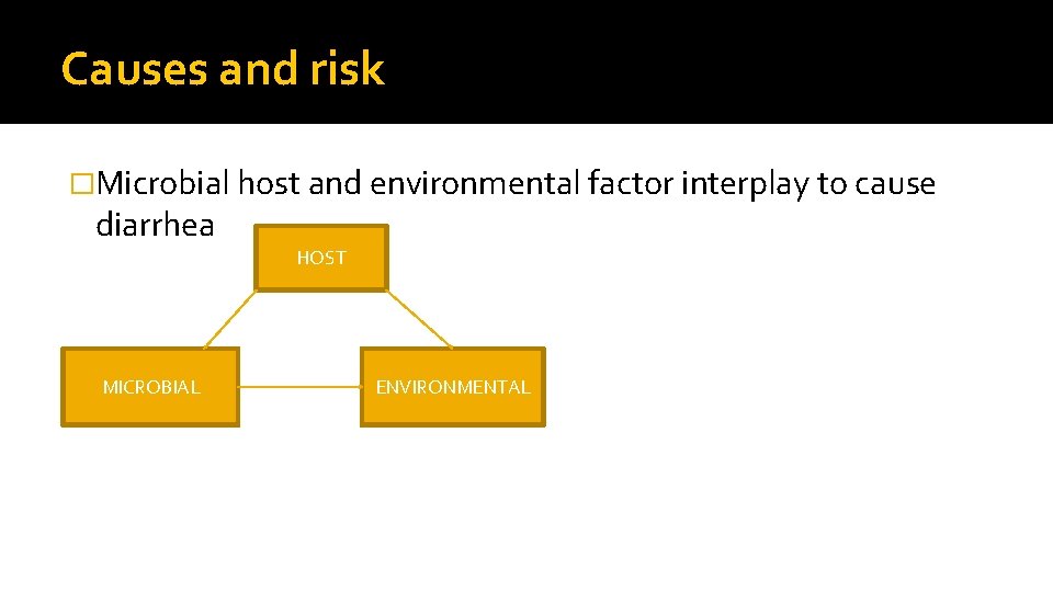
Causes and risk �Microbial host and environmental factor interplay to cause diarrhea HOST MICROBIAL ENVIRONMENTAL
Mode of infection �Acquired through the fecal–oral route or by ingestion of contaminated food or water. �Associated with poverty, poor environmental hygiene, and development indices. �Enteropathogens that are infectious in a small inoculum (shigella, enterohemorrhagic escherichia coli, campylobacter jejuni, noroviruses, rotavirus, giardia lamblia, cryptosporidium parvum, entamoeba histolytica) can be transmitted by personto-person contact, �Whereas others, such as cholera, are generally a consequence of contamination of food or water supply
DIARRHEA CLASSIFICATION �According to pathogen �According to duration �According to mechanism of diarrhea �According to clinical types of diarrhea
Diarrhea according to duration �Acute diarrhea last< 14 days �Chronic diarrhea > 14 days
Mechanisms of diarrhea �Osmotic eg lactose intolerance �Secretory eg cholera �Mixed secretory- osmotiv eg rota virus �Mucosal inflammation �Motility disturbance
Clinical types of diarrhea �Acute watery diarrhea. Most common, usually self limiting and most episodes resolve within 7 days an the main complication is dehydation eg rota virus and cholera �Acute bloody diarrhea- dysentry. Damage to the intestinal mucosa by invasive organisms. Complicatons are sepsis , HUS. Eg shigella. E. Coli
According to pathogens �Infective and non infective cause �Viral, bacterial, parasitic �Side effects of antibiotics �Infectons not related GIT system
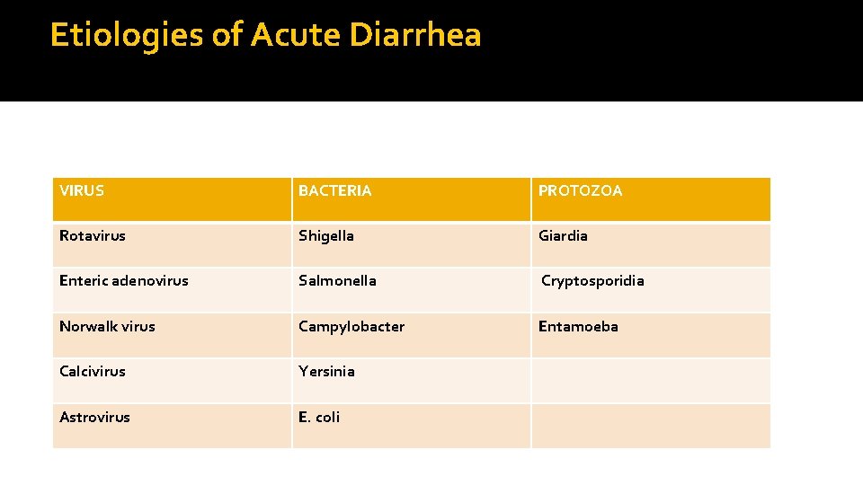
Etiologies of Acute Diarrhea VIRUS BACTERIA PROTOZOA Rotavirus Shigella Giardia Enteric adenovirus Salmonella Cryptosporidia Norwalk virus Campylobacter Entamoeba Calcivirus Yersinia Astrovirus E. coli
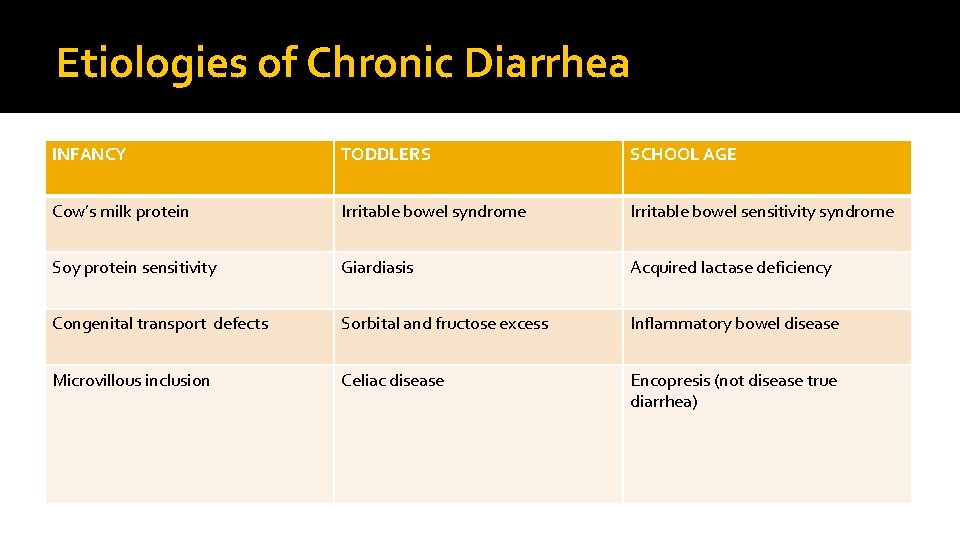
Etiologies of Chronic Diarrhea INFANCY TODDLERS SCHOOL AGE Cow’s milk protein Irritable bowel syndrome Irritable bowel sensitivity syndrome Soy protein sensitivity Giardiasis Acquired lactase deficiency Congenital transport defects Sorbital and fructose excess Inflammatory bowel disease Microvillous inclusion Celiac disease Encopresis (not disease true diarrhea)
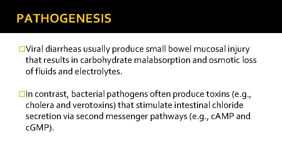
PATHOGENESIS �Viral diarrheas usually produce small bowel mucosal injury that results in carbohydrate malabsorption and osmotic loss of fluids and electrolytes. �In contrast, bacterial pathogens often produce toxins (e. g. , cholera and verotoxins) that stimulate intestinal chloride secretion via second messenger pathways (e. g. , c. AMP and c. GMP).
�In osmotic diarrheas, fasting eliminates the osmotic load from luminal carbohydrates and thus reduces fluid loss. �In secretory diarrheas, fasting does not eliminate toxins that act as secretagogues and fluid loss continues unchanged.
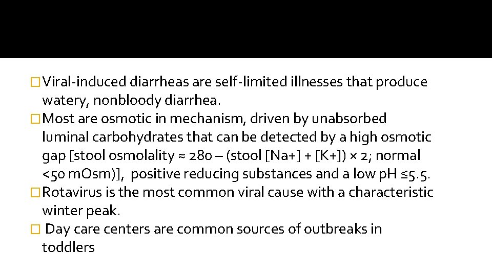
�Viral-induced diarrheas are self-limited illnesses that produce watery, nonbloody diarrhea. �Most are osmotic in mechanism, driven by unabsorbed luminal carbohydrates that can be detected by a high osmotic gap [stool osmolality ≈ 280 – (stool [Na+] + [K+]) × 2; normal <50 m. Osm)], positive reducing substances and a low p. H ≤ 5. 5. �Rotavirus is the most common viral cause with a characteristic winter peak. � Day care centers are common sources of outbreaks in toddlers
�Bacterial diarrheas usually cause a colitis manifested by bloody, mucousy diarrhea associated with cramping and tenesmus. �Because the yield from stool cultures is often low some advocate screening with stool leukocyte smears or fecal lactoferrin assays.
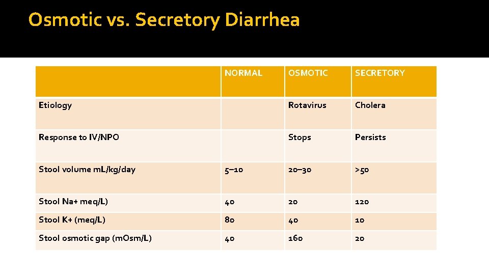
Osmotic vs. Secretory Diarrhea NORMAL OSMOTIC SECRETORY Etiology Rotavirus Cholera Response to IV/NPO Stops Persists Stool volume m. L/kg/day 5– 10 20– 30 >50 Stool Na+ meq/L) 40 20 120 Stool K+ (meq/L) 80 40 10 Stool osmotic gap (m. Osm/L) 40 160 20
�BACILLUS CEREUS �Short incubation period (median incubation period; emesis: 2 hours, diarrhea: 9 hours). �illness due to ingestion of a preformed, heat-stable toxin. �Patients are afebrile, stool is nonbloody. Emesis more significant than diarrhea. �Diagnosis: History (reheated fried rice), stool culture. �Treatment: Supportive; no indication for antibiotic therapy.
� CAMPYLOBACTER JEJUNI � Mean incubation period: 48 hours. � Symptoms include bloody diarrhea, fever, severe cramping, and emesis. � Patients may develop mesenteric adenitis, mimicking acute appendicitis. � Diagnosis: History (poultry, raw milk), fecal leukocytes, stool culture. � Treatment: Antibiotics are usually unnecessary in immunocompetent children. � Consider antibiotics in patients who have prolonged bloody diarrhea associated with fever and a large number of stools, or in immunosuppressed patients. • When antibiotic therapy is indicated, a 5– 7 -day course of erythromycin is recommended (clinical benefit is only seen if erythromycin is given early in illness).
�ESCHERICHIA COLI (NONHEMORRHAGIC) �There are five categories of diarrheagenic E. coli; each has a distinct clinical picture and different management recommendations. �ETEC (enterotoxigenic E. coli): Enterotoxin elaborated within the small bowel causes an increased secretion of fluid and electrolytes from the intestine. Stool is watery and nonbloody. Treatment of choice is a 5 -day course of trimethoprimsulfamethoxazole or cefixime. � EPEC (enteropathogenic E. coli): Organism causes a secretory watery diarrhea. Toxin formation is not involved. Antibiotic of choice is a 5 -day course of neomycin or trimpethoprimsulfamethoxazole.
�EAEC (enteroaggregative E. coli): Enterotoxin is formed; results in persistent watery diarrhea (can last more than 2 weeks). Antibiotic of choice is unknown. �EIEC (enteroinvasive E. coli): Organism invades the colonic enterocytes, then releases enterotoxins. Stool is watery in nature. Treatment of choice is a 5 -day course of trimethoprim -sulfamethoxazole.
�EHEC (ENTEROHEMORRHAGIC E. COLI)-↑ M&M �Median incubation period: 96 hours. �EHEC isolates are almost all serotype O 157: H 7. �Toxins (known as Shiga-like toxins) are elaborated, resulting in colitis with bloody diarrhea Patients are usually afebrile. � 2 – 20%of patients may develop hemolytic uremic syndrome (HUS).
� HUS-microangiopathic hemolytic anemia, thrombocytopenia, and acute renal dysfunction. �Diagnosis: History (undercooked beef and many other vehicles), �Stool culture, Shiga-like toxin detection assay, and O 157: H 7 serotyping with agglutination assay. �Treatment: Correction of dehydration, fluids, and electrolytes �Antibiotic therapy for EHEC is contraindicated, as it increases the likelihood of developing HUS; �all antimotility agents are also contraindicated.
�SALMONELLA SPECIES �Mean incubation period: 24 hours. �Symptoms include bloody diarrhea, fever, abdominal cramping, myalgias, and headache. � Reactive arthritis can occur in 2% of cases. �Diagnosis: History (poultry, pork, eggs, dairy products, vegetables, fruit), fecal leukocytes, stool culture. �Treatment: Supportive care. . �If bacteremia is suspected or documented, patients should be treated with third-generation cephalosporin (e. g. , ceftriaxone) while cultures and susceptibilities are pending.
�SHIGELLA SPECIES � Mean incubation period: 24 hours; as few as 10 organisms can cause diarrheal symptoms. �Symptoms include bloody diarrhea, fever, abdominalcramping, neurologic manifestations (seizure, confusion, hallucinations). �Diagnosis: History (egg salad, lettuce), fecal leukocytes, stool culture. �Treatment: Supportive therapy and a 5 -day course of antibiotic therapy (cefixime, ceftriaxone, trimethoprimsulfamethoxazole, ampicillin). �Must check sensitivities;
�STAPHYLOCOCCUS AUREUS �Mean incubation period: 3 hours. �Illness is due to ingestion of preformed enterotoxins; �Toxin binds to intestinal receptors which stimulate emetic center in the brain. �Symptoms include acute, forceful emesis, and diarrhea (emesis predominates); patients are afebrile. �Diagnosis: History (ham, poultry, potato, and egg salad); isolate organism in culture of vomitus, food. �Treatment: Supportive care; antimicrobial therapy not indicated.
�VIBRIO CHOLERAE � Mean incubation period: 48 hours. � Toxin-producing strains O 1 and O 139 are responsible for epidemics; the toxin causes a severe secretory diarrhea. � Symptoms include voluminous diarrhea (rice-water stools), emesis, and low-grade fever. �Shock due to volume depletion can occur in 12 hours; electrolyte derangement is common. �Diagnosis: History (shellfish), stool, or rectal swab culture. �Treatment: Rehydration and electrolyte replacement � Antimicrobials for treatment include oral doxycycline, tetracycline, or trimethoprimsulfamethoxazole.
�Toxins can lead to a broad spectrum of disease, from �mild diarrhea to severe pseudomembranous enterocolitis. � Diagnosis: Definitive diagnosis can be made only by endoscopic examination. �Stool assay (enzyme immunoassay) for toxin or stool culture are the usual diagnostic tests; �However, organism and toxin may be present in colon without disease. � It is important to note that 25– 65% of children <12 months of age without diarrhea are colonized withtoxin-producing C. difficile.
Diarrheal evaluation �History �Ask the mother or caretaker about �Duration of diarrhea �Presence of blood �No of watery stools/day �Number of episodes of vomiting �Presence of fever, cough, convulsions, recent measels �Pre illness feeding practice �Type and amount of fluids ( including breast milk) and food taken during the illness. �Drugs or other remedies taken �Immunization history
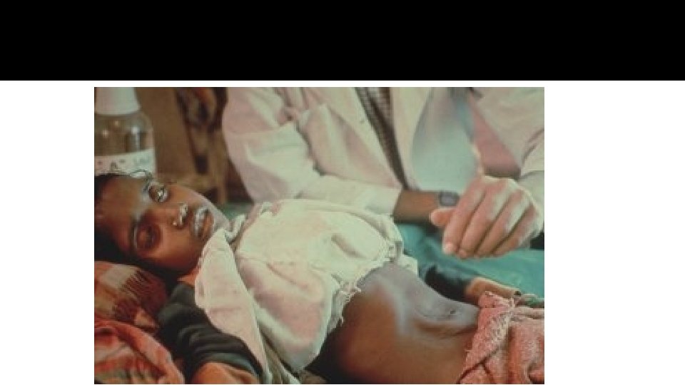
Clinical asssesment �Physical examination �General appearance �Hydration status �Systemic examination �Extra intestinal manifestation
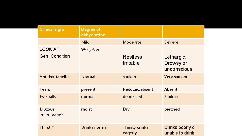
Clinical signs Degree of dehydration Mild Moderate Sev ere Restless, Irritable Lethargic, Drowsy or unconscious LOOK AT: Gen. Condition Well, Alert Ant. Fontanelle Normal sunken Very sunken Tears present Reduced/absent Absent Eye balls normal depressed Sunken Mucous membrane* moist Dry parched Thirst * Drinks normal Thirsty drinks eagerly Drinks poorly or unable to drink
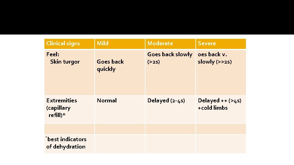
Clinical signs Feel: Skin turgor Extremities (capillary refill)* best indicators of dehydration Mild Goes back quickly Normal Moderate Severe Goes back slowly oes back v. (>2 s) slowly (>>2 s) Delayed (2 -4 s) Delayed ++ (>4 s) +cold limbs
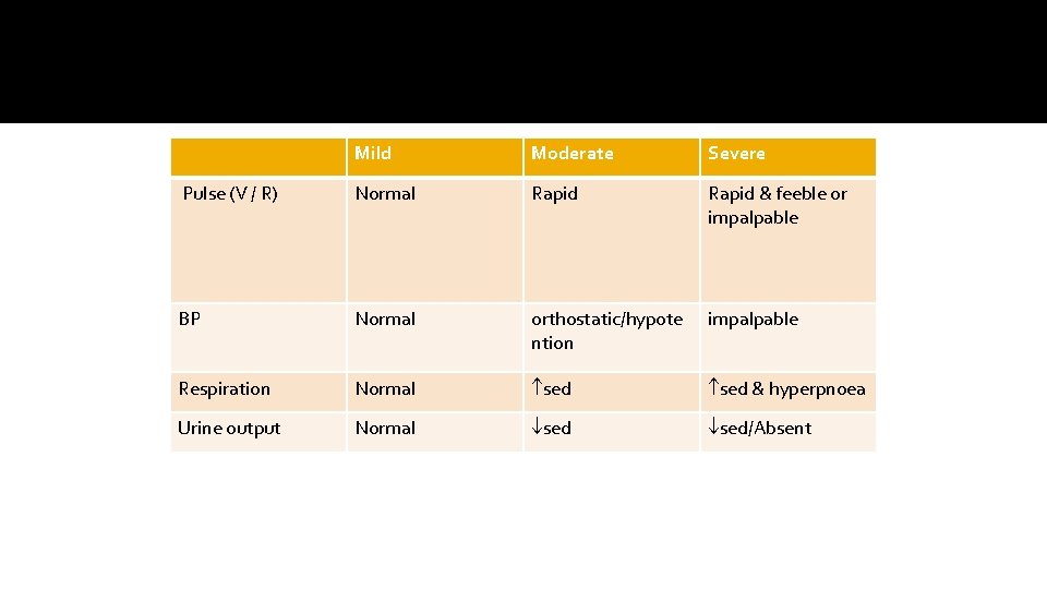
Mild Moderate Severe Pulse (V / R) Normal Rapid & feeble or impalpable BP Normal orthostatic/hypote ntion impalpable Respiration Normal sed & hyperpnoea Urine output Normal sed/Absent
Laboratory investigation �Stool microscopy: low sensitivity and specificity �-leucocyte(>10/hpf)- invasive diarrhae �-RBC, ova, trophozite or cyst �Blood test- FBC, E/U/CR �Others- -Blood culture - lactose breath hydrogen test or lactose restriction to rule out lactose intolerance -Serum antibody test to rule out celiac disease or colonoscopy for suspected ulcerative colitis
Management �Treat dehydration- conerstone in managing diarrhea �Feeding ; continue breast feeding and routine normal diet and energy dense feeds. �Hand washing after defecation and before meal alone can reduce 40% of water and excreta related disease. �Drug therapy has very little place -Antibiotic -Antisecretory ( contraindicated in children) -Antimotility ( contraindicated in children) �Follow up to ensure recovery
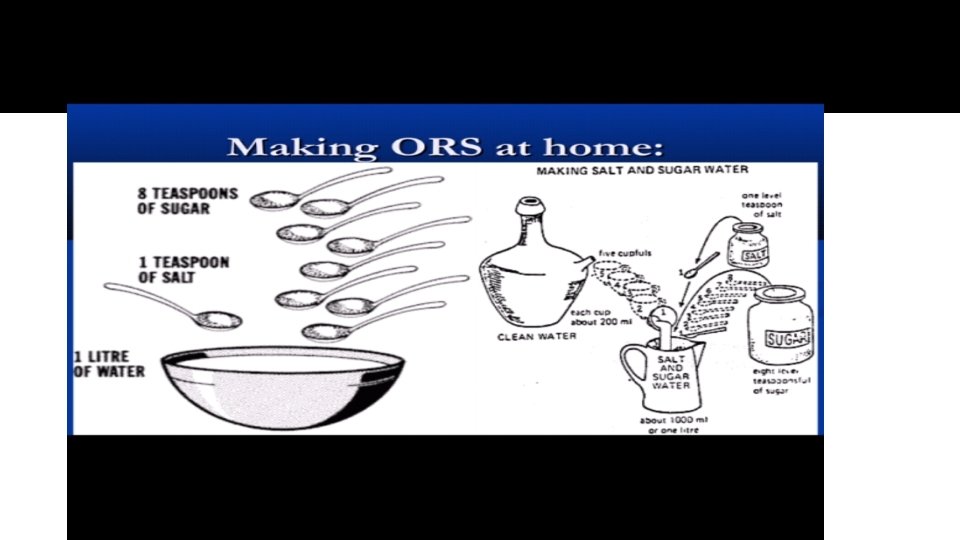
ORAL REHYDRATION THERAPY � The cheapest way to treat diarrhea- to prevent dehydration � Adequate glucose electrolyte solution � WHO recommendation: ORT+guidance on appropriate feeding • • • practices Main strategy to achieve reduction in diarrhea related morbidty and mortality 1 st introduced in 1969 1984 - trisodium citrate comp. introduced instead of hydrogen carbonate To produce stability of ORS in hot and humid climate Original ORS- contains 90 meq/l of sodium and total osmolarity 311 m. Osm/l
Why reduced osmolarity ORS �Pharmacokinetics and therapeutics values �- glucose facilitates absorption of sodium(hence water) on 1: 1 molar basis in small intestine �Sodium and potassium are needed to replace body loss in diarrhea �Citrate corrects acidosis that may occur as a result of diarrhea and dehydration
�Other clinical benefits. �-Reduces stool output /volume by ~ 25% when compared to original WHO- UNICEF ORS solution. �-Reduces vomiting by ~30% �-Reduces need for unscheduled IV therapy>30%( less hospitalization).
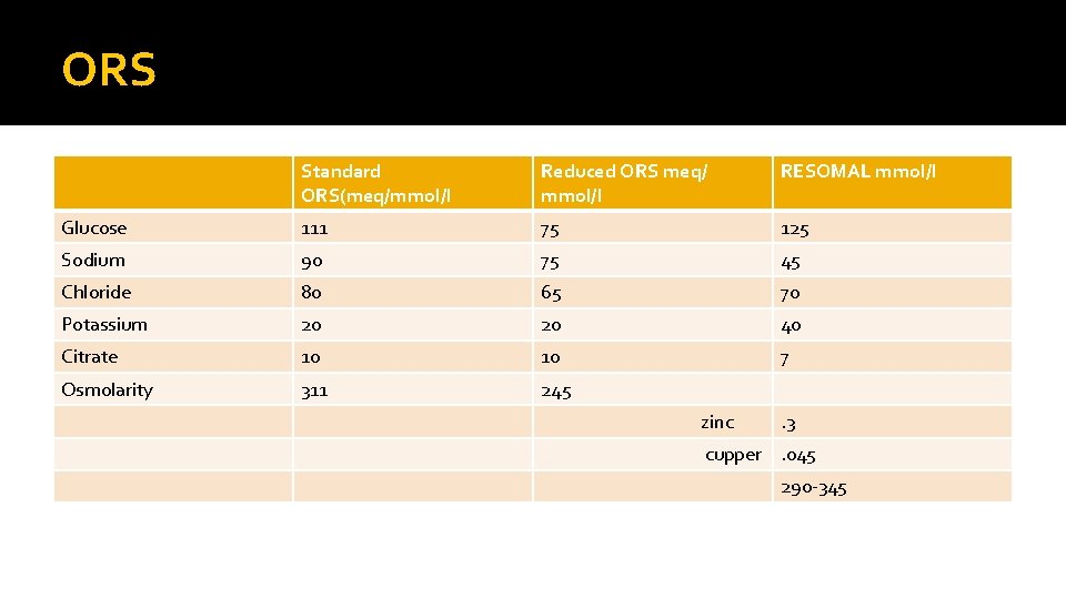
ORS Standard ORS(meq/mmol/l Reduced ORS meq/ mmol/l RESOMAL mmol/l Glucose 111 75 125 Sodium 90 75 45 Chloride 80 65 70 Potassium 20 20 40 Citrate 10 10 7 Osmolarity 311 245 zinc . 3 cupper. 045 290 -345
Indications for iv therapy �Depreesed level of consciousness �Moderate dehydration when there is no improvement after the first 4 hours of treatment with ORS �Severe dehydration �Uncontrolled vomiting. �Poor urine output �Patient unable to drink from extreme fatigue, stupor or coma �Patient with abdominal distension.
Preparation of resomal by the bed side � 2 l of water � 1 satchet of standard ors � 10 cubes of sugar= 50 grams � 40 mm 0 l of kcl �Mineral mix- 40 ml
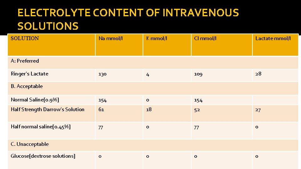
ELECTROLYTE CONTENT OF INTRAVENOUS SOLUTION Na mmol/l K mmol/l Cl mmol/l Lactate mmol/l 130 4 109 28 Normal Saline[0. 9%] 154 0 154 Half Strength Darrow’s Solution 61 18 52 27 Half normal saline[0. 45%] 77 0 0 0 A: Preferred Ringer’s Lactate B. Acceptable C. Unacceptable Glucose[dextrose solutions]
Management of mild and moderate dehydration �This can be done successfully by ORT 1. Deficit �Mild -50 ml/kg/4 hr �Moderate -100 ml/kg/4 hr �Maintenance; when hydration is complete, maintenance therapy should be started, using 100 ml of ORS/kg/24 hr until diarrhea stops.
Management of mild and moderate dehydration �Ongoing losses �Supplementary ORS is given to replace ongoing losses from diarrhea or emesis and additional 10 ml/kg of ORS is given for each stool.
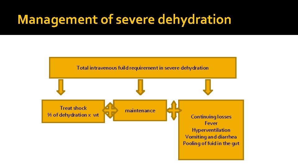
Management of severe dehydration Total intravenous fuild requirement in severe dehydration Treat shock % of dehydration x wt maintenance Continuing losses Fever Hyperventilation Vomiting and diarrhea Pooling of fuid in the gut
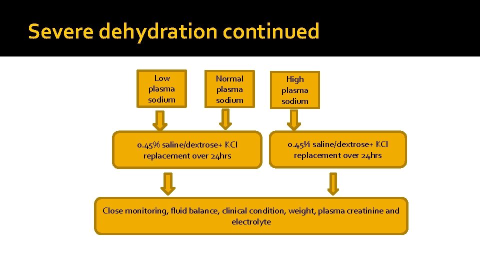
Severe dehydration continued Low plasma sodium Normal plasma sodium 0. 45% saline/dextrose+ KCl replacement over 24 hrs High plasma sodium 0. 45% saline/dextrose+ KCl replacement over 24 hrs Close monitoring, fluid balance, clinical condition, weight, plasma creatinine and electrolyte
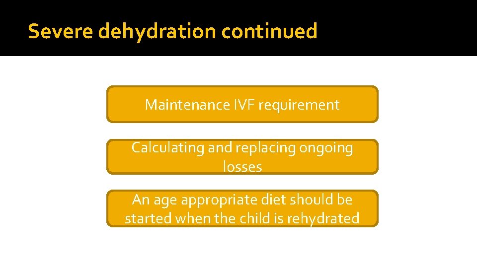
Severe dehydration continued Maintenance IVF requirement Calculating and replacing ongoing losses An age appropriate diet should be started when the child is rehydrated
Management of isonatremic dehydration �Restore intravascular volume (treat shock): 20 ml/kg of NS or rinders lactate over 20 min. (repeat if nessesary). �Calculate 24 hr maintenance water and electrolytes. �Calculate 24 hr deficit water and electrolytes. �Total amount of water and electrolytes are added together, then an appropriate fluid is selected. �Cautious use of potassium.
Management of isonatremic dehydration �Total correction is done within the first 24 hrs. Administer half of the calculated fluid during the first 8 hrs, first subtracting any boluses from this amount, then administer the reminder over the next 16 hr. �Replace ongoing losses as they occur
Management of hyponatremic dehydration �Most patient do well with the same basics �D 5 ½ NS with 20 m. Eq/L kcl �In general its not necessary to increase the sodium beyond 135 m. Eq/L, because over correction is associated with an increased risk of central pontine myelinosis. The risk of CPM also occurs with rapid correction of serum sodium conc. To avoid this avoid increasing the sodium by more than 12 m. Eq/L each 24 hr.
Management of hyponatremic dehydration �Patients with neurologic symptoms(eg seizure) from hyponatremia need to receive an acute infusion of hypertonic (3%) saline to rapidly increase the serum sodium concentration. �Each milliliter of (3%) Na. Cl increases the serum sodium by approx. 1 m. Eq/L. a child with active symptoms often improves after receiving 4 -6 ml/kg of 3% Na. Cl �The patients sodium concentration is monitored to ensure appropriate correction and the sodium concentration of the fluid is adjusted appropriately.
Management of hypernatremic dehydration �Restore intravascular volume: 20 ml/kg NS over 20 min( repeat until intravascular volume is restored). Ringers lactate is not preferred because it is more hypotonic than NS. �Determine the time for correction based on the initial sodium conc. [Na]: 145 -157 m. Eq/L: 24 hr [Na]: 158 -170 m. Eq/L: 48 hr [Na]: 171 -183 m. Eq/L: 72 hr [Na]: 184 -196 m. Eq/L: 84 hr
Management of hypernatremic dehydration �Administer fluid at a constant rate over the time for correction. �Return serum sodium levels towards the reference range by not more than 10 m. Eq/L/24 hr) �Rapid correction can have disastrous neurologic consequences, including cerebral odema and death.
Management of hypernatremic dehydration �Following adequate intravascular volume expansion, rehydration fluids should be initiated with 5% dextrose in 0. 9% Na. Cl. �Serum sodium levels should be assessed every 4 hrs. �If the sodium has decreased by less than 0. 5 m. Eq/L, then the sodium content of the rehydration fluid is decreased to D 5 in ½ NS. This allows for a slow controlled correction of the hypernatremic state. �Replace ongoing losses as they occur.
Fluid management in a severely malnourished child �Children with severe acute malnutrition and who have some or severe dehydration but no shock should receive 5 m. L/kg Re. So. Mal every 30 min for the first 2 h. � Then, if the child is still dehydrated, 5– 10 m. L/kg/h Re. So. Mal should be given in alternate hours with F-75, up to a maximum of 10 h; �Signs of improved hydration status and over hydration should be checked every half hour for the first 2 h, then hourly
Fluid management in a severely malnourished child �Children with severe acute malnutrition and signs of shock or severe dehydration and who cannot be rehydrated orally or by nasogastric tube should be treated with intravenous fluids, either: �Half-strength darrow’s solution with 5% dextrose, or ringer’s lactate solution with 5% dextrose. �If neither is available, 0. 45% saline + 5% dextrose should be used.
Fluid management in a severely malnourished child �In case of shock with lethargy or unconsciousness, intravenous rehydration should begin immediately, using 15 ml/kg/h of one of the recommended fluids; �If a child with severe acute malnutrition presenting with shock does not improve after 1 h of intravenous therapy, a blood transfusion (10 ml/kg slowly over at least 3 h) should be given; �It is important that the child is carefully monitored every 5– 10 min for signs of over hydration and signs of congestive heart failure. �If signs of over hydration and congestive heart failure develop, intravenous therapy should be stopped immediately;
Zinc in diarrhea �Zinc deficiency is common in developing countries and zinc is lost during diarrhea. �Zinc deficiency is associated with impaired electrolyte and water absorption, decreased brush border enzyme activity and impaired cellular and humeral immunity. �Treatment with zinc reduces the duration and severity of AD and also reduces the frequency of further episodes during the subsequent 2 -3 months �WHO- recommends 10 mg dly for 10 -14 days for children<6 mths 20 mg daily for those >6 mths
Probiotics in the treatment of diarrhea �Mechanism � 1. protects the intestine by competing with pathogens for attachment �Strengthening tight junctions between enterocytes �Enhancing the mucosl immune response to pathogens
Antibiotics in acute diarrhea �Indicated only in �Acute bloody diarrhea with gross blood �Severe invasive bacterial diarrhea eg shigella �Cholera �Associated systemic infection �Severe malnutrition �Giardiasis, Entamoeba histolytica �Suspected or proven sepsis �Immuno-compromised children
�Contraindicated in �E. coli o 157 -H 7 – risk of HUS �Uncomplicated salmonella enteritis because they prolong bacteria shedding
SIADH( complication of treatment) �Inappropriate high secr of ADH in presence of concn urine & euvolemic Hyponatraemia �Causes: CNS disorders, Resp, Tumors, Drugs �Labs: ↓serum Na, ↑urine Na, ↑ Urine SG, ↓Urine output. �Rx: Fluid Restriction - In chr SIADH, try Lithium, or Demeclocycline
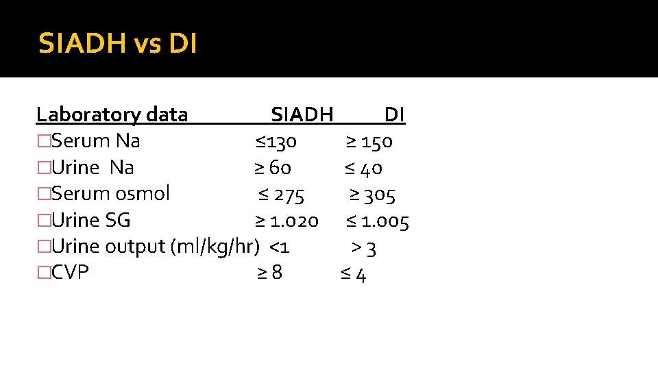
SIADH vs DI Laboratory data SIADH DI �Serum Na ≤ 130 ≥ 150 �Urine Na ≥ 60 ≤ 40 �Serum osmol ≤ 275 ≥ 305 �Urine SG ≥ 1. 020 ≤ 1. 005 �Urine output (ml/kg/hr) <1 > 3 �CVP ≥ 8 ≤ 4
Complications and consequences of watery diarrhea �Dehydration �Electrolyte disturbance �Base deficit �Malnutrition �Persistent diarrhea �Toxic ieus �Renal failure �HUS �DIC �Convulsion �Cerebral damage and cerebral venous thrombosis
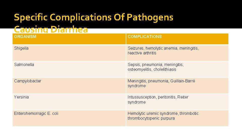
Specific Complications Of Pathogens Causing Diarrhea ORGANISM COMPLICATIONS Shigella Seizures, hemolytic anemia, meningitis, reactive arthritis Salmonella Sepsis, pneumonia, meningitis, osteomyelitis, cholelithiasis Campylobacter Meningitis, pneumonia, Guillain-Barré syndrome Yersinia Intussusception, peritonitis, Reiter syndrome Enterohemorragic E. coli Hemolytic uremic syndrome, thrombotic thrombocytopenic purpura
Prevention �GENERAL HEALTH PROMOTON -Health education -Hand washing -Environmental sanitation -Safe drinking water -Good personal hygeine -Good nutrition �SPECIFIC PROTECTION -Hand washing -Exclusive breastfeeding -Immunization against rota virus and measles
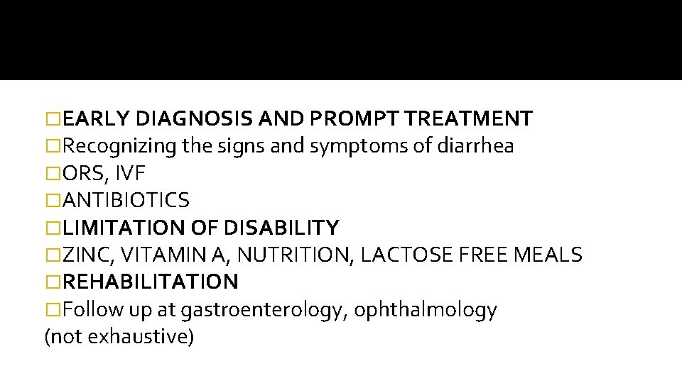
�EARLY DIAGNOSIS AND PROMPT TREATMENT �Recognizing the signs and symptoms of diarrhea �ORS, IVF �ANTIBIOTICS �LIMITATION OF DISABILITY �ZINC, VITAMIN A, NUTRITION, LACTOSE FREE MEALS �REHABILITATION �Follow up at gastroenterology, ophthalmology (not exhaustive)