Management of Cervical Spine Disease in Downs Syndrome

 Anon Am J Med Genetics")


 NR <5 mm • Neural canal width")
 NR <5 mm • Neural canal width")


 Neurol. Deterioration Osseous fusion")


- Slides: 70
Management of Cervical Spine Disease in Down's Syndrome: A Neurosurgical perspective Dominic Thompson Department of Paediatric Neurosurgery Great Ormond Street London
Flemish (1515) Anon Am J Med Genetics
Cervical spinal pathology in Downs syndrome -The controversies • • Aetiology Diagnosis Screening Treatment
Aetiology
Aetiology • Primary features • Ligamentous laxity • Hypotonia • Hypermobility
Aetiology • Primary features • Skeletal consequences • Ligamentous laxity • Hypotonia • Hypermobility • • Craniovertebral instability Scoliosis Hip instability Patellar instability
Cervical spinal pathology in Downs syndrome • • Occipito-atlantal subluxation Atlantoaxial subluxation Os Odontoideum Incomplete C 1 ring Cervical stenosis Segmentation anomaly Premature cervical spondylarthrotic myelopathy
Cervical spinal pathology in Downs syndrome • • Occipito-atlantal subluxation Atlantoaxial subluxation Os Odontoideum Incomplete C 1 ring Cervical stenosis Segmentation anomaly Premature cervical spondylarthrotic myelopathy
Os Odontoideum – congenital or acquired? OS C 1
Os odontoideum – developmental anatomy • Sclerotomes cranial and caudal moieties – scleromites Cranial – hypocentrum - disc Caudal – centrum - body
Os odontoideum – developmental anatomy • Sclerotomes cranial and caudal moieties – scleromites Cranial – hypocentrum - disc Caudal – centrum - body
Os odontoideum – developmental anatomy • Sclerotomes cranial and caudal moieties – scleromites • Cranial – hypocentrum - disc • Caudal – centrum - body • C 1 hypocentrum • C 1 centrum
Os odontoideum – developmental anatomy • Sclerotomes cranial and caudal moieties – scleromites • Cranial – hypocentrum - disc • Caudal – centrum - body • C 2 hypocentrum -regresses • C 2 centrum
Os odontoideum – developmental anatomy • Congenital theory of the Os. = Failure of incorporation of the C 1 centrum • Implications Large os (entire C 1 centrum) Base below joint
Os odontoideum – developmental anatomy • Congenital theory of the Os. = Failure of incorporation of the C 1 centrum • Absence of C 1 centrum rare
Os odontoideum – developmental anatomy Acquired theory • Trauma to odontoid due to instability • Implications Os relatively small Base at or above joint line
Os odontoideum – developmental anatomy
Radiological findings in 12 symptomatic cases • • Atlanto-dental interval Neural canal width Atlanto-occipital instability Os odontoideum Posterior spina bifida Anterior spina bifida Pars defect C 4 (Nader-Sepathi et al 2005) 5 -13 mm (av. 7 mm) 6 -11 mm (av. 8 mm) 2 cases 10 cases 3 cases 2 cases 1 case
Radiological findings in 12 symptomatic cases • • Atlanto-dental interval Neural canal width Atlanto-occipital instability Os odontoideum Posterior spina bifida Anterior spina bifida Pars defect C 4 (Nader-Sepathi et al 2005) 5 -13 mm (av. 7 mm) 6 -11 mm (av. 8 mm) 2 cases 10 cases 3 cases 2 cases 1 case
Radiological findings in 12 symptomatic cases • • Atlanto-dental interval Neural canal width Atlanto-occipital instability Os odontoideum Posterior spina bifida Anterior spina bifida Pars defect C 4 (Nader-Sepathi et al 2005) 5 -13 mm (av. 7 mm) 6 -11 mm (av. 8 mm) 2 cases 10 cases 3 cases 2 cases 1 case
Craniovertebral instability Os Odontoideum Delayed Atlantal ossification
Diagnosis
Diagnosis of craniovertebral instability in Downs Syndrome Aims • To identify children with or at risk of spinal cord damage due to craniovertebral instability • To instigate effective treatment
What to measure?
Defining atlantoaxial subluxation • Atlanto-dental interval (ADI) NR <5 mm • Neural canal width (NCW) NR >14 mm ADI
Defining atlantoaxial subluxation • Atlanto-dental interval (ADI) NR <5 mm • Neural canal width (NCW) NR >14 mm NCW
Defining atlantoaxial subluxation in Downs syndrome Author N ADI Pueschel 1986 404 4. 5 mm Morton 1995 90 4 mm Semine 85 4. 5 mm Elliot 1988 67 4 mm Van Dyke 1988 34 5 mm
Defining atlantoaxial subluxation in Downs syndrome Author N ADI % Pueschel 1986 404 4. 5 mm 14. 6 Morton 1995 90 4 mm 7. 8 Semine 85 4. 5 mm 12 Elliot 1988 67 4 mm 10 Van Dyke 1988 34 5 mm 9
Defining atlantoaxial subluxation in Downs syndrome Author N ADI % Symptoms Pueschel 1986 404 4. 5 mm 14. 6 1. 5 Morton 1995 90 4 mm 7. 8 0 Semine 85 4. 5 mm 12 1. 2 Elliot 1988 67 4 mm 10 0 Van Dyke 1988 34 5 mm 9 0
Acute vs chronic deterioration • Nader-Sepathi et al 2005 • 5/12 acute deterioration • Preceeding symptoms in 4 • Davidson 1988 • 31 Downs with neurological symptoms/signs • 29 symptoms for >1 mnth
• • 12 years old Downs syndrome Gait deterioration Clinical signs of myelopathy
Extension Flexion
ADI Changes over time N=67 Measurements 5 years apart Morton et al 1995
Poor clinical and radiological correlation 84 Patients with Downs syndrome Subluxation 17 Symptomatic 5 Asymptomatic 12 Non subluxation 67 Symptomatic 18 Asymptomatic 49 Ferguson 1987
Poor clinical and radiological correlation • Gait abnormality as a measure of AAS • Specificity 81% • Sensitivity 50% Gait Radiographs Total Normal Abnormal 92 (91) 21 (70) 9 (9) 9 (30) 101 30 Selby 1991
Poor clinical and radiological correlation • Gait abnormality as a measure of AAS • Specificity 81% • Sensitivity 50% Gait Radiographs Total Normal Abnormal 92 (91) 21 (70) 9 (9) 9 (30) 101 30 • Poor reproducibilty in repeat films Selby 1991
Defining Occipito-atlantal subluxation • Coexisting occipitoatlantal instability is common 40 -50% • Treadwell 1990 • Taggart 2000 • Difficult to diagnose
Defining Occipito-atlantal subluxation • Powers ratio
Defining Occipito-atlantal subluxation • Basion-Axial Interval • Harris et al
Defining Occipito-atlantal subluxation • Wiesel et al
Defining Occipito-atlantal subluxation • “Measurement of atlantooccipital translation by any of these methods is not reproducible” • Lori et al 1996
Limitations of Radiographic Measurements in Downs Syndrome • • ADI No consensus on measurement Poor reproducibility Poor correlation with symptoms O-C instability, present but difficult to measure
Screening
Should Children With Down Syndrome be Screened for Atlantoaxial Instability?
Should Children With Down Syndrome be Screened for Atlantoaxial Instability? • Special Olympics 1983 – All patients with Downs syndrome should have lateral cervical spine X rays – ADI>4. 5 mm mandates exclusion from high risk sporting activities • American Academy of Pediatrics 1984 • Department of Health Standing Medical Advisory Committee 1986
Should Children With Down Syndrome be Screened for Atlantoaxial Instability? • Special Olympics 1983 – All patients with Downs syndrome should have lateral cervical spine X rays – ADI>4. 5 mm mandates exclusion from high risk sporting activities • American Academy of Pediatrics 1984 • Department of Health Standing Medical Advisory Committee 1986
Screening • Incidence must be common • Screening test should be sensitive and specific • Asymptomatic AAS must be a risk factor for symptomatic AAS • A safe and effective intervention should be available
British Guidelines 2009 Downs Medical Interest Group of UK Cervical spine X rays are not recommended for the asymptomatic child with Downs syndrome , Medical personnel and carers need to be aware of the potential for neurological symptoms to develop
British Guidelines 2009 Downs Medical Interest Group of UK Warning Signs Neck pain, Abnormal head posture, Torticollis, (Wry neck) Reduced neck movements, Deterioration of gait and/or frequent falls Increasing fatigability on walking, Deterioration of manipulative skills,
British Guidelines 2009 Downs Medical Interest Group of UK Action Full physical and neurological examination Specialist Referral
British Gymnastics Association • Guidelines for the participation of individuals with Downs syndrome in gymnastics and trampolining • For all applicants to: – Registered clubs – Competitive events • Screening by medical practitioner • Approved by • • British Gymnastics Medical Commission Gymnastics and movement for people with disabilities Technical Committee
Clinical screening criteria A. No signs of myelopathy – Increasing muscle weakness, alteration in muscle tone – Loss of sensation – Decreasing co-ordination – Diminishing kinaesthetic awareness – Change in walking pattern – New incontinence B. Neck flexion – Flex to chin on chest without difficulty C. Neck muscle control – Pulled to sitting position maintaining head neck alignment
Results • 1995 -2009 • 399 applications from Individuals with Down syndrome • 6 positive responses – 4 Poor head neck muscle control – 2 Flex to chin on chest without difficulty – No further follow up
Results • 393 granted membership • No injuries referable to the cervical spine reported during the time period • 3 in National Squad
Treatment
Treatment • “A safe an effective intervention should be available”
Surgery for AAS in Down Syndrome Author Number Re-op (%) Neurol. Deterioration Osseous fusion Doyle 15 6 (40) 4 (27) 12 (80) Thompson 15 6 (40) 1 (8) 15 (100) Segal 10 3 (30) 2 (20) (both died) 4 (40) Taggard 25 0 0 23 (96)
Reasons for failure N=15, 6 failures • • Inadequate immobilisation Inadequate reduction Inadequate fixation Inadequate age
Inadequate immobilisation Reduce and immobilise prior to surgery Maintain immobility post operatively in non instrumented fixation
Inadequate fixation • Non union associated – Resorption of rib graft – Limited C 1 -C 2 Nader-Sepathi et al 2005 Doyle et al 1996
Inadequate reduction • • 5 years Attempted posterior fixation Inadequate reduction Construct failure
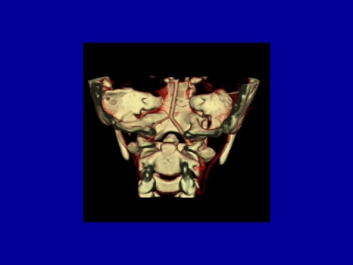
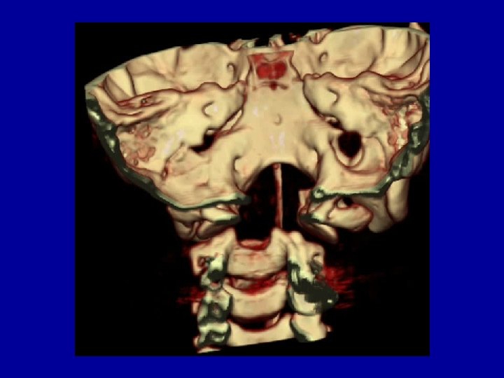
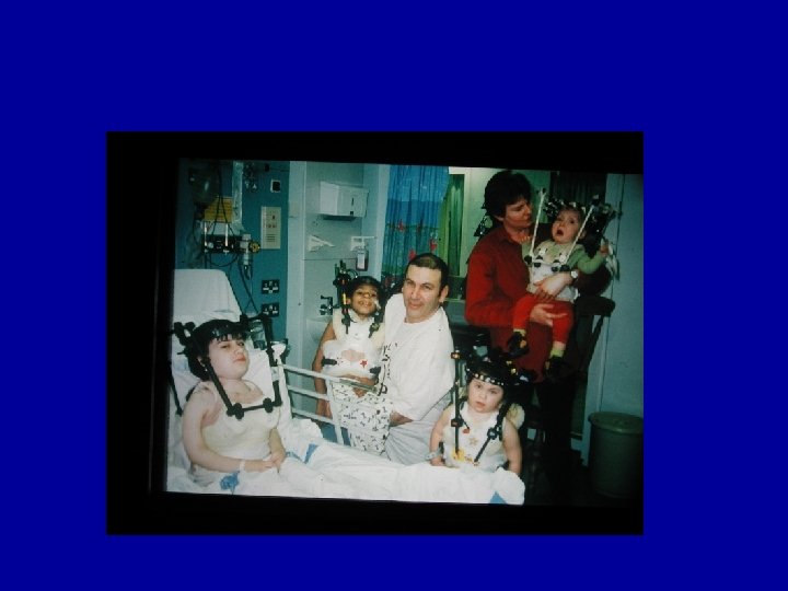
• Stage I – MUA, halo body jacket • Stage II – Transoral odontoidectomy • Stage II – Posterior decompression and revision of instrumentation
Case 1: Downs syndrome • Stage I – MUA, halo body jacket • Stage II – Transoral odontoidectomy • Stage II – Posterior decompression and revision of instrumentation
Case 1: Downs syndrome • Stage I – MUA, halo body jacket • Stage II – Transoral odontoidectomy • Stage II – Posterior decompression and revision of instrumentation
Inadequate reduction • Transoral decompression – 6 out of 15 cases – All had os odontoideum
Conclusions • Osseous anomalies at the CCJ may be consequence rather than cause of instability • Morphometric measures of AAI and OCI lack sufficient sensitivity and specificity to justify screening • Education rather than screening
Conclusions • Early investigation and treatment of symptomatic patients • Beware the child with Downs! • Ensure – Effective immobilisation – Adequate reduction – Rigid fixation