MANAGEMENT OF CATARACT AND COMPLICATIONS Dr P Pradeep


 Extracapsular cataract extraction (ECCE) Conventional ECCE")

 Corneoscleral Iridectomy section")

 Closure of incision- 5")

 suture Conjunctival flap (fornix based) Partial thickness groove/gutter Entry")
 Continuous Removal circular capsulorrhexis (CCC)")


 Superior rectus suture Conjunctival flap and exposure of")
 Hydrodissection")


, iris and lens Iris injury and iridodialysis")


")


- Slides: 43
MANAGEMENT OF CATARACT AND COMPLICATIONS Dr. P Pradeep Professor Dept of Ophthalmology
NON SURGICAL MANAGEMENT TREATMENT OF CAUSE Adequate control of diabetes mellitus, Removal of cataractogenic drugs such as corticosteroids, phenothiazenes and strong miotics Removal of irradiation (infrared or X-rays) Early and adequate treatment of ocular diseases like uveitis
MEASURES TO DELAY PROGRESSION Commercially available preparations containing iodide salts of calcium and potassium are being prescribed in abundance in early stages of cataract Vit E and aspirin Conclusive results for use are not found MEASURES TO IMPROVE VISION IN INCIPIENT AND IMMATURE CATARACT Refraction corrected at frequent intervals Use of mydriatics in small axial cataracts Arrangement of illumination. Peripheral opacities - brilliant illumination
INDICATIONS FOR SURGERY Visual improvement Medical indications: -Lens induced glaucoma -Phacoanaphylactic endophthalmitis -Retinal diseases like diabetic retinopathy or retinal detachment Cosmetic indication in mature cataract with no hope of vision
PREOPERATIVE WORKUP General examination Ocular examination Anterior segment evaluation Intraocular pressure Examination of lids, conjunctiva and lacrimal apparatus Fundus examination Macular function tests Evaluation of retina Keratometry and biometry
Preoperative preparations Consent Topical antibiotics 3 days before surgery Scrub bath and marking of eye IOP lowering Mydriasis ANAESTHESIA Local anesthesia is preferred 2% Lignocaine with 0. 75% Bupivacaine
Surgical cataract extraction TYPES Intracapsular cataract extraction (ICCE) Extracapsular cataract extraction (ECCE) Conventional ECCE SICS Phacoemulsification
INTRACAPSULAR CATARACT EXTRACTION It is an important landmark in the history of cataract surgery The entire cataractous lens along with the intact capsule is removed It is presently obsolete Steps A. Superior rectus (bridle) suture B. Conjunctival flap
Partial thickness groove/gutter (150 o) Corneoscleral Iridectomy section
Lens delivery methods Indian smith method - by tumbling technique Cryoextraction Capsule forceps method – using arruga’s forceps Irisophake method - obsolete Wire Vectis method – in subluxated and dislocated lens
Formation of Anterior Chamber Implantation of anterior chamber IOL(ACIOL) Closure of incision- 5 -7 interrupted sutures (nylon) Conjunctival flap reposited and secured Subconjunctival injection-dexamethasone 0. 25 ml and gentamicin 0. 5 ml given Patching of the eye
EXTRACAPSULAR CATARACT EXTRACTION Major portion of anterior capsule with epithelium, nucleus and cortex are removed; leaving behind intact posterior capsule. Posterior chamber IOL can be placed in the remaining capsular bag Types Conventional ECCE Manual SICS Phacoemulsification
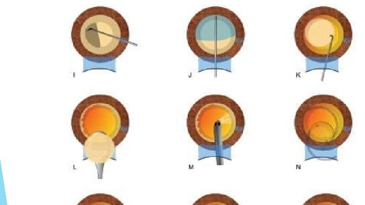
CONVENTIONAL ECCE Superior rectus (bridle) suture Conjunctival flap (fornix based) Partial thickness groove/gutter Entry into anterior chamber Injection of viscoelastic substance in anterior chamber - 2% Methyl. Cellulose or 1% Sodium Hyaluronate (Maintains anterior chamber and protects endothelium)
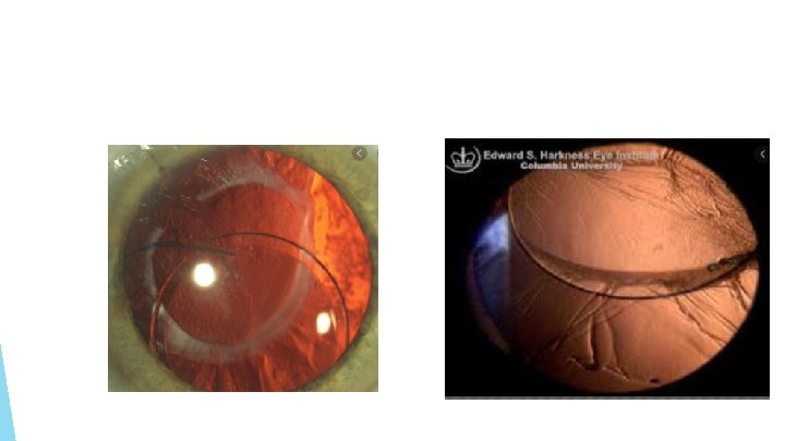
Anterior capsulotomy Can-opener's Linear technique capsulotomy (Envelope technique) Continuous Removal circular capsulorrhexis (CCC) of anterior capsule Completion of corneoscleral section Hydrodissection
Removal of nucleus Pressure and counter-pressure method Irrigating wire vectis technique Aspiration of the cortex Implantation of IOL
Closure of the incision - 3 -5 interrupted sutures Removal of viscoelastic substance Conjunctival flap is reposited and secured Subconjunctival Patching of eye injection
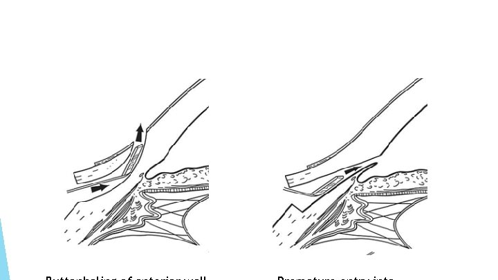
MANUAL SMALL INCISION CATARACT SURGERY (SICS) Superior rectus suture Conjunctival flap and exposure of sclera Haemostasis Sclero corneal tunnel incision: External scleral incision - 5. 5 mm to 7. 5 mm Sclero corneal tunnel - 1 -1. 5 mm Internal corneal incision
Anterior capsulotomy - can be can-openers, envelope or continuous circular capsulorrhexis (CCC) Hydrodissection Nuclear management prolapse of nucleus into ant chamber delivery of nucleus through corneoscleral tunnel Aspiration of cortex IOL implantation
PHACOEMULSIFICATION STEPS Corneoscleral incision-very small 3 mm Continuous curvilinear capsulorrhexis of 4 -6 mm Hydrodissection Nucleus is emulsified and aspirated Remaining cortical lens matter is aspirated IOL Implantation
ECCE FOR CHILDHOOD CATARACT Surgical techniques employed for childhood cataract are essentially of two types: Irrigation and aspiration of lens matter Lensectomy Irrigation and aspiration of lens matter: Steps are same as SICS In addition, Posterior capsulorrhexis of about 3 -4 mm size recommended in children to avoid posterior capsule opacification and Anterior vitrectomy done
LENSECTOMY Lensectomy in children is performed under general anaesthesia Most of the lens including anterior and posterior capsule along with anterior vitreous are removed with the help of a vitreous cutter, infusion and suction machine In very young children primary IOL implantation is not planned
Complications of cataract surgery They are enumerated as A. Preoperative complications B. Operative complications C. Early postoperative complications D. Delayed (late) postoperative complications E. IOL-related complications
PREOPERATIVE COMPLICATIONS Anxiety § Subconjunctival haemorrhage Nausea & gastritis § Spontaneous dislocation of lens Allergic conjunctivitis Corneal abrasion Local anaesthesia complications § Retrobulbar haemorrhage § Oculocardiac reflex § Perforation of globe
OPERATIVE COMPLICATIONS Superior rectus muscle laceration and/or haematoma Excessive bleeding during conjunctival flap or incision Incision related complications Irregular incision in conventional ECCE In manual SICS and phacoemulsification Button holing of tunnel – abandon and dissect at a deeper plane
Injury to the cornea (Descemet's detachment), iris and lens Iris injury and iridodialysis Complications Escaping related to anterior capsulorrhexis Small capsulorrhexis – predisposes to Posterior Capsule tear, Nuclear drop, zonular dehiscence. Managed with relaxing incisions Very large capsulorrhexis
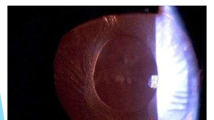
Posterior Zonular capsular rupture dehiscence Vitreous loss Prevented preoperatively by decreasing aqueous, orbita and vitreous volume and better ocular akinesia Managed Nucleus by partial anterior vitrectomy drop into the vitreous cavity Posterior loss of lens fragments Expulsive choroidal hemorrhage
Nucleus drop into vitreous cavity Zonular dehiscence
EARLY POSTOPERATIVE COMPLICATIONS HYPHAEMA – mostly absorbs spontaneously. IOP should be lowered IRIS PROLAPSE – should be reposited and wound sutured STRIATE KERATOPATHY – due to mild corneal oedema with Descemet’s folds FLAT ANTERIOR CHAMBER With wound leak is associated with hypotony Ciliochoroidal detachment – cured with presuure bandage
Hyphema Iris prolapse
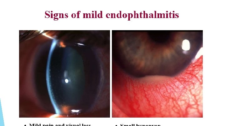
POST OPERATIVE ANTERIOR UVEITIS managed with topical steroids, cycloplegics and NSAIDS TOXIC ANTERIOR SEGMENT SYNDROME BACTERIAL ENDOPHTHALMITIS – most dreaded complication generally presents within 48 -72 hrs Intravitreal antibiotics are advised
LATE POSTOPERATIVE COMPLICATION • CYSTOID MACULAR OEDEMA • DELAYED CHRONIC POSTOPERATIVE ENDOPHTHALMITIS • PSEUDOPHAKIC BULLOUS KERATOPATHY • RETINAL DETACHMENT
CYSTOID MACULAR OEDEMA Honeycomb Flower Use appearance on fundus petal pattern on fluorescein angiography of antiprostaglandin eyedrops as prophylaxis PSEUDOPHAKIC BULLOUS KERATOPATHY is an indication for penetrating keratoplasty EPITHELIAL INGROWTH lines the back of cornea and trabecular meshwork as a membrane causing intractable glaucoma FIBROUS DOWNGROWTH causes secondary glaucoma
Cystoid Macular Edema - FFA
AFTER CATARACT It is the opacity persists or develop after ECCE Causes – a) residual opaque lens matter b) proliferative type from left-out anterior epithelial cells Clinical types Soemmering’s ring Elshnig’s pearls Dense membranous
After cataract is managed as follows Best treated by Nd-YAG capsulotomy Discission with cystitome or zeigler’s knife Dense membranous type needs surgical membranectomy Soemmering’s ring with clean central PC needs no treatment Elschnig’s pearls – Nd-YAG laser or cystitome discission
IOL RELATED COMPLICATIONS MALPOSITION OF IOL Sun-set syndrome Sun-rise syndrome Lost lens syndrome Windshield wiper syndrome PUPILLARY CAPTURE OF THE IOL following postoperative iritis TOXIC ANTERIOR SEGMENT SYNDROME - excited by either ethylene gas used for sterilising IOLs or by the lens materials
THANK YOU