Management of Burn Wasan AlDalabeeh Aamal Al Hami

Management of Burn Wasan Al-Dalabeeh Aamal Al. Hami Saba’ Abu. Shamma

Skin is the largest organ

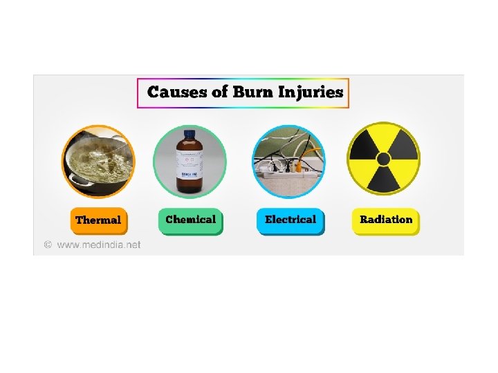
Burn injury to skin

Burn complications Airway obstruction Inhalation injuries Fluid loss Hypovolemic shock Compartment syndrome Limb ischemia Infection. Intestinal changes. Bad scar.

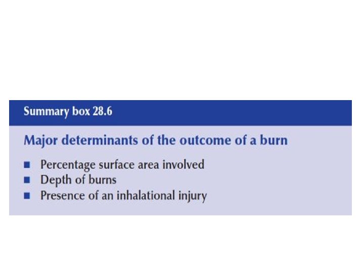
Burn care requires a specialized multidisciplinary team
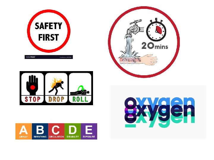

pre-hospital care • Ensure rescuer safety. • Stop the burning process. Stop, drop and roll • Check for other injuries, • Pain relief ABC + rapid secondary survey. • Cool the burn wound should occur for a minimum of 10 minutes and is effective up to 1 hour after the burn injury. • oxygen consciousness Anyone involved in a fire in an enclosed space should receive oxygen, especially if there is an altered level of . • Elevate Sitting a patient up Elevation of burned limbs will reduce swelling and discomfort

At the. Hospital

Initial Assessment 1. Mechanism of Injury 2. Associated Injuries 3. Patient Age 4. State of Health

Airway Endotracheal tube until the swelling subside << 48 h • Supraglottic tissue edema progresses over the fi rst 12 hours and can obstruct the airway rapidly • The larynx protects subglottic tissue from direct thermal injury but not from injury due to inhaled toxic gases. • Physical signs include hoarseness, stridor, facial burns, singed facial hair, expectoration of carbonaceous sputum, and presence of carbon in the oropharynx.

HISTORY & EARLY SIGNS : HX of inhalation of hot gases such as in a house or car fire (enclosed spaces). PE - blisters on the hard palate - burned nasal mucosa and loss of all the hair in the nose. the most valuable signs are the - presence of deep burns around the mouth and in the neck. Acute cricothyroidotomy

Breathing • Effort • Depth of respiration • Auscultation of breath sounds Wheezing - Inhalation Injury – Aspiration of gastric contents

Breathing 1 - Inhalation injuries Physiotherapy , nebulizers and warm humidified oxygen Monitor respiratory rate , blood gases Deteriorate !! positive pressure , ICU. 2 - thermal injuries to the lower airway steam

3 - metabolic poisoning Carboxyhaemoglobin levels raised above 10% must be treated with high inspired oxygen for 24 hours to speed its displacement from haemoglobin 4 - Mechanical block Escharatomy

Circulation ** Aggressive and prompt fluid resuscitation ** Risk of hypovolemic shock due to dynamic fl uid shifts from the intravascular compartment to the interstitium, and exudative and evaporative water loss from the burn injury. Exposure Remove all clothing and jewelry

CRITERIA ? ?

The criteria for acute admission to a burns unit.

ASSESSMENT OF THE BURN WOUND

Assessing size 1 - the size of the patient’s whole hand for small areas Equals 1% of BSA TBSA= total body surface areas The palm rule = the patients palm accounts for approximately 1% of TBSA, used for estimating small burns

2 -Draw the burn on a Lund and Browder chart which maps out the percentage TBSA of sections of our anatomy. It also takes into account different proportional body surface area in children according to age

3 -The rule of nines : - each upper limb is 9 - each lower limb 18 - The torso 18 each side - head and neck 9

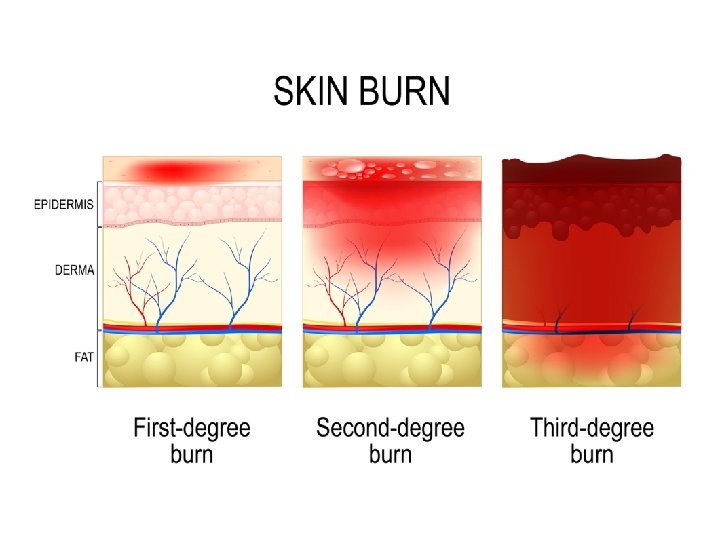
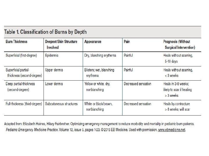
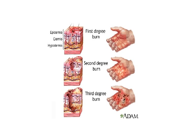
Assessing depth: Burns should be classified and managed based on which layer of skin to which they extend

DEPTH VS CAUSE

MANAGEMENT A. Resuscitation 1. Oxygen. should be provided to patients with all but the most minor injuries. A 100% oxygen high-humidity facemask for those with possible inhalation injury assists the patient’s expectoration from dry airways and treats carbon monoxide poisoning. 2. 2. Intravenous access. All adult patients with burns of 15% or greater or children with burns over 10% TBSA require intravenous fluids. Two large peripheral venous catheters should be started immediately to provide circulatory volume support. 3. Fluid. Improved survival in the era of modern burn care is largely attributable to early and aggressive volume resuscitation. Fluid resuscitation based on standard formula.

Parkland Formula : The estimated crystalloid requirement for the first 24 hours after injury is calculated on the basis of patient weight (determined early after the burn as a baseline) and BSA burn percentage. Lactated Ringer solution volume in the first 24 hours = 4 m. L × %BSA (second-, third-, and fourth-degree burns only) × body weight (kg). One-half of the calculated volume is given in the first 8 hours after injury, and the remaining volume is infused over the next 16 hours with adjustments being made as clinical conditions and size/depth of burns evolve.

How?

Example ; adult 100 kg , has 20% of his body surface are burn , how much fluid he need ? • 4*100*20 = 8000 ml gives him 4 liter in first 8 hour

TYPES OF FLUID USED 1 -Crystalloid resuscitation: Ringer’s lactate is the most commonly used crystalloid. 2 -Hypertonic saline *Hypertonic saline has been effective in treating burns shock. *It produces hyperosmolarity and hypernatraemia. This reduces the shift of intracellular water to the extracellular space. *Advantages include less tissue oedema and a resultant decrease in escharotomies and intubations 3 -Colloid resuscitation: Some studies show they should be avoided as an intravenous therapy until after the first 24 hours postburn, at which time capillary leak diminishes. Next 24 hours: Begin colloid infusion of 5% albumin 0. 3– 1 ml/kg/% burn/16 per hour

• In children, maintenance fluid must also be given to compensate for ongoing evaporative losses and quickly depleted glycogen stores This is normally dextrose–saline given as follows (maintenance in children) • 100 ml/kg for 24 hours for the first 10 kg; • 50 ml/kg for the next 10 kg; • 20 ml/kg for 24 hours for each kilogram over 20 kg body weight.

Monitoring of resuscitation 1 -The key to monitoring of resuscitation is urine output. should be between : 0. 5 -1 ml/kg/hr Adults 1 -1. 5 ml/kg/hr Children 2 -Normal pulse rate 3 -Normal Blood Pressure If the urine output is inadequate and the patient is showing signs of hypoperfusion (restlessness with tachycardia, cool peripheries and a high hematocrit), then a bolus of 10 ml /kg should be given. *It is important that patients are not over resuscitated, and urine output in excess of 2 ml/kg/ hour should signal a decrease in the rate of infusion.
")
Treatment depending on the degree • First degree burns: keep clean + topical antibiotic(neomycin) + pain meds. • Second degree burns: Remove blisters, apply antibiotic ointment (usually Silvadene®) and dressing, pain meds, Most second-degree burns do not require skin grafting (epidermis grows from hair follicles and from margins). Newer treatment can be used as well: • 1. Biobrane® (silicone artificial epidermis—temporary) • 2. Silverlon® (silver ion dressings) • Third degree burns: Early excision of eschar (within first week postburn) and STSG.
 Early irrigation and Debridement with N. S and")
Management of the Burn Wound 1) Early irrigation and Debridement with N. S and sterile instrument. 2) Analgesia : All burn patients need good analgesia (IV morphine). Apply cold saline soaks for analgesia if burns are < 25% BSA (watch for hypothermia). 3) Topical antimicrobial agent 4) Dressing 5) +/- tetanus +/- H 2 blocker (curling’s ulcer)

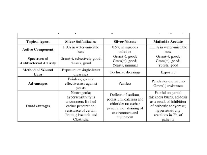
Topical antimicrobial agent The most common organisms causing burn wound infection are : staph aureus , strep group A , Pseudomonas a. and Fungi
 of")
Nutrition • Any adult with a burn greater than 15% (10% in children) of TBSA has an increased nutritional requirement. • All Patients with burns of 20% of TBSA or greater should receive a Nasogastric tube. ü (early feeding should start within 6 hours of the Injury to reduce gut mucosal damage. ) ü (to prevent aspiration pneumonia caused by paralytic ileus and vomiting) .
 Monitoring and control of infection Burns patients are immunocompromised. Swabs should be taken")
6) Monitoring and control of infection Burns patients are immunocompromised. Swabs should be taken regularly A rise in WBC count, thrombocytosis and increased catabolism are warnings of infection , most common discoloration of burn eschar.

• Escharotomy if needed : • It is a surgical incision through the eschar into the subcutaneous tissues to allow the extremity to continue to swell without compressing the underlying blood vessels. • Areas of concern: Circumferential burns of extremities or thorax. • Indications: • Impending or established vascular compromise of the extremities or digits. • Impending or established respiratory compromise due to circumferential torso burns

• Fasciotomy • Escharotomy may fail, especially when the burn is from highvoltage electrical injury or is associated with soft tissue, bone, or vascular injury. • If compartment syndrome persists after escharotomy, incision of the fascia is also required. • General anesthesia is required.

Operative Treatment -Any deep partial-thickness and fullthickness burns, except those that are less than about 4 cm 2 , need surgery. ■ 1 -The anaesthetist needs to be ready for significant blood loss ■ 2 -Topical adrenaline reduces bleeding ■ 3 - All burnt tissue needs to be excised 4 -Stable cover, permanent (stsg)or temporary, should be applied at once to reduce burn load

MANAGEMENT OF ELECTRICAL INJURY • Don’t become the next victim • Wounds are greater than surface burn • ABC’s • Arrhythmias >> {cardiac monitoring if history of LOC or ECG abnormalities} • Rhabdomyolysis >> ARF

TREATMENT OF CHEMICAL BURNS Wear gloves and protective clothing Remove saturated clothing Brush skin if agent is a powder Irrigate, irrigate! copious lavage with water *Then identify the chemical and assess the risks of absorption

So • Abcd • Fluid • Tetanus • H blocker • Wound care 1 - dressing 2 - topical antibiotic 3 - assessment for compartment or infection.
- Slides: 47