MANAGEMENT OF AGE RELATED MACULAR DEGENERATION CHAIRPERSON DR


 6 mg Verteporfin/metre 2 Diode laser, 689 nm,")

 28 base pegylated RNA aptamer Binds to VEGF")
 Recombinant humanized monoclonal antibody Fab fragment 0. 3 -0.")


 Complete monoclonal antibody to VEGF 1. 25 mg/0. 05 ml intraocularly")


- Slides: 47
MANAGEMENT OF AGE RELATED MACULAR DEGENERATION CHAIRPERSON : DR. KRISHNENDU SARKAR SPEAKER : KUMAR SAURABH
APPROACH TO THE PATIENT HISTORY CLINCAL EXAMINATION DIAGNOSTIC TESTS TREATMENT FOLLOW UP
HISTORY Symptoms Age of the patient History of smoking History of eye ailments in family History of use of glasses History of drug intake History of cardiovascular diseases
CLINICAL EXAMINATION Visual acuity Contrast sensitivity Amsler grid test Slit lamp indirect biomicroscopy Fundus photography Systemic examination
Visual acuity Dimness of vision Gradual with nonexudative AMD Sudden with exudative AMD Worse on pinhole testing
Contrast sensitivity Reduced contrast sensitivity
Amsler grid test
Slit lamp indirect biomicroscopy Usually sufficient to establish the diagnosis Contact lenses 60 Diopter or 90 Diopter lenses
Fundus photography Used for grading of drusen Graticules over 35 mm colour fundus photograph
Inner macula 1500 micron Outer macula 1500 – 3000 micron
DRUSEN SIZE Compared to the width of a major vein at the edge of disc (125 micron) Types Size Smallest Less than one-half vein width (63 micron) Small One-half to one vein width (63 - 124 micron) Medium One to two vein width (125 – 249 micron) Large Two to four vein width (250 – 499 micron) Drusenoid PED More than four vein width ( > 500 micron)
NUMBER OF DRUSEN DENSITY OF DRUEN TYPE OF DRUSEN Less than 20 More than 20 Scattered Sub-confluent Confluent Hard Soft Equal to choroidal fluorescence Slightly brighter Brightest
Systemic examination Rule out diabetes mellitus Screen for cardiovascular diseases
DIAGNOSTIC TESTS Digital Fluorescein Angiography Indocyanine Green Angiography Optical Coherence Tomography
Digital Fluorescein Angiography Less useful in non-exudative disease DRUSEN Hyperfluorescent: Hydrophilic = High risk for CNV Hypofluorescent: Hydrophobic = High risk for PED
Classic CNV Well defined area of hyperfluorescence in early phase Bright hyperfluorescence in peak phase Diffuse leakage and blurring of margins around 1 -2 minutes.
Occult CNV Poorly defined area Late leakage
Predominantly Classic CNV Minimally Classic CNV occupies > 50% of entire lesion including contiguous blood, pigment, scar and staining. Classic CNV occupies 1 - 49% of entire lesion including contiguous blood, pigment, scar and staining. Occult With No Classic CNV
Fibrovascular PED Stippled hyperfluorescence Less bright and less well defined than classic CNV Late leakage or persistent staining
RPE Tear Sharp well defined area of hyperfluorescence Adjacent area of blocked fluorescence due to torn RPE Late staining No late leakage
Indocyanine Green Angiography INDICATIONS : Occult CNV Distinguishing serous from fibrovascular PED Suspected CNV with overlying hemorrhage Recurrent CNV adjacent to photocoagulation scar
Hot spot Occult CNV Less than 1 disc diameter Relatively well defined
Placoid hyperfluorescence Occult CNV More than one disc diameter Not well defined
Combined lesion Hot spot Placoid hyperfluorescence
Classic CNV
Optical Coherence Tomography Pr. ONTO Study: OCT detects recurrent neovascularization before vision loss CNV
Fibrovascular PED
Macular scar
TREATMENT Non-exudative AMD Patient education and follow up Low vision aids Nutritional supplementation AREDS Criteria Vit E 400 IU Beta Carotene 15 mg Extensive intermediate drusen At least one large druse Noncentral geographic atrophy Advanced AMD in one eye Vit C 500 mg Cupric Oxide 2 mg Zinc Oxide 80 mg
TREATMENT Exudative AMD Laser Photocoagulation Extra-foveal CNV Juxta-foveal CNV 200 micron spot size; 0. 2 -0. 5 second duration Krypton laser or Argon laser
ICG Assisted Feeder Vessel Photocoagulation Sub-foveal CNV Aimed at closure of extra-foveal feeder vessel Preserves the foveal tissue
Transpupillary Thermotherapy Proposed for sub-foveal occult CNV 810 nm diode laser Single large spot is used TTT 4 CNV has shown no statistically significant benefit over sham treatment
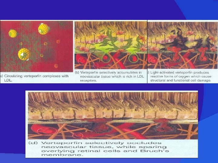
Photodynamic Therapy Definite Indications Predominantly classic sub-foveal CNV Occult sub-foveal CNV with recent disease progression (lesion size < 5400 micron) Possible Indication Minimally classic occult CNV approaching 50%
PDT Agents & Methods Verteporfin (Visudyne) 6 mg Verteporfin/metre 2 Diode laser, 689 nm, Rostaporfin (Photrex) Energy 50 J/cm 2 Intensity 600 m. W/cm 2 Motexafin lutetium (Optrin) Talaporfin sodium Spot size 1000 micron Time 83 seconds
Receptor Targeted PDT Verteporfin + VEGF Receptor 2 antagonist Aimed at reducing collateral damage of adjacent retina Combination PDT Verteporfin + Intravitreal Triamcinolone acetonide Aimed at reducing the increased post-PDT VEGF expression
Anti- VEGF Aptamer Pegaptanib Sodium (Macugen) 28 base pegylated RNA aptamer Binds to VEGF 165 0. 3 mg intravitreal every 6 weeks for 2 years All types of CNV
Anti- VEGF Antibody Ranibizumab (Lucentis) Recombinant humanized monoclonal antibody Fab fragment 0. 3 -0. 5 mg(0. 05 ml) intravitreal injections monthly for 2 years
MARINA Trial Minimally Classic/Occult Trial Of The Anti-VEGF Antibody Ranibizumab In Treatment Of Neovascular AMD Goal Outcome measure To assess the safety and efficacy of Ranibizumab in a sham injection controlled study Sham (n=238) Ranibizumab 0. 5 mg (n= 240) Month 12 62% 95% Month 24 53% 90% Month Loss of < 15 letters in visual acuity (%)
ANCHOR Trial Anti-VEGF Antibody For The Treatment Of Predominantly Classic Choroidal Neovascularization In AMD Goal To compare the safety and efficacy of Ranibizumab to Verteporfin PDT Outcome measure Verteporfin PDT (n=143) Ranibizumab 0. 5 mg (n= 140) Loss of < 15 letters in visual acuity (%) 64% 96% 6% 40% Gain of more than 15 letters in visual acuity (%)
Trial Goal better visual outcomes. PIER than sham-injected controls Phase IIIb, Multicenter Randomized, Double Masked, Sham Injection Controlled Study Of The Safety And Efficacy Of Ranibizumab To assess whether fewer injections of Ranibizumab can result in
Bevacizumab (Avastin) Complete monoclonal antibody to VEGF 1. 25 mg/0. 05 ml intraocularly
Steroid Compounds Triamcinolone Acetonide Single 4 mg intravitreal injection Maximum antiangiogenic effect at 3 months Little or no effect at 1 year post-injection Anecortave Acetate Another potent steroid being studied for subtenon injection
Macular Surgery Surgical Removal Of CNV Macular Translocation Limited Macular Translocation RPE Transplantation SST did not show any benefit with submacular surgery over observation alone for subfoveal CNV
Future Trends Small Interfering RNA Therapy Combretastatin A-4 Phosphate Prodrug Squalamine Pigment Epithelium Derived Factor Inducer
Thank You