MAMMOGRAPHY Pt 2 EQUIPMENT LECTURE more RTEC 255


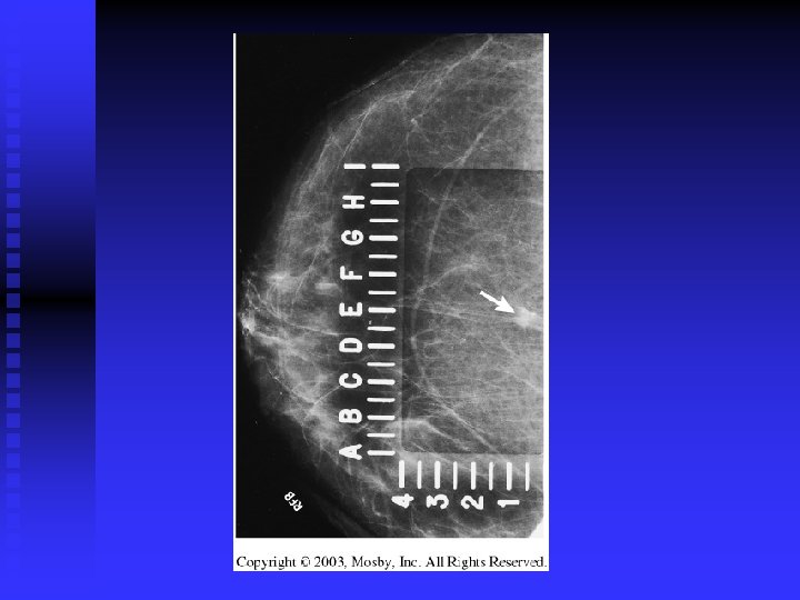


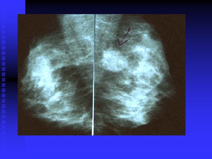
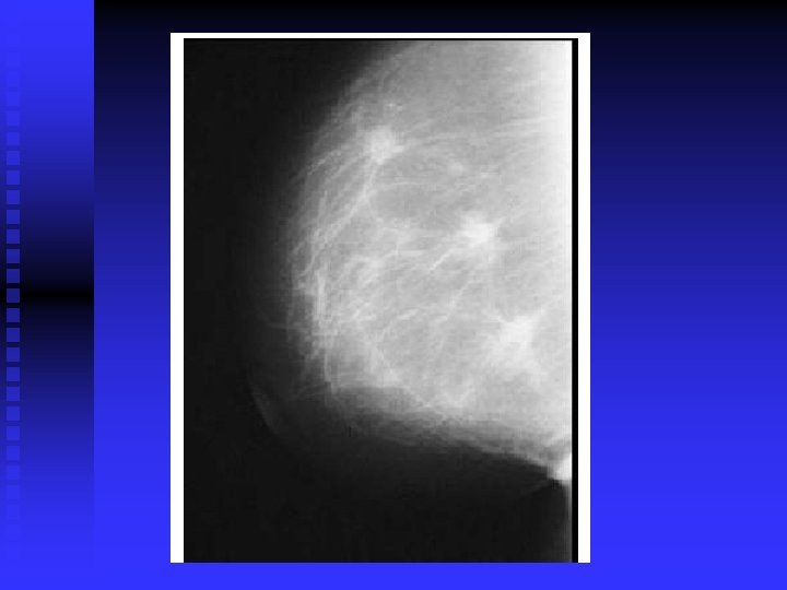


 contains an irregular carcinoma that is producing considerable spiculation, nipple retraction")


- Slides: 80
MAMMOGRAPHY - Pt 2 EQUIPMENT LECTURE & more…. . RTEC 255 Week # 3 D. CHARMAN, M. Ed. , R. T. (R, M)
MAMMOGRAPHY RADIOGRAPHIC IMAGING OF THE BREAST
Man – o - gram
Mammograms don’t look fun but they can save a life!
A mammogram can find breast cancer when it is very small -- 2 to 3 years before you can feel it. n No screening tool is 100% effective. Good quality mammograms can find 85 -90% of cancers n
Do it for those you love….
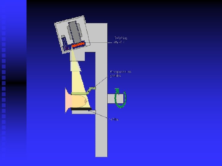
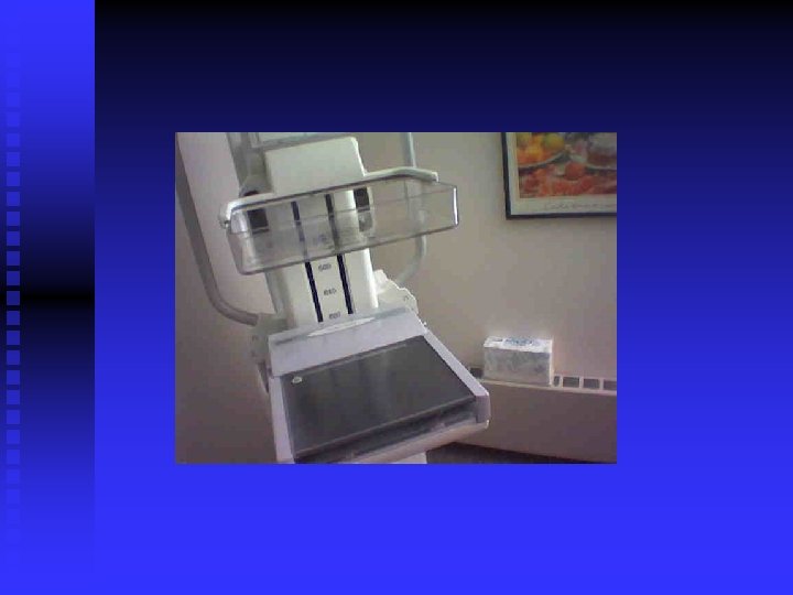
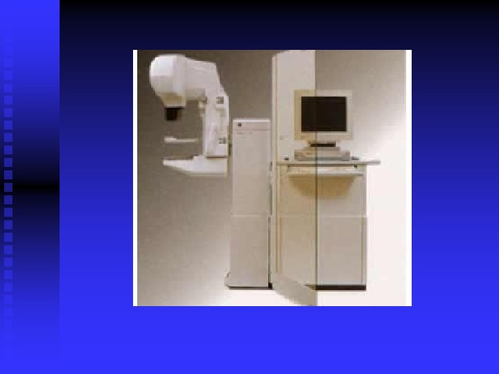
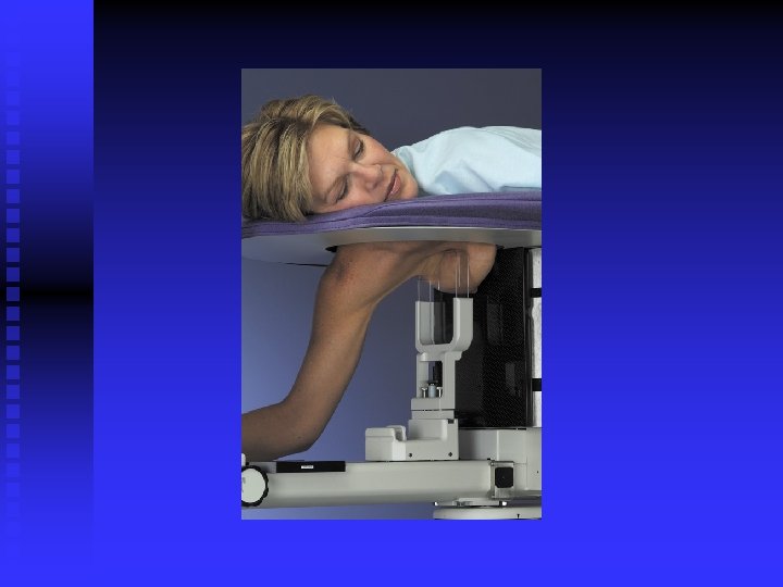
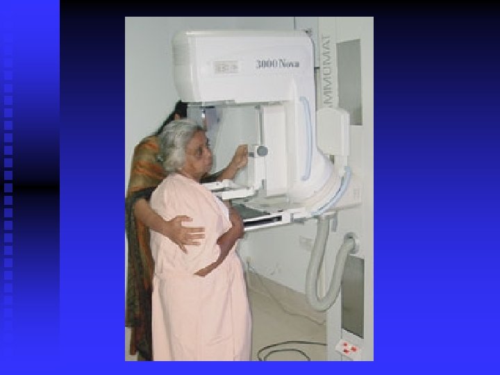
Mammography Equipment Bushong – Ch
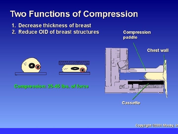
Mammogram Soft Tissue Radiography n low subject contrast u low kvp – more PE n
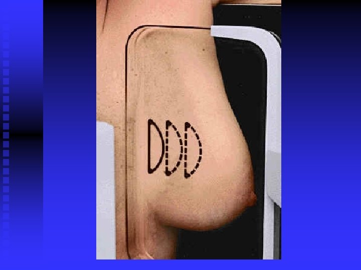
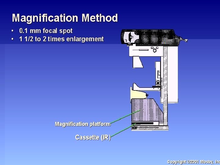
Magnification = increase OID
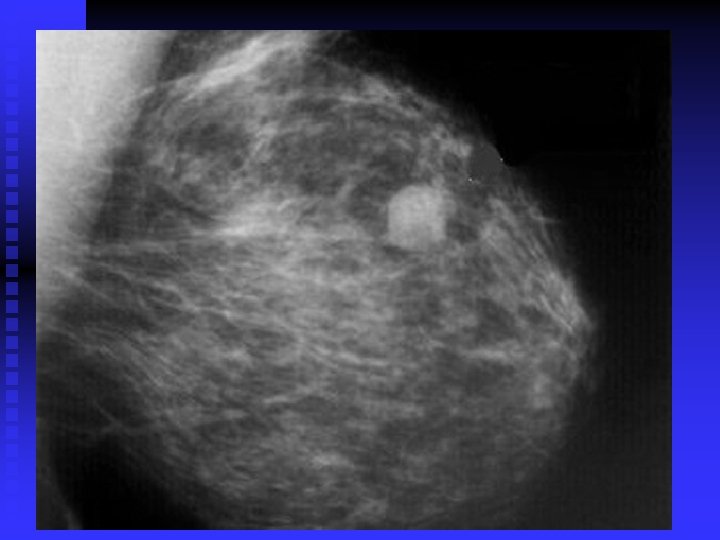
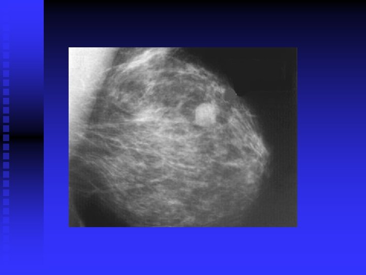
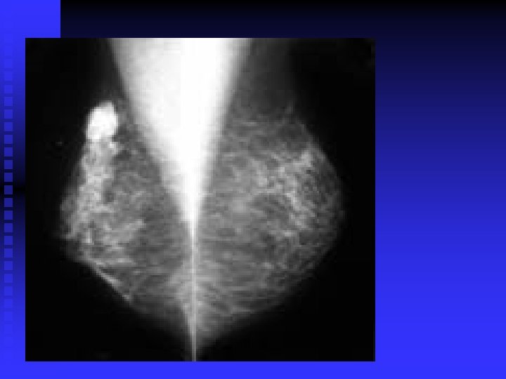
Cysts
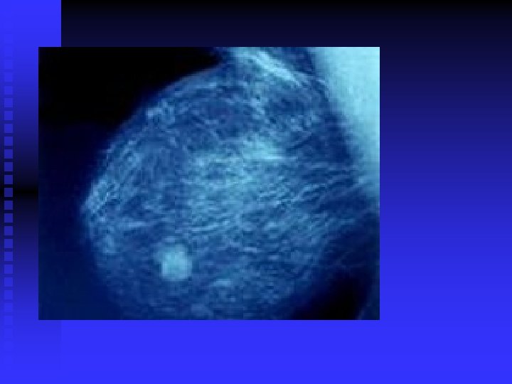
Infiltrating Ductal Cancer
Infiltrating Ductal Cancer
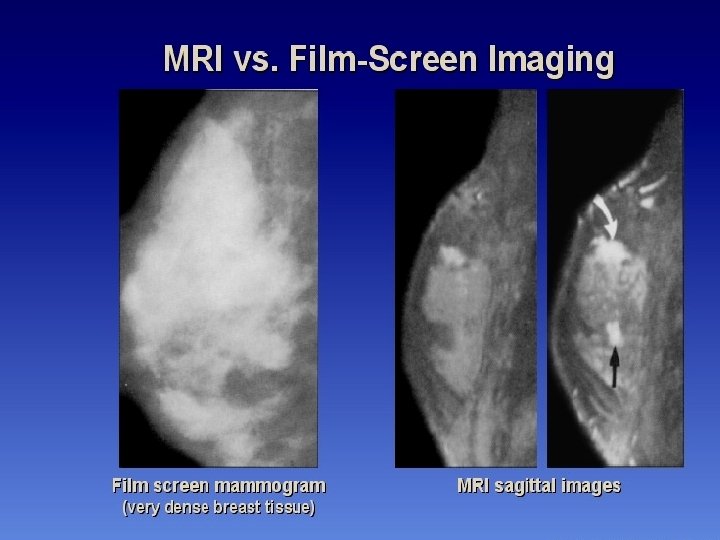
MRI vs. Mammo
Digital vs. Conventional
THE AUGMENTED BREAST Difficulty with IMPLANTS
Difficulty with Lateral Sternum may need to take 2 laterals – 1 upper, 1 lower
OTHER CHALLENGES TO MAMMOGRAPHY
A little more about… MAMMOGRAPHY PT 3 255 09 wk 3
Sentinel Lobe – Nuclear Med
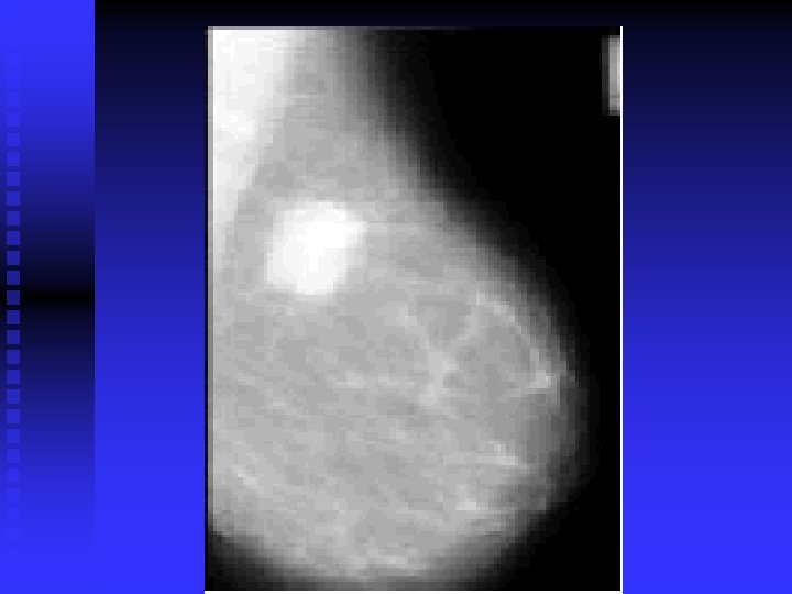
Pathology? + or - FIBROADENOMA CYST
DCIS– CELLS HAVEN’T BROKEN OUT SO WON’T SPREAD
INFILTRATING DUCTAL CA
DCIS
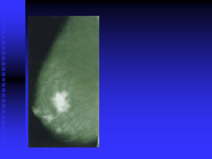
CLUSTER CALCIFICATIONS
year old woman with normal post-op mammogram MRI shows 5 mm invasive ductal carcinoma
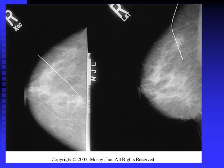
Spot compression
POSITONING COUNTS!
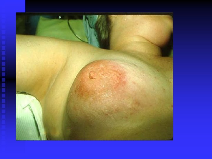
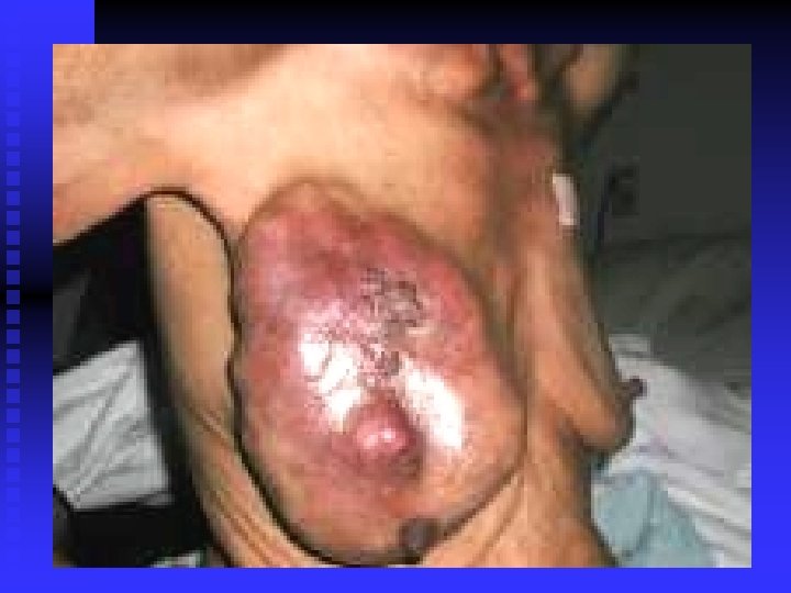
PATHOLOGY LOOKING OUTSIDE HOW CAN IT GO THIS FAR?
Pagets disease of the nipple
47 yo breast cancer
Abnormal axillary lymph nodes Breast Cancer
normal
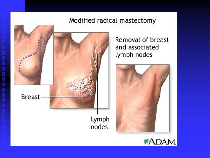
male mastectomy
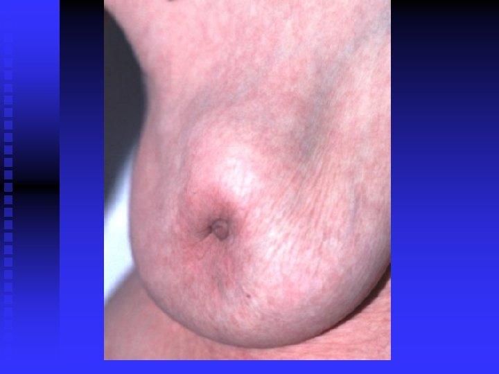
Male breast cancer and gynecomastia
MRI for dense breast tissye
Spot Compression Especailly good for dense breast
Surgical scars can mimic characteristics of breast cancer.
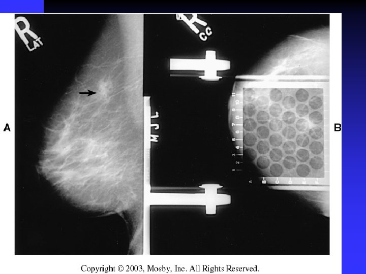
Left breast (L) contains an irregular carcinoma that is producing considerable spiculation, nipple retraction (arrow), and skin thickening. Right breast (R) contains fibroadenoma.
27 -year-old woman who stopped breastfeeding 2 months before having this mammogram.
42 -year-old woman with fibrocystic condition
28 -year-old woman 4 months postpartum and not breast-feeding. The right breast contains a large mass (arrow) palpable on physical examination. The left breast contains two smaller nonpalpable masses (arrows) with microcalcifications. All three lesions were breast cancers.
1. Abnormal axillary lymph nodes 2. Breast 3. Breast Cancer
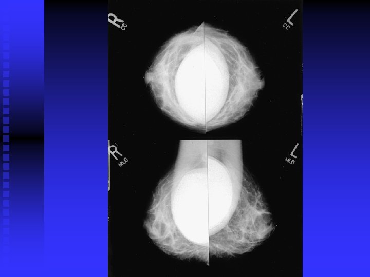
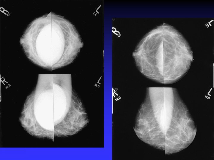
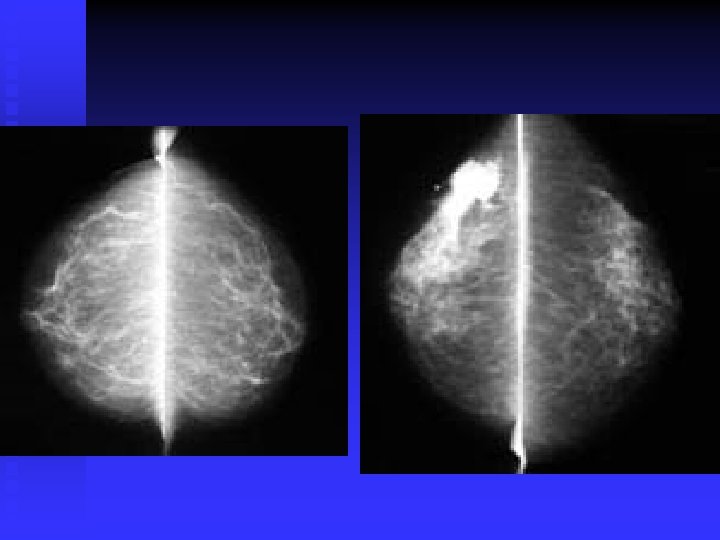
importance of high-quality mammograms is evident in the two images of the same breast
FINAL THOUGHTS n