Mammography Cases 1 12 Case directory 1 2


, invasive")

 n n n Uncommon benign breast tumor Composed of lipid, glandular,")
")
")

 n n n Round, smoothly bordered mass")


 n Case findings: n n US: multiple solids, hyperechoic nodules")
 n Differentiation between benign and malignant phyllodes tumors is not")
 Predominantly circumscribed malignant phyllodes tumor")
 US: smooth margined lobulated noncompressible lesion with internal cystic spaces")
")
 Fat necrosis")

 Punctate: small grains of calcium, round, regular, dense,")

 Jagged: coarse, dense, large, branching calcium deposits following inflammation or irradiation")


: spherical capsules of fibroadenoma or as depicted here, calcified cystic capsules")


- Slides: 58
Mammography Cases 1 -12
Case directory 1 2 3 4 5 6 7 8 9 10 11 12
Case 1
Silicone injection n Innumerable high density rim-calcified masses are seen, characteristic of silicone granulomas
Breast implant n Retropectoral, single lumen implant containing high density silicone
Breast implant n n Single lumen, subglandular implants Capsular contracture in left breast implant lost normal tear-drop shape
Case directory Saline breast implant
Case 2
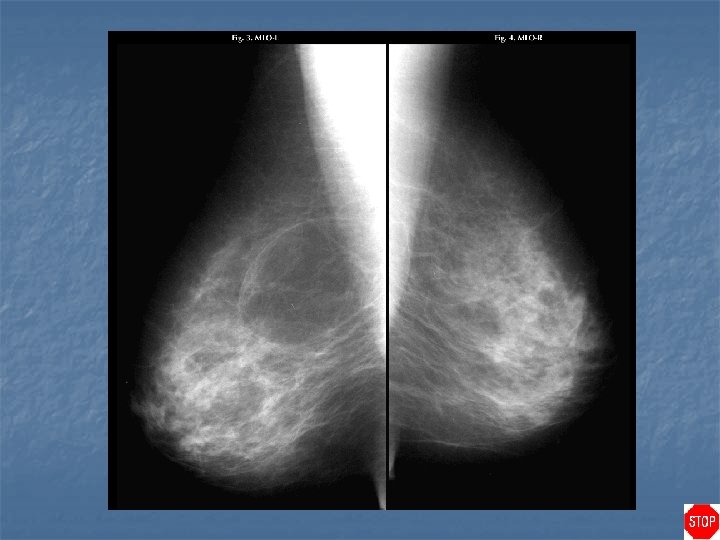
Gynecomastia n n Proliferation of fibroglandular tissues resulting in breast enlargement Hyperplasia of the ductal elements, fibroepithelium, and stroma (lobules do NOT develop) Dense tissues fan out from retroareolar region rather than forming a mass Bimodal age distribution: MC puberty, 5 th decade
Case directory Gynecomastia n Etiology: excessive estrogens or deficient androgens n n n Idiopathic Physiologic: newborn, puberty, senescence Pathologic: Klinefelter's syndrome, congenital adrenal hyperplasia, adrenal neoplasms, testicular tumors, paraneoplastic syndromes, liver failure, thyrotoxicosis, testicular failure Drugs: cannabinoids, psychotropics, antihypertensives (reserpine, spironolactone), digitalis, cimetedine, hormonal (estrogens, HCG, antiandrogens) No increased risk for male breast cancer Male breast cancer presents at a later stage so worse prognosis at presentation
Case 3
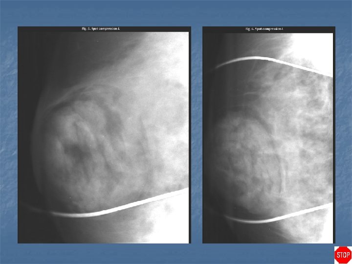
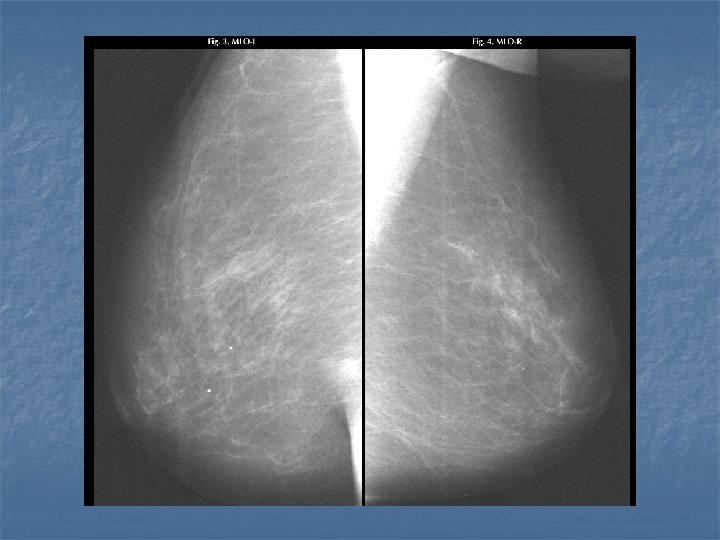
Radial scar n Case findings: n n n 7 mm density in retroareolar location in the left breast, with radiating linear strands surrounding it Best seen on the CC view Spiculated lesion, central nidus with internal radiolucencies ( white star) Predominantly radiolucent spiculations (rather than radiodense) Requires open surgical biopsy malignancy present in 25% of cases (some are invasive carcinomas, but others are radial scars that have small internal or adjacent areas of carcinoma) Open surgical biopsy: less subject to sampling error than are percutaneous biopsy procedures (aspiration or core biopsy)
Case directory Spiculated lesion n Malignant: n n n MC invasive ductal (90%), invasive lobular (10%) Malignant spicules: extend in all directions from a central tumor mass Benign: n n n MC radial scar, sclerosing adenosis Post-surgical scar: change in shape and density on different projections, contain central lucency, and regress over time, follow-up within 6 months Benign spicules: bundled in parallel
Case 4
Hamartoma (lipofibroadenoma, fibroadenolipoma) n n n Uncommon benign breast tumor Composed of lipid, glandular, and fibrous tissues May appear encapsulated, but does not possess a true capsule Surrounded by a thin layer of fibrous tissue Cut sausage and breast within a breast: terms used to describe the classic mammographic appearance of a breast hamartoma
Hamartoma (lipofibroadenoma, fibroadenolipoma)
Case directory Hamartoma (lipofibroadenoma, fibroadenolipoma)
Case 5
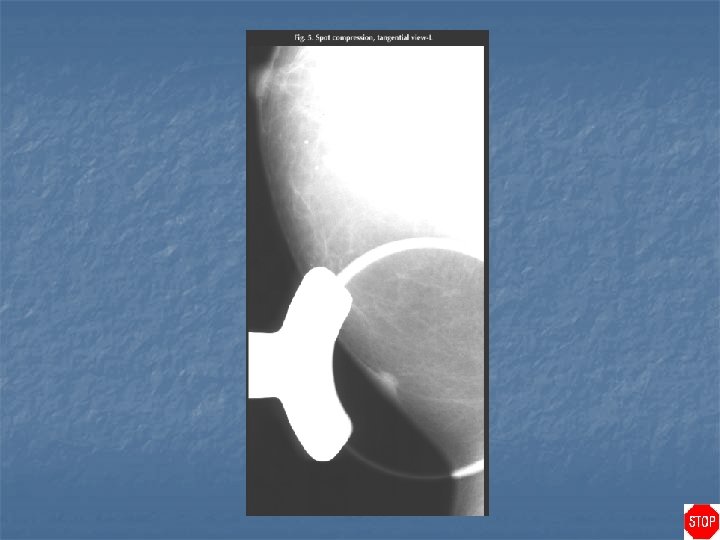
Case directory Sebaceous cyst (epidermal inclusion cyst) n n n Round, smoothly bordered mass that often abuts the skin surface Mass projects into the subcutaneous tissues rather than out from the skin surface Tangential view of the mass demonstrates that it is located within the skin
Case 6
Case directory Neurofibromatosis
Case 7
Case directory Lipoma n Case findings: n n n n very thin radiopaque border surrounding the mass is its fibrous capsule BI-RADS 2: mass is fatty in density, and therefore benign Fat containing mass, circumscribed radiolucent mass, with a thin capsule Encapsulated mature adipose tissue Soft lesion: can grow to substantial size before they become palpable Can calcify, either within the capsule, or within the mass itself if there is central infarction MC seen in postmenopausal women
Case 8
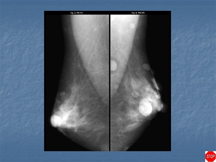
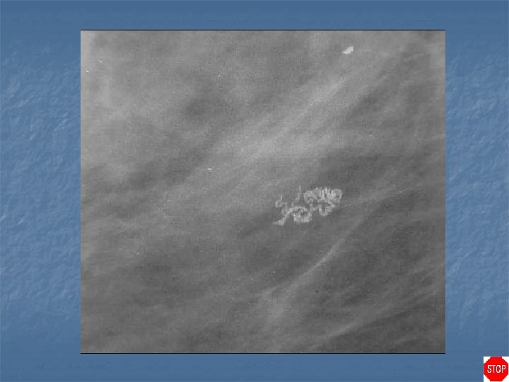
Filariasis n Case findings: n n n Mammograms showed unusual serpiginous, tubularwormlike calcifications without an accompanying mass Serpiginous calcifications represent calcified degenerating parasite tissue (filarial granulomas of the breast) Parasite: Wuchereria bancrofti, Brugia malayi
Case directory Case 9 Ultrasound of right breast
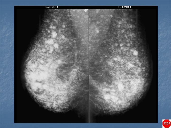
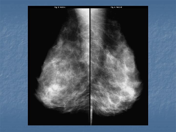
Phylloides tumor (cystosarcoma phyllodes) n Case findings: n n US: multiple solids, hyperechoic nodules with central cystic components CT: well-circumscribed tumor without obvious enhancement Age: 45 years-old Features: n n n Circumscribed round, oval or lobulated tumor, sometimes with partly indistinct borders No spiculation on mammography US: circumscribed hypoechoic lesions with varying degrees of inhomogeneity and small cystic spaces
Phylloides tumor (cystosarcoma phyllodes) n Differentiation between benign and malignant phyllodes tumors is not possible on imaging Locally invasive tumor which rarely metastasizes n DDX rapidly enlarging masses: n n n Phylloides tumor Juvenile giant fibroadenoma Abscess Hematoma
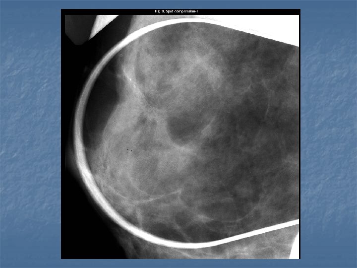
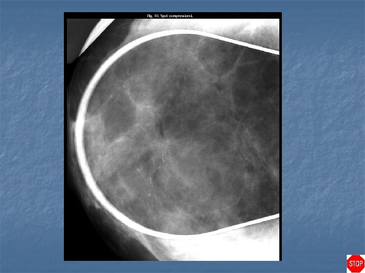
Phylloides tumor (cystosarcoma phyllodes) Predominantly circumscribed malignant phyllodes tumor
Phylloides tumor (cystosarcoma phyllodes) US: smooth margined lobulated noncompressible lesion with internal cystic spaces and varying echogenicity
Case directory Phylloides tumor (cystosarcoma phyllodes)
Case 10 Benign microcalcification Plasma cell mastitis (secretory calcifications) Fat necrosis
Fibroadenoma Vascular Benign calcifications
Case directory Benign calcification n (a) Punctate: small grains of calcium, round, regular, dense, fairly equal in size, benign microcalcification n (b) Linear: long, dense, smooth, calcifications, outlining the ductal system, usually the result of ductal inflammatory changes n (c) Spherical: round or oval, having lucenters, may be the result of fat necrosis as exemplified here, but may also represent small calcified cysts n (d) Coarse (popcorn): large, irregular, very high density, pathognomonic for calcified fibroadenoma n (e) Cylindrical: calcium deposits within walls of tubular structures might be vascular as illustrated here or past periductal inflammatory changes n (f) Smooth: dense, round, smoothly bordered, medium sized, isolated, benign grains of calcium
Case 11
Calcification n (a) Jagged: coarse, dense, large, branching calcium deposits following inflammation or irradiation therapy (dermatomyositis this case) n (b) Heterogeneous: very small < 0. 5 mm variable sizes, shapes and densities, highly suspicious (DCIS this case) n (c) Regular: small, dense, round, benign microcalcification located at the periphery of a small calcifying cyst n (d) Branching: heterogeneous microcalcification, filling the ductal system with its dichotomic distribution, highly suspicious (high grade DCIS, comedo type this case)
Skin calcifications, polihedric, annular and dense Tangential view: show that the calcifications are located in the skin
Rod shaped calcifications: stick-like, solid or hollow concretions, sometimes branching, usually the result of previous ductal inflammatory changes Secretory calcification, or plasma cell mastitis
Acinar calcifications are concretions formed within acini of dilated lobules. Calcifications depicted here revealed a foci of lobular hyperplasia
Rim calcifications (eggshell): spherical capsules of fibroadenoma or as depicted here, calcified cystic capsules
Milk of calcium: precipitated amorphous calcium particles, floating within a cyst as seen on the left picture True lateral position: particles may show the “tea cup” phenomenon as illustrated on the right
Suture calcification: knot-like, calcium deposits on suture materials seen in a postoperative, post-irradiated breast
Case directory Heterogeneous calcification: variable shape, variable size and variable density, may might show a branching pattern and are usually arranged in clusters or distributed according to the segmental partition of the breast (comedocarcinoma these 2 cases)
Case 12
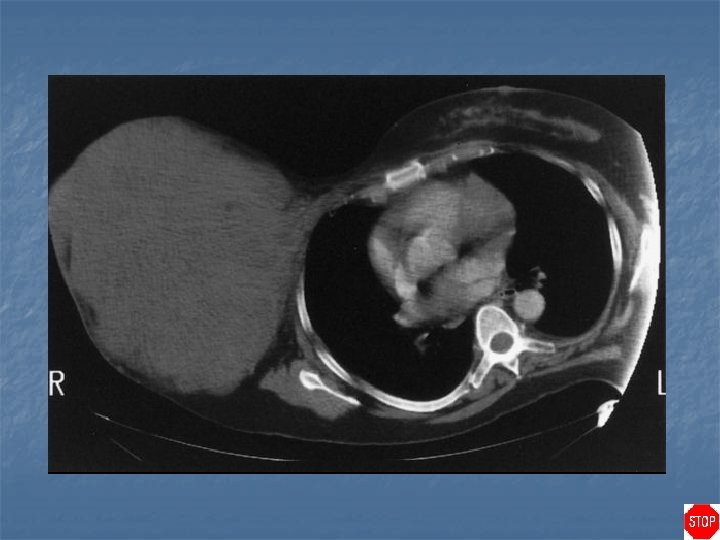
Rheumatoid arthritis n Case findings: n n Enlarged dense axillary lymph nodes DDX of enlarged axillary lymph nodes: n n n BCA with lymph node metastasis Lymphoma HIV RA TB, sarcoidosis Benign reactive nodal hyperplasia
Metallic deposits in axillary nodes from RA gold injections
Case directory Rheumatoid arthritis n DDX of calcified axillary lymph nodes: MC metastasis (ovarian, mucinous tumors) n RA with gold treatment n Treated lymphoma n