Mammary Gland It is fully developed in adult


 axillary lymph nodes. The")


- Slides: 13
Mammary Gland It is fully developed in adult females It is rudimentary: – In female before puberty – In males Site: in the superficial fascia of the pectoral region. Shape: it is conical or hemispherical. Extent: – upwards: 2 nd rib – downwards: 6 th rib – medially: lateral margin of the sternum – laterally: midaxillary line (axillary tail)
Relations: – supero-medial 2/3: rest on pectoral fascia covering pectoralis major muscle (separated by retromammary space. – inferlateral 1/3: rests on the serratus anterior muscle & external oblique muscle
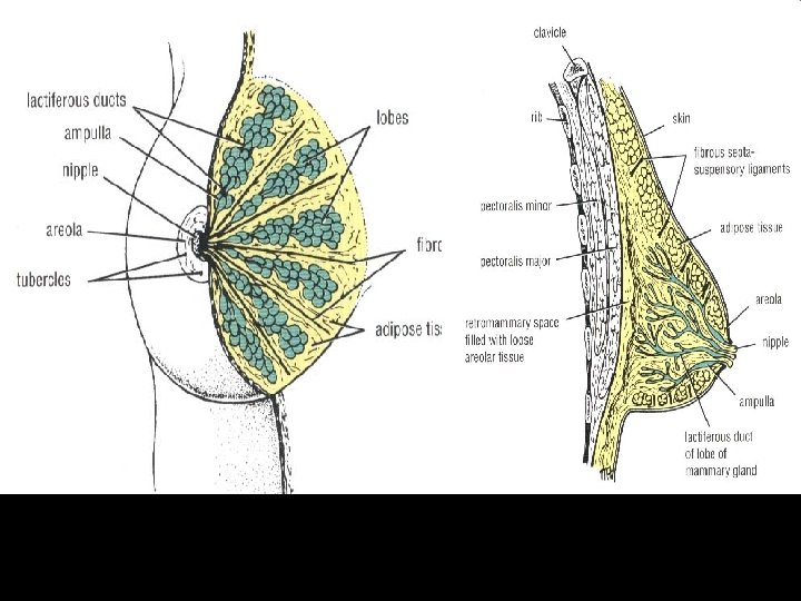
The skin of the breast shows: The nipple: a conical projection lying opposite to the 4 th intercostal space in males and in females before pregnancy The areola: – area of pigmented skin surrounding the nipple – it is pink in nulligravida but change to dark brown in the 2 nd month of pregnancy (permanent change) NB: - The breast has no capsule (to allow its distension during pregnancy). - No subcutaneous fat beneath the nipple and areola.
Internal structure of the breast: the mammary gland is formed of 15 -20 lobes. each lobe drains into a lactiferous duct. the lactiferous ducts (15 -20) converge toward the nipple & dilate to form lactiferous sinuses. the lactiferous sinuses open separately on the nipple. NB: the lobes of the breast are separated by suspensory ligaments of Cooper (these are fibrous septa stretched between the skin & pectoral fascia).
Axillary Tail the supero-lateral part of the breast extends upwards and laterally along the lower border of pectoralis major pierces deep fascia to reach the axilla tail of the breast
Arterial supply: Mammary branches of internal thoracic artery. Mammary branches of intercostal arteries lateral thoracic artery (the main supply). pectoral branch of thoraco-acromial artery.
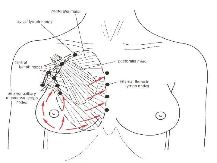
Lymph Drainage The lateral quadrants drain into the anterior (pectoral) axillary lymph nodes. The medial quadrants drain into internal thoracic lymph nodes. A few vessels drain into the posterior intercostal lymph nodes. Some vessels drain to the opposite lymph nodes. Some vessels drain into the abdominal lymph nodes.
Clavipectoral fascia it is a well-defined membranous fascia filling the gap between subclavius & pectoralis minor muscles. attachment: – superiorly: it splits to enclose subclavius, then attached to the lips of subclavian groove. – inferiorly: it splits to enclose pectoralis minor, then extends down as the suspensory ligament of axilla which is attached to axillary fascia hollow of axilla. – medially: is attached to 1 st & 2 nd costal cartilages. – laterally: is attached to coracoid process. N. B. : • the upper part is thickened and is called costo-coracoid ligament • structures pierce clavipectoral fascia: – – Cephalic vein Lateral pectoral nerve Thoraco-acromial artery Lymphatic vessels
THANK YOU