MALPRESENTATION And CORD PROLAPSE MALPRESENTATION n Malpresentation is


, fetal abnormalities (eg, CNS malformations,")

 - Hips flexed, knees extended n")


")

 is the inability to deliver the fetal shoulders after")


 1. Postpartum hemorrhage 11% 2. Vaginal laceration 19% 3. Perineal tears")


.")


- Slides: 80
MALPRESENTATION And CORD PROLAPSE
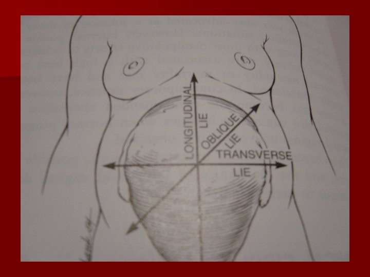
MALPRESENTATION n. Malpresentation is the situation where a fetus within the uterus is in any position that is not cephalic
Etiologic factors in malpresentation n Maternal Great parity Pelvic tumors Pelvic contracture Uterine malformation Fetal Prematurity Multiple gestation Hydramnios Macrosomia Hydrocephaly Trisomies Anencephaly Myotonic dystrophy Placenta previa n
Breech Presentation
Introduction Breech presentation occurs in 3 -4% of all deliveries. The occurrence of breech presentation decreases with advancing gestational age. Breech presentation occurs in 25% of births that occur before 28 weeks’ gestation, in 7% of births that occur at 32 weeks, and 1 -3% of births that occur at term. . Perinatal mortality is increased 2 - to 4 -fold with breech presentation, regardless of the mode of delivery. Deaths most often are associated with malformations,
Predisposing factors n prematurity, uterine abnormalities (eg, malformations, fibroids), fetal abnormalities (eg, CNS malformations, neck masses, aneuploidy), and multiple gestations. AF abnormality. Abnormal placentation. Contracted pelvis. MG. Pelvic tumor.
n Perinatal mortality is increased 2 - to 4 -fold with breech presentation, regardless of the mode of delivery. n Congenital malformation 6%
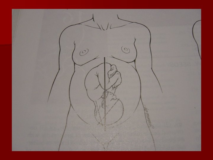
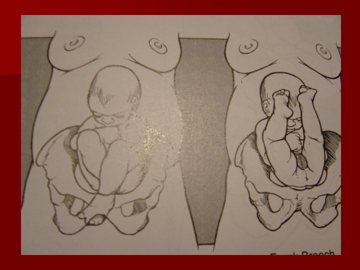
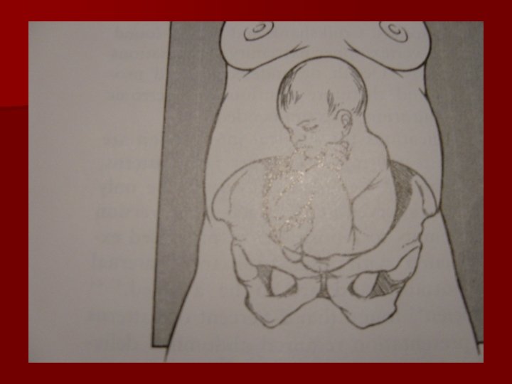
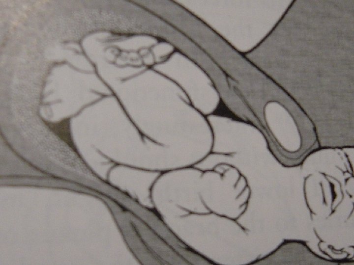
Types of breeches n Frank breech (50 -70%) - Hips flexed, knees extended n Complete breech (5 -10%) - Hips flexed, knees flexed n Footling or incomplete (10 -30%) - One or both hips extended, foot presenting
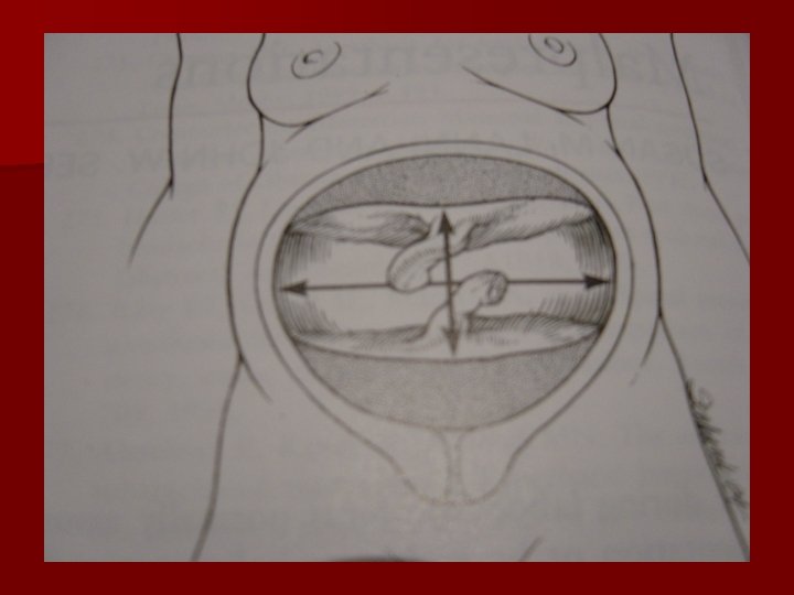
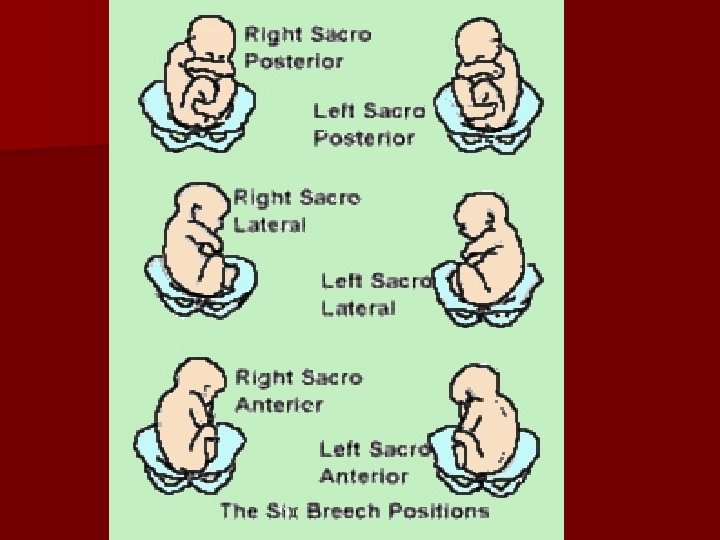
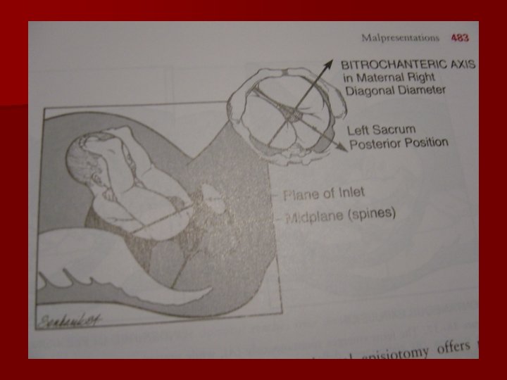
position SA, SP, LST, RST LSP, RSP. LSA, RSA
STATION
DIAGNOSIS n. Palpations and ballottement n. Pelvic exam n. X-ray studies n. Ultrasound
MANAGEMENT n. Antepartum n. During labor n. Delivery
Criteria for VD or. CS n VD Frank GA>34 w FW=2000 -3500 gr Adequate pelvis Flexed head Nonviable fetus No indication Good progress labor n CS FW<1500 or> 3500 gr Footling Small pelvis Deflexed head Arrest of labor GA 24 -34 w Elderly PG Inf or poor history Fetal distress
VAGINAL BREECH DELIVERY n. Three types of vaginal breech deliveries: 1. Spontaneous breech delivery 2. Assisted breech delivery 3. Total breech extraction
Footling breech presentation : Once the feet have delivered, there . may be temptation to pull on the feet. However, this should never be done with a singleton gestation because it may precipitate an entrapped head in an incompletely dilated cervix or it may precipitate nuchal arms. As long as the fetal heart rate is stable and no physical evidence of a prolapsed cord exists, expectant management may be followed, awaiting full cervical dilatation.
Assisted vaginal breech delivery n Thick meconium passage is common as the breech is squeezed through the birth canal. This usually is not associated with meconium aspiration because the meconium passes out of the vagina and does not mix with the amniotic fluid.
n Picture 3. Assisted vaginal breech delivery: The Ritgen maneuver is applied to take pressure off the perineum during vaginal delivery. Episiotomies often are cut for assisted vaginal breech deliveries, even in multiparous women, to prevent soft-tissue dystocia.
n Picture 4. Assisted vaginal breech delivery: No downward or outward traction is applied to the fetus until the umbilicus has been reached.
Picture 5. Assisted vaginal breech delivery: With a towel wrapped around the fetal hips, gentle downward and outward traction is applied in conjunction with maternal expulsive efforts until the scapula is reached. An assistant should be applying gentle fundal pressure to keep the fetal head flexed.
Picture 6. Assisted vaginal breech delivery: After the scapula is reached, the fetus should be rotated 90° in order to delivery the anterior arm.
Picture 7. Assisted vaginal breech delivery: The anterior arm is followed to the elbow, and the arm is swept out of the vagina.
Picture 8. Assisted vaginal breech delivery: The fetus is rotated 180°, and the contralateral arm is delivered in a similar manner as the first. The infant is then rotated 90° to the back-up position in preparation for delivery of the head.
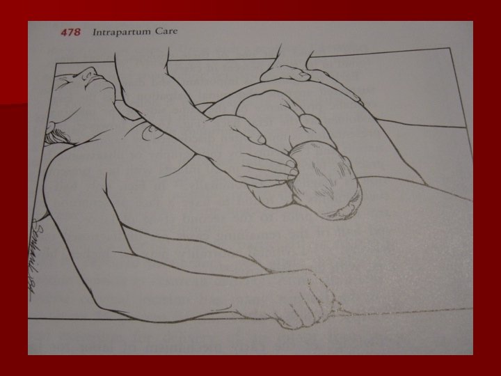
Picture 9. Assisted vaginal breech delivery: The fetal head is maintained in a flexed position by using the Mauriceau-Smellie. Veit maneuver, which is performed by placing the index and middle fingers over the maxillary prominence on either side of the nose. The fetal body is supported in a neutral position with care to not overextend the neck.
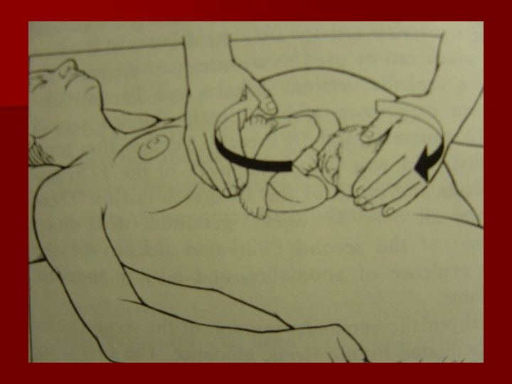
Picture 10. Piper forceps application: Pipers are specialized forceps used only for the aftercoming head of a breech presentation. They are used to keep the head flexed during extraction of the fetal head. An assistant is needed to hold the infant while the operator gets on one knee to apply the forceps from below.
Picture 11. Assisted vaginal breech delivery: Low 1 -minute Apgar scores are not uncommon after a vaginal breech delivery. A pediatrician should be present for the delivery in the event that neonatal resuscitation is needed.
Picture 12. Assisted vaginal breech delivery - The neonate after birth
Risks n. Lower Apgar scors n. An entrapped head n. Nuchal arms , n. Cervical spine injury n. Cord prolapse
PROGNOSIS
Table 1. Zatuchni-Andros Breech Scoring Add 0 Points Add 1 Point Add 2 Points 0 1 2 39+ 38 <37 EFW (lb) 8 7 -8 <7 Previous breech 0 1 2 Dilatation 2 3 4 Station -3 -2 -1 Parity Gestational age (wk) If the score is 0 -4, cesarean delivery is recommended
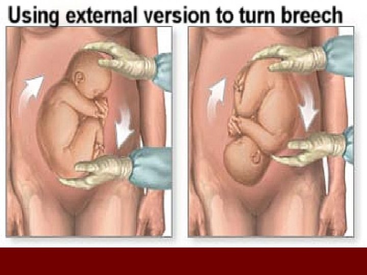
VERSION n. External n. Internal
Internal podalic version
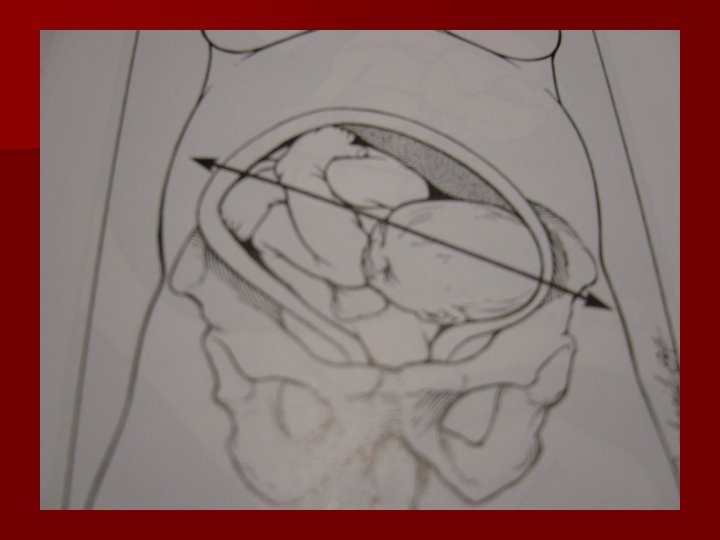
COMPOUND PRESENTATION
COMPLICATION SD n. Immediate neonatal; birth asphyxia , traumatic injury n. Maternal; PPH, lacerations
SHOULDER DYSTOCIA (Sh. D)
Shoulder dystocia will still the obstetric nightmare
Definition: Shoulder dystocia (Sh. D) is the inability to deliver the fetal shoulders after delivery of the head, without the aid of specific maneuvers (ie. other than gentle downward traction on the head).
Definition Objective definition : Mean head-to-body delivery time > 60 seconds
PATHOPHYSIOLOGY Shoulder dystocia results from a size discrepancy between the fetal shoulders and the pelvic inlet when: The bisacromial diameter is large relative to the biparietal diameter 2. Pelvic prim is flat rather than gynecoid 1. .
SHOULDER DYSTOCIA n 0. 15 -1. 7%, n Risk factor; macrosomia, diabetes, histo ry of SD, prolonged 2 th stage of labor, maternal obesity, multiparity, postterm. n 50%SDnorisk factor n Sono
Release techniques Complications of Sh D 1. Maternal 2. Fetal
Maternal Complications (25%) 1. Postpartum hemorrhage 11% 2. Vaginal laceration 19% 3. Perineal tears 2 nd&3 rd 4% 4. Cervical laceration 2%
Fetal. Release techniques Complications of Sh D
Fetal Complications of Sh D Brachial plexus injuries, Fractures of the humerus, and Fractures of the clavicle are the most commonly reported injuries associated with shoulder dystocia
Fetal Complications of Sh D Traction combined with fundal pressure has been associated with a high rate of brachial plexus injuries and fractures
Fetal Complications of Sh D Fewer than 10% of deliveries complicated by shoulder dystocia will result in a persistent brachial plexus injury.
Fetal Complications Release techniques Head –shoulder interval > 7 min. Brain injury (sensitivity & specificity : 70 %) n With hypoxic fetus it is much shorter
Can shoulder dystocia be predicted ?
RISK FACTORS FOR SHOULDER DYSTOCIA PRECONCEPTIONAL: 1. Maternal birth weight 2. Prior shoulder dystocia 12% 3. Prior macrosomia Pre-existing diabetes Obesity Multiparity Prior gestational diabetes Advanced maternal age 4. 5. 6. 7. 8.
RISK FACTORS FOR SHOULDER DYSTOCIA Antenatal: n Excessive maternal weight gain n Macrosomia n G. diabetes n Short stature n Post term
RISK FACTORS FOR SHOULDER DYSTOCIA Intrapartum: Protracted or arrested active phase 2. Protracted or failure of descent of head 3. Need for midpelvic assisted delivery 1.
RISK FACTORS FOR SHOULDER DYSTOCIA Most of the prenatal and antenatal risk factor are interrelated with fetal macrosomia. So the main risk factor is: Fetal Macrosomia
MANAGEMENT (Within 5 - 7 minutes).
Management 1 -Suprapubic pressure 2 -Mc. Robert manoeuver 3 - Woods corkscrew. 4 -Rubens manoeuver 5 -Delivery of P. shoulder 6 -Zavanelli 7 -All fours 8 -Cleidotomy 9 -symphysiotomy
ACOG Issues Guidelines Recommendation 1991 1 -Call for help: assistants, anesthesiologist 2 -Initial gentle attempt of traction. 3 -Generous episiotomy. 4 -Suprapubic pressure.
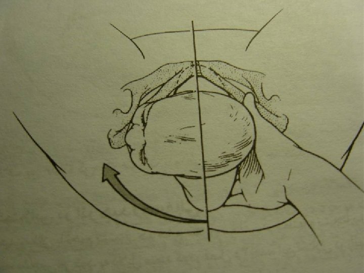
ACOG Issues Guidelines Recommendation 1991 5 -The Mc Roberts manoeuvre (Exaggerated hyper flexion of the thighs upon the abdomen. ) & Suprapubic pressure in the direction of the Foetal face .
Mc. Roberts manoeuvre: X ray pelvimetry study No increase in pelvic dimensions. Decrease in the angle of pelvic inclination P=0. 001 Straightening of the sacrum P= 0. 04% Tends to free the impacted anterior shoulder Gherman et al Obstet Gynecol 95: 43 , 2000
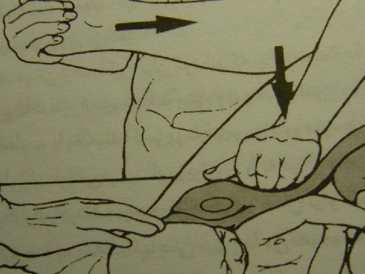
ACOG Issues Guidelines Recommendation 1991 If Mc Roberts failed: 6 -Woods manoeuvre: • The hand is placed behind the posterior shoulder of the fetus. • The shoulder is rotated progressively 180 d in a corkscrew manner so that the impacted anterior shoulder is released. .
ACOG Issues Guidelines Recommendation 1991 7 -Delivery of the posterior arm : .
By inserting a hand into the posterior vagina and ventrally rotating the arm at the shoulder delivery over the perineum
UMBILICAL CORD PROLAPSE
Umbilical Cord Prolapse n Etiology – 1 -275 deliveries n Classification – Complete: cord is seen or palpated ahead of presenting part (OB Emergency) – Fundic: cord felt through intact membranes ahead of presenting part – Occult: hidden or not visible at any time during course of labor n Definition: umbilical cord that lies below/beside presenting part
Umbilical Cord Prolapse n Precipitating factors: – Long umbilical cord – Abnormal location on placenta – Small or preterm infant – Polyhydramnios – Multiple gestation n Precipitating factors: – Amniotomy before fetal head is engaged – IUPC placement – External cephalic version
Umbilical Cord Prolapse n Clinical Manifestations: – Cord observed or palpated – Bradycardia following ROM – Repetitive, variable decelerations that do not respond to medical intervention (e. g. amnioinfusion) – Prolonged decelerations (>15 bpm lasting 2 mins or longer yet <10 mins)
Umbilical Cord Prolapse n Nursing interventions: – – – Assess fetal viability Call for assistance Relieve pressure from cord (usually presenting part) § § § § Continuous manual relief of pressure from presenting part Avoid excessive manipulation of cord Re-position client: Trendelenburg, modified Sim’s, or kneechest Prepare for emergency delivery Administer oxygen by mask 10 -12 L/min Fill maternal bladder with 500 -700 cc NS Continuous fetal monitoring Possible neonatal resuscitation (notify neonatal team per hospital protocol)
Umbilical Cord Prolapse n Aim of Medical management: – Immediate delivery of viable infant – Hallmark treatment: C-section