MALIGNANT TROPHOBLASTIC DISEASE Done by Mayar Alatrash GTD

MALIGNANT TROPHOBLASTIC DISEASE Done by : Mayar Alatrash
 Complete")
GTD Maligna nt Invasive mole Choriocarcinom a Benign Placental site trophoblastic tumor (PSTT) Complete molar pregnancy Partial molar pregnancy

EPIDEMYOLOGY - 25% of all GTD are malignant: - 75% of them are invasive mole - 24 -25% are choriocarcinoma - Less than 1% for PSTT ** most of them will follow complete molar pregnancy , and minority will follow the partial one

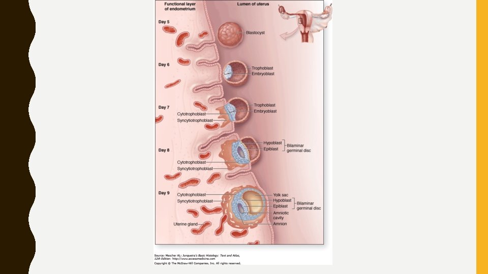
LET’S HAVE A TALK … What is trophoblast mean ? are cells forming the outer layer of a blastocyst, which provide nutrients to the embryo and develop into a large part of the placenta. proliferates and differentiates into 2 cell layers: cytotrophoblast & syncytiotrophoblast (which implant into uterus , come into contact with maternal blood and form chorionic villi and secrete h. CG ) What do you know about h. CG? a hormone produced by the placenta after implantation It helps in secreting progesterone and sustain pregnancy h. CG-positive indicates an implanted blastocyst and mammalian embryogenesis.

BUT WHAT IS THE DIFFERENCE IN THIS SITUATION


**The More abnormal pregnancy the more likely to develop MGTD** 50% of MGTD occurs months to a year after a Molar pregnancy 25% after miscarriage / elective abortion / ectopic pregnancy 25% after a normal pregnancy

STAGING Stage 1 : confined to uterus Stage 2 : metastases to pelvis , vagina Stage 3 : metastases to lung Stage 4 : distant metastases (brain , live , etc)

PERSISTENT /INVASIVE MOLE Almost always follow the evacuation of a molar pregnancy ( complete more than partial ) Clinical presentation Hx of molar pregnancy w/ h. CG rise or plateau on follow up Abnormal uterine bleeding!! (not specific) -- the lesion may penetrate the entire myometrium , rupture through the uterus and result in bleeding into broad ligament and peritoneal cavity Physical examination Usually normal Diagnosis Persistent increase in h. CG Based on h. CG levels and pelvic sono CXR at least , CT MRI for head , pelvis and abdomen

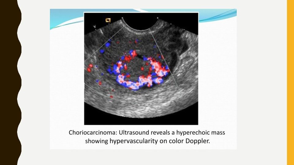
CHORIOCARCINOMA Trophoblastic disease following a normal pregnancy is always choriocarcinom a - Frank malignant form - The tumor has a tendency to disseminate hematogenously, particularly to the lungs, vagina, brain, liver, kidneys, and gastrointestinal tract.

SYMPTOMS Most patients with choriocarcinoma present with symptoms of metastatic disease. - Vaginal bleeding is a common symptom of uterine choriocarcinoma or vaginal metastasis. - Amenorrhea Because of the gonadotropin excretion - Hemoptysis, cough, or dyspnea may occur as a result of lung metastasis. - In the presence of central nervous system metastases, headaches, dizzy spells, “blacking out, ” or other symptoms referable to a space-occupying lesion in the brain. - Rectal bleeding or “dark stools” could represent disease that has metastasized to the gastrointestinal tract.

SIGNS Uterine enlargement may be present, with blood coming through the os, as seen on examination with a speculum. A tumor metastatic to the vagina may present with firm ‘ discolored mass. Occasionally, the patient presents with an acute abdomen because of rupture of the uterus, liver, or theca lutein cyst. Neurologic signs, such as partial weakness or paralysis, dysphasia, aphasia, or unreactive pupils, indicate probable central nervous system involvement

DIAGNOSIS choriocarcinoma is a great imitator of other diseases, so unless it follows a molar pregnancy, it may not be suspected. In females of reproductive age, a β-h. CG measurement to screen for choriocarcinoma should be performed when any unusual symptoms or signs develop

INVESTIGATION ** CT scan of the abdomen, pelvis, and head. ** lumbar puncture should be performed if the CT scan of the brain is normal, -- Because simultaneous evaluation of the β-h. CG level in the cerebrospinal fluid and serum may allow detection of early cerebral metastases. -- Because the β-subunit does not readily cross the blood–brain barrier, a ratio of serum to cerebrospinal fluid β-h. CG levels of less than 40 : 1 suggests central nervous system involvement, with secretion of the β-h. CG directly into the cerebrospinal fluid.

Before talking about treatment We should meet FIGO score

Treatment chemotherapy low risk : methotrexate or actinomycin D - Methotrexate is usually given as a daily dose for 5 consecutive days or every other day for 8 days - alternating with folinic acid (leucovorin). This folinic acid “rescue” regimen is associated with significantly less bone marrow, gastrointestinal, and liver toxicity. - Actinomycin D is given for 5 consecutive days intravenously or every other week as a single dose.

CONT… High risk : combination chemotherapy is always used. A regimen that has been successfully employed is the modified “Bagshawe” regimen The drugs used include etoposide, actinomycin D, vincristine, cyclophosphamide, meth- otrexate, and folinic acid. For patients whose disease fails to improve with these agents, combinations of cisplatin and etoposide or vinblastine, with or without bleomycin, have been used.

CONT… In patients with disease metastatic to the brain or liver, radiation is often employed to these areas in conjunction with chemotherapy. Surgery plays a role in selected cases, especially hysterectomy and pulmonary resection for chemotherapy resistant disease

FOLLOW UP All patients should have weekly β-h. CG level measurements until three normal levels have been measured. For patients with GTN who have a good prognosis, monthly measurements should be done until 12 normal levels have been recorded. Patients with GTN who have a poor prognosis should have monthly levels until 24 normal measurements have been recorded. Patients should use effective contraception during follow-up, following which they may attempt pregnancy.

PROGNOSIS About 95 -100% of patients with GTN who have a good prognosis are cured of their disease. Patients with poor prognostic features can be expected to be cured in only 50 -70% of cases. The majority of the patients who die have brain or liver metastases
 - Uncommon but important variant of GTD - Consists")
PLACENTAL SITE TROPHOBLASTIC TUMOR (PSTT) - Uncommon but important variant of GTD - Consists predominantly of an intermediate trophoblast and a few syncytial elements. - These tumors produce small amounts of h. CG and human placental lactogen relative to their mass, - Tend to remain confined to the uterus, and metastasize late in their course. In contrast to other trophoblastic tumors, - Placental site tumors are relatively insensitive to chemotherapy, so

THANK YOU
- Slides: 24