MALE REPRODUCTIVE SYSTEM Corpus spongiosum Ductuli TESTIS TESTIS


")


 Stroma: 1 - Tunica vaginalis. 2 - Tunica albuginea. 3 - Tunica")


 Seminiferous tubules. (2) Interstitial cells of Leydig.")
 Are rounded or polygonal. Are found singly or in")
 Abundant s. ER. Mitochondria: Numerous, With tubular cristae. Numerous")


")
 Are tall columnar cells (or pyramidal cells). Have ill-defined lateral cell")
 s. ER: ++++ r. ER: limited Golgi apparatus: well-developed Mitochondria: Numerous")


 Spermatogonia: - Type A Spermatogonia: Dark type")


")
. 3")


 • Head + Body + Tail. • Structure: (1) Epithelium: Ps.")
 Deferens")
 (1) Epithelium: Ps. Str. Col. E with stereocilia (2) Basal")
 Epithelium: Simple columnar epithelium. (2) Subepithelial C. T. : is folded.")
. 2 - Prostate. 3")


 Mucosa: is highly convoluted. -Epithelium: Ps. Str. Col. E. -Subepith. C.")
 * (Submucosal Zone) ** (Mucosal Zone) Most benign prostatic hyperplasia* Major")


")
")


 Stroma: 1 - Capsule: (thin fibromuscular capsule): Dense irregular collagenous C.")
 Parenchyma: 1 - Prostatic acini: - Lining: Simple Col. or Ps.")
 • Spherical or oval bodies of glycoproteins in the lumen")
 (1) Capsule: Fibroelastic C. T. + SMFs+ Sk. MFs. (2)")


- Slides: 65
MALE REPRODUCTIVE SYSTEM
(Corpus spongiosum)
Ductuli
TESTIS
TESTIS (A) Stroma: 1 - Tunica vaginalis. 2 - Tunica albuginea. 3 - Tunica vasculosa. 4 - Septa. 5 - Interstitial tissue. (B) Parenchyma: 1 - Seminiferous tubules. 2 - Interstitial cells of Leydig.
STROMA OF THE TESTIS
TUNICA VAGINALIS • It is the visceral layer of serous sac. • It is formed of mesothelial cells. • It is found in the anterior & lateral surfaces of the testis.
TUNICA ALBUGINEA • Dense irregular collagenous C. T.
TUNICA VASCULOSA • It is formed of loose vascular C. T. • Lininig The Tunica albuginea • And surrounding the septa.
SEPTA OF THE TESTIS • • Dense irregular collagenous C. T. Incomplete non-branching septa. Radiate from mediastinum testis. Divide the testis into about 250 intercommunicating compartments (testicular lobules= lobuli testis).
INERSTITIAL TISSUE • Loose vascular C. T. in between the seminiferous tubules. • Contents: 1 - Loose vascular C. T. (mention). 2 - Interstitial cells of Leydig.
INTERSTITIAL TISSUE
PARENCHYMA OF THE TESTIS (1) Seminiferous tubules. (2) Interstitial cells of Leydig.
INTERSTITIAL CELLS OF LEYDIG (L/M) Are rounded or polygonal. Are found singly or in groups. Nucleus: Central, rounded, vesicular With prominent nucleolus. Some cells are binucleated. Cytoplasm: Pale, acidophilic & vacuolated.
INTERSTITIAL CELLS OF LEYDIG (E/M) Abundant s. ER. Mitochondria: Numerous, With tubular cristae. Numerous lipid droplets. Some r. ER. Crystals of Reinke. FUNCTION: Secrete testosterone.
SEMINIFEROUS TUBULES • 1 -4 seminiferous tubules in each testicular lobule. • Each is lined with seminiferous epithelium. • Seminiferous epithelium contains 2 types of cells: 1 - Spermatogenic cells (are germ cells). 2 - Sertoli cells (are somatic cells). • Each is surrounded by: 1 - Basement membrane. 2 - Tunica propria: C. T. layer (collagen fibers + fibroblasts) which contains 1 -2 layers of Myoid cells are not found in man.
SEMINIFEROUS TUBULE
SERTOLI CELLS
SERTOLI CELL (E/M)
SERTOLI CELL (L/M) Are tall columnar cells (or pyramidal cells). Have ill-defined lateral cell boundaries. “ “ “ apex. Nucleus: Basal, Vesicular, Irregular (why? Infoldings), With prominent nucleolus. Cytoplasm: Pale basophilic.
SERTOLI CELLS (E/M) s. ER: ++++ r. ER: limited Golgi apparatus: well-developed Mitochondria: Numerous Lysosomes: Numerous Cytoskeletal elements: Abundant Crystalloids of Charcott-Boettcher Occluding junctions (Zonula type)
SERTOLI CELLS Functions: 1 - Support & Nutrition of spermatogenic cells. 2 - Phagocytosis. 3 - Secretion: Testicular fluid, ABP, Inhibin H. 4 - Formation of blood-testis barrier.
SERTOLI CELLS Dividability: Can not divide in reproductive period.
BLOOD-TESTIS BARRIER • It is composed by attachment of processes of the lateral borders of adjacent Sertoli cells by zonula occluding (tight) junctions. • It divides spermatogenic cells into 2 compartments: 1 - Basal compartment: contains spermatogonia. 2 - Adluminal compartment: contains the other spermatogenic cells. • Function: 1 - It prevents autoimmune infertility. 2 - It protects spermatocytes from drugs and toxic materials.
SEMINIFEROUS TUBULE
SEMINIFEROUS TUBULE
SEMINIFEROUS TUBULE
• • • SPERMATOGENIC CELLS (1) Spermatogonia: - Type A Spermatogonia: Dark type A Pale type A - Type B Spermatogonia. (2) 1 ry Spermatocytes. (3) 2 ry Spermatocytes. (4) Spermatids: early & late. (5) Spermatozoa.
SPERMIOGENESIS
SPERMIOGENESIS • Definition: Transformation of spermatids into spermatozoa. • Features: 1 - Formation of acrosome. 2 - Nucleus: Condensation, elongation, slight flattening, acrosomal cap (head cap). 3 - Development of flagellum. 4 - Formation of mitochondrial sheath. 5 - Loss of much cytoplasm: Cytoplasmic residual bodies→ Are phagocytosed by Sertoli cells.
INTRATESTICULAR GENITAL DUCTS Ductuli
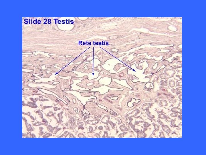
INTRATESTICULAR GENITAL DUCTS 1 - Tubuli recti. 2 - Rete testis. 3 - Ductuli efferentes (Vasa efferentia).
TUBULI RECTI • Epithelium: Initial segment: Sertoli-like cells. Distal segment: simple cuboidal epith.
RETE TESTIS • Epithelium: Simple cuboidal epithelium.
DUCTULI EFFERENTES • No: 10 -20. • Structure: 1 - Epithelium: Patches of simple cuboidal cells (Absorptive) alternating with regions of ciliated columnar cells. 2 - C. T. layer with few circularly-arranged SMFs.
EXTRATESTICULAR GENITAL DUCTS (Corpus spongiosum)
EXTRATESTICULAR GENITAL DUCTS 1 - Ductus epididymis. 2 - “ deferens (Vas deferens). 3 - Ejaculatory duct.
EPIDIDYMIS
EPIDIDYMIS
EPIDIDYMIS (DUCTUS EPIDIDYMIS) • Head + Body + Tail. • Structure: (1) Epithelium: Ps. Str. Col. E. with stereocilia. (E/M of stereocilia: Long, branched, non-motile microvilli). (2) Basal lamina. (3) Loose C. T. (4) Layer of circularly-arranged SMFs.
V Vas (ductus) Deferens
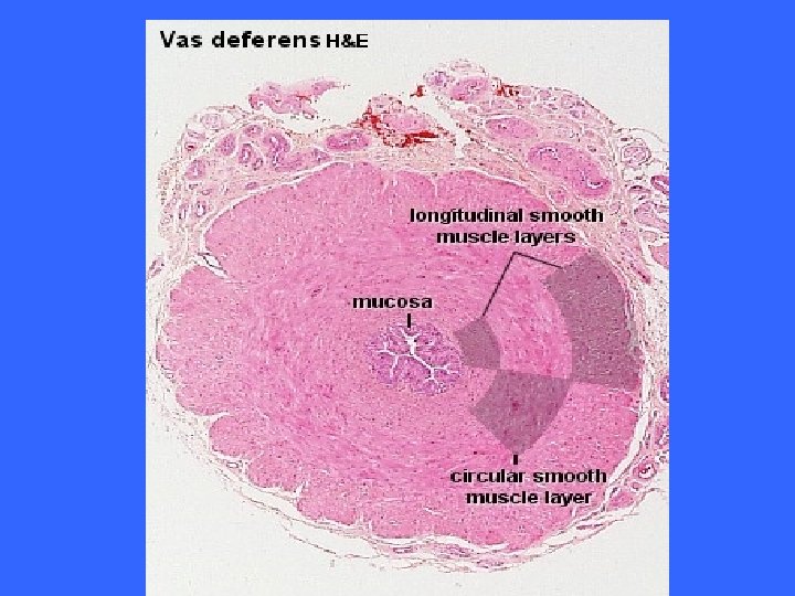
DUCTUS DEFERENS (VAS DEFERENS) (1) Epithelium: Ps. Str. Col. E with stereocilia (2) Basal lamina. (3) Loose C. T. (Loose fibroelastic C. T. ). N. B. Lumen is irregular- why? Mucosa has longitudinal folds. (4) Thick smooth muscle coat ( 3 layers): Inner longitudinal muscle layer. Middle circular “ “. Outer longitudinal “ “. (5) Adventitia: Loose fibroelastic C. T. N. B. Ampulla has highly folded, thickened epithelium.
EJACULATORY DUCT (1) Epithelium: Simple columnar epithelium. (2) Subepithelial C. T. : is folded. N. B. No smooth muscle in its wall.
ACCESSORY GENITAL GLANDS 1 - Seminal vesicles (No. : 2). 2 - Prostate. 3 - Bulbourethral glands ( No. : 2).
SEMINAL VESICLE
SEMINAL VESICLE
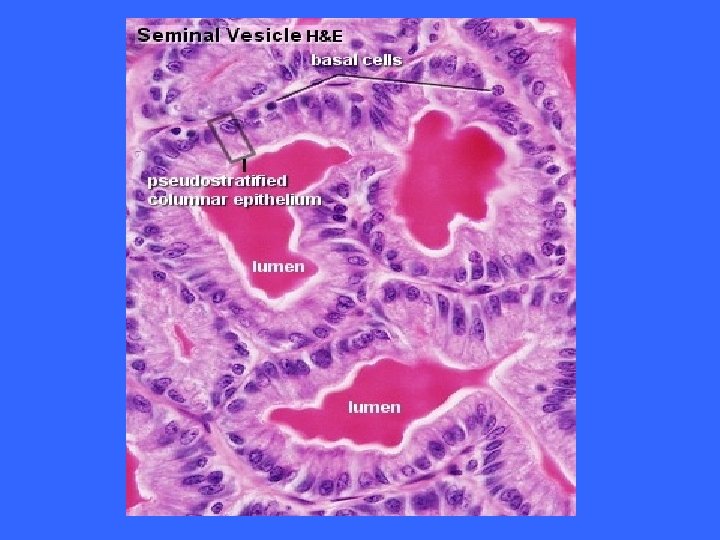
SEMINAL VESICLE (1) Mucosa: is highly convoluted. -Epithelium: Ps. Str. Col. E. -Subepith. C. T. : Fibroelastic C. T. (2) Muscle coat: -Inner circular layer. -Outer longitudinal layer. (3) Adventitia: Fibroelastic C. T. N. B. Seminal fluid produced by seminal vesicles: -Is fructose-rich fluid. -Is viscous fluid. -Is pale yellow ( due to lipochrome pigments). -Represent 70% of the semen (ejaculate).
PROSTATE (Main Zone) * (Submucosal Zone) ** (Mucosal Zone) Most benign prostatic hyperplasia* Major site of prostatic cancer**
PROSTATE
PROSTATE
PROSTATIC CONCRETIONS (CORPORA AMYLACEA)
PROSTATIC CONCRETIONS (CORPORA AMYLACEA)
PROSTATE
PROSTATE *It is formed of 30 -50 compound tubuloalveolar glands, which are arranged in 3 discrete, concentric layers (zones): 1 - Mucosal glands. 2 - Submucosal glands. 3 - Main glands. It secretes: serous, white fluid rich in: 1 - Acid phosphatase. 2 - Proteolytic enzymes. 3 - Citric acid. 4 - Fibrinolysin.
PROSTATE L/M: (A) Stroma: 1 - Capsule: (thin fibromuscular capsule): Dense irregular collagenous C. T. + SMFs. 2 - Septa: (Indistinct in adult men): Are thick complete fibromuscular septa. 3 - Stroma in between acini: (fibromuscular stroma): Richly vascular Dense irregular collagenous C. T. + SMFs.
PROSTATE L/M: (B) Parenchyma: 1 - Prostatic acini: - Lining: Simple Col. or Ps. Str. Col. E. - Prostatic concretions. 2 - Duct system.
PROSTATIC CONCRETIONS (CORPORA AMYLACEA) • Spherical or oval bodies of glycoproteins in the lumen of some prostatic acini. • Often are calcified. • Their No. increases with aging.
BULBOURETHERAL GLANDS (COWPER’S GLANDS) (1) Capsule: Fibroelastic C. T. + SMFs+ Sk. MFs. (2) Septa: “” “”. Septa divide each gland into lobules. (3) Epithelium: Mucus-secreting simple cuboidal epith. or simple columnar epith. N. B. They are compound tubuloalveolar glands. They empty into the membranous urethra.
PENIS xxxxxxxxxxxx x Spongiosumx
PENIS • • Two corpora cavernosa. One corpus spongiosum. Glans penis. Prepuce: is lined with mucous membrane. (Epith. : moist str. Sq. non-ker. E. ). • Erectile tissue of penis: Numerous vascular spaces (variably-shaped) separated by trabeculae of C. T. & SMFs.
BEST WISHES