Major types of nutrients and digestion Lecture 12


 • Has two catalytic sites 1 - The lactase catalytic")


 •")

 • Glucose moves via the")


, transport (Hb), immune")


 stimulates")


- Slides: 60
Major types of nutrients and digestion Lecture 1+2 Dr. Jehad Al-Shuneigat
• The 7 types of nutrients are Carbohydrates, Protein, Fats, Vitamins, Minerals, Water and Fibers
examples of serving size: * Grain products— 1 slice of white bread or 1⁄2 cup of cooked rice; * Vegetable group— 1⁄2 cup cooked vegetables * Fruit group— 1 apple or banana; * Milk Group— 1 cup of milk or 60 g processed cheese; * Meat and Beans Group— 2– 3 oz (6080 g) cooked lean meat or fish or 1 egg or 2 tbsp peanut butter.
1. Carbohydrates Functions • Energy • Fibers • Recognition and adhesion between cells Carbohydrate covalently attached to proteins or lipids examples: glycoproteins, glycolipids, these classes of molecules are called glycoconjugates.
• Most foods derived from animals, such as meat or fish, contain very little carbohydrate except for small amounts of glycogen. The major dietary carbohydrate of animal origin is lactose. • Although all cells require glucose for metabolic functions, neither glucose nor other sugars are specifically required in the diet. Oxidation • 1 g carbohydrates produces about 4 kcal/g, • 1 g proteins produces 4 kcal/g • 1 g fats produces 9 kcal/g. • Energy is also expressed in joules. One kilocalorie equals 4. 18 kilojoules (k. J).
Carbohydrates are classified into three groups: 1 - Monosaccharide's: =Also called simple sugars =Have the formula (CH 2 O)n =Cannot be broken down into smaller sugars. 2 - Oligosaccharides: = Consist of from 2 to 10 monosaccharides molecules joined by a linkage called glycosidic bonds (covalent bond). = The most abundant are the disaccharides = Trisaccharides also occur frequently. = All common monosaccharides and disaccharides have names ending with the suffix “-ose. ” 3 - Polysaccharides: are polymers of monosaccharide’s. They may be either linear like cellulose or branched like glycogen. Polysaccharides may contain hundreds or even thousands of monosaccharide units.
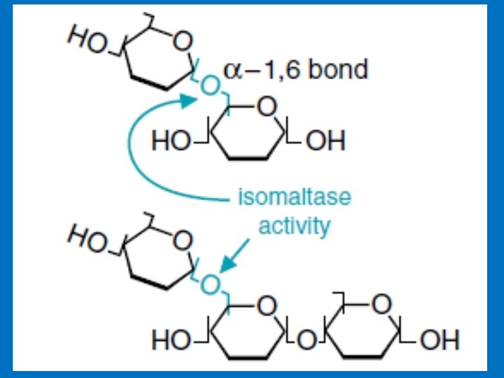
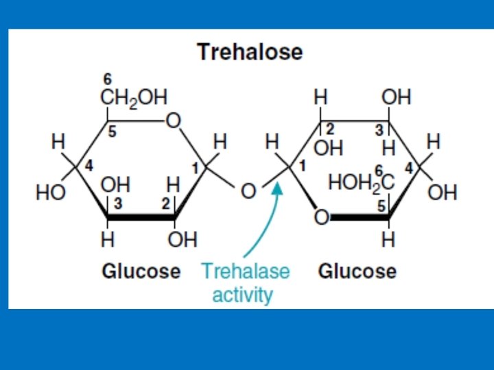
Disaccharides 1 - Maltose Consist of two glucose units joined by α-1, 4. Comes from the hydrolysis of starch 2 - Cellobiose, is a two glucose disaccharide joined by β-1, 4 obtained from the acid hydrolysis of cellulose 3 - Isomaltose consists of two glucose units linked by α-1, 6. obtained from the hydrolysis of some polysaccharides like dextran 4 - Trehalose two glucose units linked by an α-bond (α-1, 1) 5 - Lactose Milk sugar, Composed of Galactose & Glucose joined by (β-1, 4) link. 6 - Sucrose Composed of (glucose-α-1, 2 -fructose) obtained from cane or beet commonly known as table sugar
Glycosidic bond in maltose Glycosidic bond the covalent bond between the anomeric carbon atom of a saccharide and some other group or molecule with which it forms a glycoside.
Reducing and non -reducing end α-1, 1
Polysaccharides • Starch represents the main type of digestible polysaccharides. Found in cereal grains (wheat, rice, corn, oats, and barley) and tubers such as potatoes. • Starch is made up of : 1 - Amylose (about 20% of starch) strait chain of glucose which is linked by α-1, 4 glycosidic bonds 2 - Amylopectin (about 80% of starch) (branched) the α -1, 4 chains contain branches connected via α-1, 6 glycosidic bonds • None starch polysaccharides are the main component of dietary fibers which include: Cellulose, hemicellulose, pectin's and gum.
Amylose Amylopectin
Glycogen structure
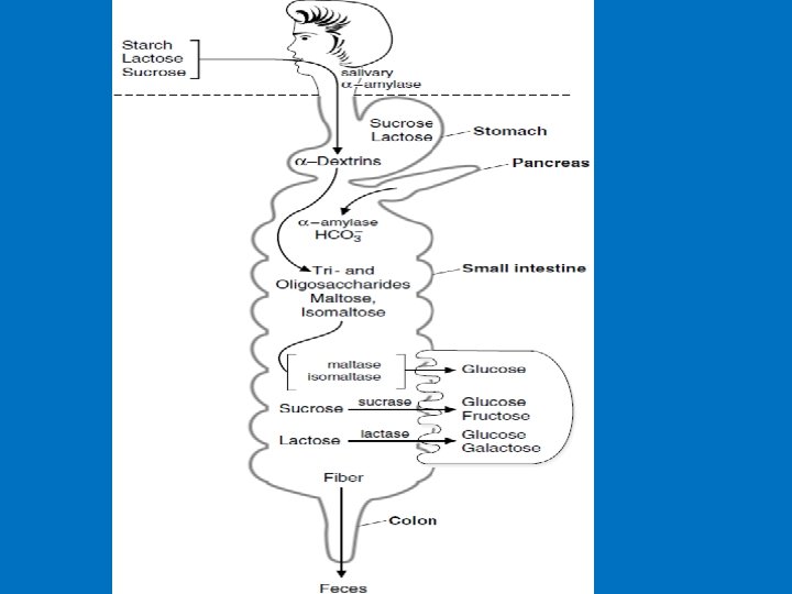
Digestion of dietary carbohydrates • Glycosidases: enzymes that hydrolyze the glycosidic bonds between the sugars • Glycosidases convert polysaccharides and disaccharides to monosaccharides • Undigested carbohydrates enter the colon, where they may be fermented by bacteria
• A. Salivary α-amylase • The digestion of starch begins in the mouth. • The salivary glands secrete approximately 1 liter of liquid per day into the mouth (p. H 6. 4 - 7) containing salivary α-amylase. • α-Amylase is an endoglucosidase, which means that it hydrolyzes internal α-1, 4 bonds between glucose residues at random intervals in the polysaccharide chains. • Requires Cl− ion for activation with an optimum p. H of 6. 7 • Digestion of starch and glycogen in the mouth gives maltose, isomaltose and α-dextrins • ln the stomach: Carbohydrate digestion stops temporarily, salivary α-amylase may be largely inactivated by the acidity of the stomach.
Action of pancreatic and α-amylase Endoglucosidase Exoglucosidase
Digestion in Small Intestine A. Pancreatic Secretions • Secretions from the exocrine pancreas (approximately 1. 5 L/day) enter the duodenum which contain 1 - Bicarbonate (HCO 3 -), which neutralizes the acidic p. H of stomach contents 2 - Digestive enzymes, including pancreatic α-amylase. Pancreatic α-amylase (optimum p. H=7. 1, also activated by chloride ions) continues to hydrolyze the starches and glycogen, forming the disaccharide maltose, isomaltose, the trisaccharide maltotriose (three glucose molecules linked with α-1, 4 glycosidic bonds), and oligosaccharides. • These oligosaccharides, are usually 4 -9 glucose units long linked by α 1 -4 and contain one or more α-1, 6 branches. • α-Amylase has no activity toward sugar containing polymers other than glucose linked by α-1, 4 bonds.
Digestion in Small Intestine B. Disaccharidases of the intestinal brush-border membrane 1. Glucoamylase • It has two catalytic sites with similar activities. • Glucoamylase is an exoglucosidase that is specific for the α– 1, 4 bonds between glucose residues. • It begins at the non-reducing end of saccharides • The glucoamylase is heavily glycosylated with oligosaccharides that protect it from digestive proteases. • α-amylase split of large polysaccharides molecule and thus supplying new substrate molecules for glucoamylase
Location of disaccharide complexes in intestinal villi • The villi are finger like projections of the mucosa into the lumen of the small intestine. • A villus has thousands of microvilli. • Collectively, microvilli known as a “brush border”.
Glucoamylase activity. Glucoamylase is an α-1, 4 exoglycosidase, which initiates cleavage at the nonreducing end of the sugar.
2. Sucrase–isomaltase complex • Sucrase–isomaltase has two catalytic sites that differs in substrate specificity from the other (Intestinal protease clips it into two separate subunits) The two catalytic sites are: A- The sucrase–maltase site = Nearly 100% of sucrose hydrolyze (glucose-α-1, 2 -fructose) = Maltase activity. B- The isomaltase–maltase site = All α-1, 6 -glucose bonds = Maltase activity. 80% of the maltase is by these complex (maltose: glucose α-1, 4 glucose) activity of the small intestine. The remainder of the maltase activity is found in the glucoamylase complex.
3. Trehalase • Has only one catalytic site. • Found in algae, mushrooms, and other fungi • Trehalase is only half as long as the other disaccharidases • Trehalose made up of two glucose units linked by an α-bond between their anomeric carbons (α-1, 1).
4. β-glycosidase complex (lactase-glucosylceramidase) • Has two catalytic sites 1 - The lactase catalytic site hydrolyzes the β-bond connecting glucose and galactose in lactose (also splits the β-1, 4 bond between some cellulose disaccharides). 2 - The β-bond between glucose or galactose and hydrophobic residues ceramide in glycolipids
Location of disaccharidases within the intestine A- Sucrase–isomaltase activity is highest in the jejunum, B- β-Glycosidase activity is also highest in the jejunum. C- Glucoamylase activity increases along the length of the small intestine and is highest in the ileum.
• Metabolism of Sugars by Colonic Bacteria • Starches high in amylose, or less well hydrated (e. g. , starch in dried beans), are resistant to digestion and enter the colon. • Colonic bacteria rapidly metabolize the saccharides, forming gases, short-chain fatty acids, and lactate. • The short-chain fatty acids are absorbed by the colonic mucosal cells and can provide a substantial source of energy for these cells. • Incomplete products of digestion in the intestines increase the retention of water in the colon, resulting in diarrhea.
Lactose Intolerance • The small intestine does not make enough of the enzyme lactase. • The lactose that is not absorbed is converted by colonic bacteria to lactic acid, methane gas (CH 4), and H 2 gas that result in: abdominal pain, gases, and diarrhea. Lactase deficiency may be (i) Congenital: complete deficiency of lactase enzyme since birth (rare). (ii) Acquired: which occurs later on in life. which include: a. Primary lactase deficiency develops over time and begins after about age 2 when the body begins to produce less lactase with a possible genetic link. b. Secondary lactase deficiency results from injury to the small intestine, gastrointestinal diseases, including exposure to intestinal parasites.
How is lactose intolerance diagnosed? 1. Hydrogen Breath Test. The person drinks a lactose-loaded beverage and then the breath is analysed at regular intervals to measure the amount of hydrogen. Normally, very little hydrogen is detectable in the breath, but undigested lactose produces high levels of hydrogen. 2. Stool Acidity Test. Undigested lactose creates lactic acid and other fatty acids that can be detected in a stool sample. Glucose may also be present in the stool as a result of undigested lactose. Management: 1 - Most people with lactose intolerance can tolerate some amount of lactose in their diet know your limit and stick to it. 2. Decreasing or removing lactose from milk and milk products.
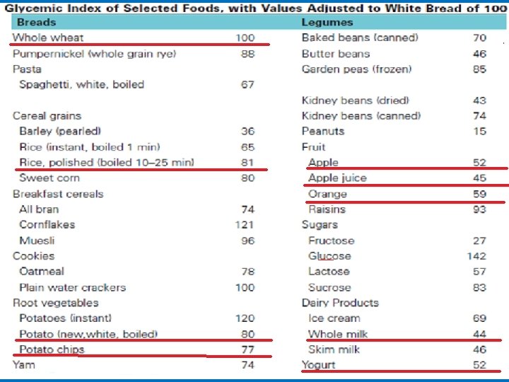
• Absorption of sugars Not all complex carbohydrates are digested at the same rate within the intestine, and some carbohydrate sources lead to a near-immediate rise in blood glucose levels slowly raise blood glucose levels The glycemic index of a food is an indication of how rapidly blood glucose levels rise after consumption. Glucose has the highest glycemic indices (142) with white bread defined as an index of 100. • The glycemic response to ingested foods depends not only on the glycemic index of the foods, but also on the fiber and fat content of the food, as well as its method of preparation.
Absorption of sugars Two mechanisms are responsible for absorption of monosaccharides: active transport and facilitated diffusion. Features Concentration gradient Energy expenditure Carrier protein/ transporter Speed Facilitated diffusion Active transport Down the concentration gradient from high to low. Against a concentration gradient from low to high none Energy expenditure is in the form of ATP required Fastest mode Note: Glucose and other monosaccharide's are polar molecule. It cannot pass through lipid bilayer of cell therefore passive diffusion doesn't apply for sugar.
• Although ions and most polar molecules cannot diffuse across a lipid bilayer, many such molecules (such as glucose) are able to cross cell membranes. These molecules pass across membranes via the action of specific transmembrane proteins, which act as transporters. Such transport proteins determine the selective permeability of cell membranes and thus play a critical role in membrane function. They contain multiple membrane-spanning regions that form a passage through the lipid bilayer, allowing polar or charged molecules to cross the membrane through a protein pore without interacting with the hydrophobic fatty acid chains of the membrane phospholipids.
• Absorption by the Intestinal Epithelium • 1. Na+-dependent transporters (Active transport) • Na+-dependent glucose transporters, which are located on the luminal side of the absorptive cells, enable these cells to concentrate glucose from the intestinal lumen. A low intracellular Na+ concentration is maintained by a Na+, K+ATPase that uses the energy from ATP cleavage to pump Na+ out of the cell into the blood. • Thus, the transport of glucose from a low concentration in the lumen to a high concentration in the cell is promoted by the cotransport of Na+ from a high concentration in the lumen to a low concentration in the cell (secondary active transport).
Na+ binds to the cotransporter, which results in a conformational change that permits glucose to bind to the glucose -binding site. The cotransporter undergoes a new conformational shift, placing Na+ and glucose near the inner surface of the membrane.
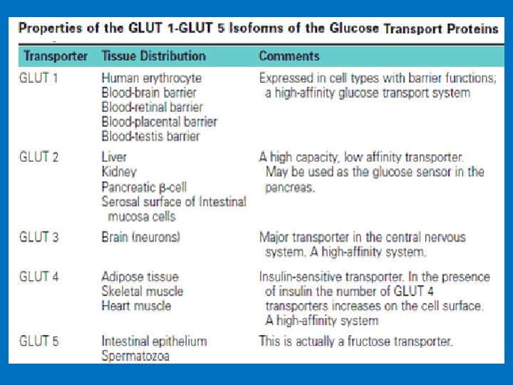
• 2. Facilitative glucose transporters (Na+ independent transporter) • Glucose moves via the facilitative transporters from the high concentration inside the cell to the lower concentration in the blood without the need of energy. In addition to the Na+dependent glucose transporters, facilitative transporters for glucose also exist on the luminal side of the absorptive cells. • The various types of facilitative glucose transporters found in the plasma membranes of cells referred to as GLUT 1 to GLUT 14. • One common structural theme to these proteins is that they all contain 12 membrane-spanning domains. • Because glucose leaves the intestine via the hepatic portal vein, the liver is the first tissue it passes through. The liver extracts a portion of this glucose from the blood.
Na-dependent and facilitative transporters in the intestinal epithelial cells. Both glucose and fructose are transported by the facilitated glucose transporters on the luminal and serosal sides of the absorptive cells. Glucose and galactose are transported by the Na-glucose cotransporters on the luminal (mucosal) side of the absorptive cells.
Facilitative transport. Transport of glucose occurs without rotation of the glucose molecule. Multiple groups on the protein bind the hydroxyl groups of glucose and close behind it as it is released into the cell (i. e. , the transporter acts like a “gated pore”). O = outside; I = inside.
Dietary fiber • Are components of food cannot be digested by human digestive enzymes they are mainly polysaccharide derivatives and lignan (Noncarbohydrate, polymeric derivatives of phenylpropane). • There are several kinds of dietary fiber. • Water insoluble fiber: Cellulose, lignin and hemicellulose are materials that stimulate regular function of the colon. • Water-soluble fiber Pectins and gums are materials that form viscous gel-like suspensions in the digestive system slowing the rate of absorption of many nutrients, including carbohydrates, and lowering serum cholesterol in many cases.
Benefits of fibers 1 - Soluble fibers are digested by bacteria and produce small chain of FA. 10% of our total calories we get from compounds produced by bacterial digestion of substances in our digestive tract. 2 - Fiber is thought to “soften” the stool, thereby reducing pressure on the colonic wall and enhancing expulsion of feces this specificlly beneficial effect to diverticular disease, in which sacs or pouches may develop in the colon because of a weakening of the muscle and submucosal structures. 3 - Disease prevention for example: pectins=may lower blood cholesterol levels by binding bile acids =beneficial for diabetes by slowing the rate of absorption of simple sugars and preventing high blood glucose levels after meals. β-glucan (obtained from oats) reduce cholesterol levels through a reduction in bile acid degradation in the intestine.
2. Fats • Functions: 1 - Cell structure, 2 - fuel storage, 3 - hormone • essential fatty acids and nonessential fatty acids • nonessential fatty acids can be synthesized in our body • essential fatty acids we get them from food =The essential fatty acids α-linoleic and α-linolenic acid are supplied by dietary plant oils.
Digestion of fat • Triacylglycerols are the major fat in the human diet because they are the major storage lipid in the plants and animals that constitute our food supply. • The remainder of the dietary lipids consists primarily of cholesterol, phospholipids, and free fatty acids. • Triacylglycerols contain a glycerol backbone to which three fatty acids are esterified. • The main route for digestion of triacylglycerols involves hydrolysis to fatty acids and 2 -monoacylglycerols in the lumen of the intestine. However, the route depends to some extent on the chain length of the fatty acids.
Triacylglycerol
Digestion of fat A. Mouth and stomach Limited digestion of lipids occurs in the mouth and stomach because of the low solubility. 1. Lingual lipase produced by cells at the back of the tongue 2. Gastric lipase produced by stomach • Same function for both enzymes: to hydrolyze short- and medium-chain fatty acids (containing 12 or fewer carbon atoms).
• B. In small intestine • 1. Action of Bile: • Emulsification (suspended in small particles in the aqueous environment) by bile salts. • The biles are amphipathic compounds • The contraction of the gallbladder and secretion of pancreatic enzymes are stimulated by the gut hormone cholecystokinin, which is secreted by the intestinal cells when stomach contents enter the intestine. • Bile act as detergents, binding to dietary fat as they are broken up by the action of the intestinal muscle. This emulsified fat, which has an increased surface area as compared with unemulsified fat, is attacked by digestive enzymes from the pancreas.
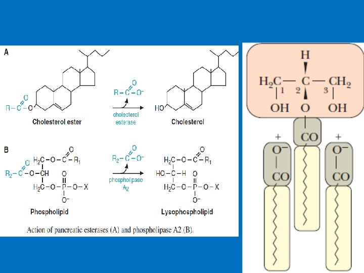
2. Action of Pancreatic secretion A- Bicarbonate (hormone secretin released from the intestine when acid enters the duodenum) raises the p. H of the contents of the intestinal lumen into a range (p. H ~ 6. 5). B-Lipase digests dietary triacylglycerols producing 2 free fatty acids and 2 -monoacylglycerol C-Colipase it binds to the dietary fat and to the lipase, thereby increasing lipase activity. D- Esterases remove fatty acids from compounds such as cholesterol esters (The ester bond is formed between the carboxylate group of a fatty acid and the hydroxyl group of cholesterol. ) E- phospholipase A 2 digests phospholipids to a free fatty acid and a lysophospholipid
• Absorption of digested fats • Short- and medium-chain fatty acids (C 4 to C 12) are absorbed directly into intestinal epithelial cells, they enter the portal blood and are transported to the liver bound to serum albumin. • The digested fatty acids and 2 -monoacylglycerols are resynthesized into triacylglycerols in intestinal epithelial cells, then packaged in lipoprotein particles chylomicrons because they are insoluble in water, and secreted by way of the lymph into the blood. • If triacylglycerols directly entered the blood, they would come together, impeding blood flow. • The fatty acids of the chylomicron triacylglycerols are stored mainly as triacylglycerols in adipose cells.
3. Protein • Function: • Cytoskeleton, movement (actin and myosin ), transport (Hb), immune protection (antibodies), receptors and as catalysts enzymes • High and low quality protein • Amino acids are the building blocks of proteins. • The sequence of amino acids in a protein is determined by the genetic code. Four levels of protein structure are commonly defined: Primary structure, Secondary structure, Tertiary structure, and Quaternary structure • There are 20 different amino acids 8 essential and 12 nonessential amino acids that form human proteins.
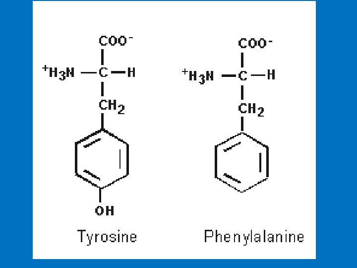
Conditionally essential amino acids • Children and pregnant women have a high rate of protein synthesis to support growth, and require more of arginine and histidine than their body synthesise. • Tyrosine is also considered conditionally essential. Tyrosine is synthesized from phenylalanine (by hydroxylation of phenylalanine), and it is required in the diet if phenylalanine intake is inadequate, or if an individual is congenitally deficient in an enzyme required to convert phenylalanine to tyrosine (the congenital disease phenylketonuria).
• Zymogens • Inactive enzymes precursors are called zymogens or proenzymes, • It only acquire full activity upon specific proteolytic cleavage of one or several of their peptide bonds. • Zymogen activation by specific proteolysis is an irreversible process. • Important to switch on processes at the appropriate time and place • The synthesis of zymogens as inactive precursors prevents them from cleaving proteins prematurely at their sites of synthesis or secretion.
Origin Zymogen/proenzymes Activator Active Protease Pancreas Trypsinogen Enteropeptidase Trypsin Pancreas Chymotrypsinogen Trypsin Chymotrypsin Pancreas Procarboxypeptidase Trypsin Carboxypeptidase Pancreas Proelastase Trypsin Elastase Stomach Pepsinogen H+ (p. H drop) Pepsin
• Activation of chymotrypsinogen • Chymotrypsinogen is a 245 -aa cross-linked by five disulfide bonds. • Chymotrypsinogen is converted to an enzymatically active form called π-chymotrypsin when trypsin cleaves the peptide bond joining Arginine 15 and Isoleucine 16. • The enzymatically active π-chymotrypsin acts upon other π-chymotrypsin molecules, removing two dipeptides, Serine 14 -Arginine 15 and Threonine 147 - Asparagine 148. • The end product of this processing pathway is the mature protease α-chymotrypsin, in which the three peptide chains. • A (residues 1 through 13), B (residues 16 through 146), and C (residues 149 through 245), remain together because they are linked by two disulfide bonds, one from A to B, and one from B to C.
Arginine 15 and Isoleucine 16 Serine 14 -Arginine 15 Threonine 147 - Asparagine 148 A disulfide bond is a covalent bond between two sulfur containing amino acids like two cysteine molecules
• Digestion of Proteins in the Stomach # Gastrin hormone (gastric mucosa) stimulates the secretion A- Pepsinogen by the chief cells of the gastric glands. B- Hydrochloric acid by the parietal cells A- Pepsinogen is activated to its active form pepsin by acidic gastric juice (p. H 1. 0 to 2. 5) that alters the conformation of so that it can cleave itself, producing the active pepsin. • Pepsin acts as an endopeptidase, cleaving peptide bonds at various points within the protein chain. B- Stomach acidity: causes dietary proteins denaturation, this serves to inactivate the proteins and partially unfolds them such that they are better substrates for proteases. • Smaller peptides and some free amino acids are produced.
• Digestion of Proteins by Enzymes from the Pancreas 1 - Secretin hormone: stimulates bicarbonate secretion 2 - Bicarbonate causes raises the p. H such that the pancreatic proteases (the hormone cholecystokinin stimulate its release), which are also present in pancreatic secretions, can be active. A- Trypsinogen is cleaved to form trypsin by enteropeptidase (a protease) secreted by the brush-border cells of the small intestine. • Trypsin is most specific that cleaves endopeptidases peptide bonds between lysine or arginine (cleaves peptide bonds of basic amino acids (+ve)). B- Trypsin catalyzes conversion of = chymotrypsinogen to chymotrypsin: that favors residues that contain hydrophobic or acidic amino acids. = proelastase to elastase: that cleaves elastin and proteins with small side chains (alanine, glycine, or serine). = procarboxypeptidases to carboxypeptidases.
• Carboxypeptidase A preferentially releases hydrophobic amino acids, whereas • Carboxypeptidase B releases basic amino acids (arginine and lysine). • These exopeptidases remove amino acids from the carboxyl ends of peptide chains. • Sequences of amino acids in a peptide are read from the amino terminal end to the carboxy-terminal end.
• Digestion of Proteins by Enzymes from Intestinal Cells • Exopeptidases produced by intestinal epithelial cells act within the brush border and also within the cell. • Aminopeptidases, located on the brush border, cleave one amino acid at a time from the amino end of peptides. • Amino acids are absorbed from the intestinal lumen through Na+- dependent transport systems and through facilitated diffusion.