Major Structures of the Forebrain Limbic System Memory

Major Structures of the Forebrain

“Limbic” System Memory and Hippocampus Saul Kassin, Psychology. Copyright © 1995 by Houghton Mifflin Company. Reprinted by permission. Hypothalamus and Amygdala

Hippocampus • Involved in other forms of learning – smaller in depressed patients – adult neurogenesis – exercise-induced neurogenesis

Hippocampus Morris Water Maze

Water Maze Results Return to Limbic
 • Suprachiasmatic nucleus (biological clock) –")
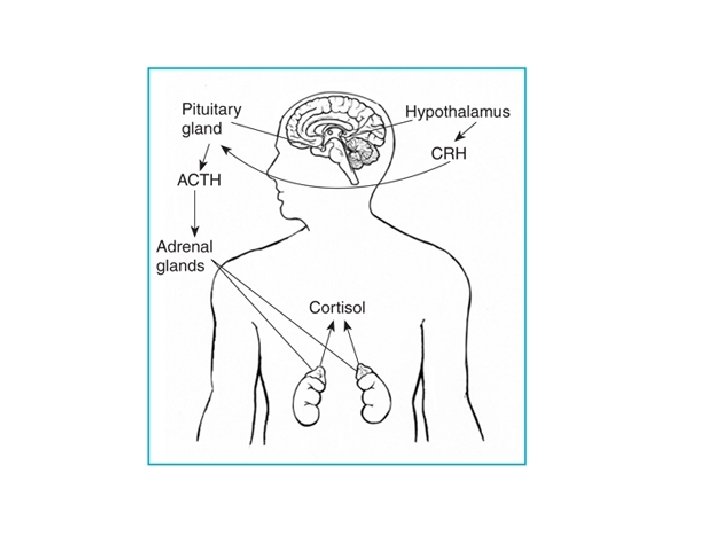
Hypothalamus • 4 Fs (fight, fleeing, feeding, …) • Suprachiasmatic nucleus (biological clock) – 24 hour process – reset by light – linked to homosexuality/bisexuality in males (much larger) • Portal to the pituitary “master” gland – CRH, corticotropic releasing hormone – ACTH, adrenocorticotropic hormone – Adrenals, cortisol

Le. Doux

High Road

Substantia Nigra Hindbrain

Parkinson’s Disease
")
Treatments • Levodopa (diminishing effects over time)

Treatments • Thalamotomy

Treatments • Deep Electrical Brain Stimulation

More Midbrain • Superior colliculus-visual switchboard – eye movements • Inferior colliculus-auditory switchboard Return
 Punch Drunk Alcoholism")
Cerebellum • Little Brain (motor programs) Punch Drunk Alcoholism

Medulla and Reticular Formation • Medulla – heart rate, breathing, blood pressure • Reticular Formation – arousal
 Encephale isolaté (normal cycles) Midpontine (always awake)")
Cerveau isolaté (comatose) Encephale isolaté (normal cycles) Midpontine (always awake)

Small Molecule Neurotransmitters

Other Classes of Neurotransmitters

Methods: Human Brain

EEG Electroencephalogram • Technique: Multiple electrodes are pasted to outside of head. • What it shows: A single line that charts the summated electrical fields resulting from the activity of billions of neurons.

EEG
 • Advantages – Detects very rapid changes in electrical activity. • Disadvantages")
EEG (cont’d) • Advantages – Detects very rapid changes in electrical activity. • Disadvantages – Very poor localization of the source of electrical activity.

PET Positron Emission Tomography • Technique: Active areas take up radioactive substances. • What it shows: What brain areas are most active during a specified period of time (e. g. , 30 seconds).

PET
 • Advantages – Allows brain in action studies (functional). – Provides visual")
PET (cont’d) • Advantages – Allows brain in action studies (functional). – Provides visual image corresponding to anatomy. • Disadvantages – Exposure to low levels of radioactivity. – Better localization than EEG, but poorer than that of MRI. – Cannot follow rapid changes (faster than 30 seconds).

MRI Magnetic Resonance Imaging • Technique: Exposes the brain to magnetic field and measures radio frequency waves. • What it shows: – MRI high resolution image of brain anatomy – f. MRI shows changes in blood flow over time. – DTI shows water flow in neural fibers. Magnetic Resonance Imaging © Dan Mc. Coy

Advantages of MRI • Provides a lovely photograph-like picture. • f. MRI gives an “action shot” Disadvantages • f. MRI “action shot” is the world’s slowest video camera

The Endocrine System: Coordinating the Internal World How can my hormones help me in a crisis?

Some Major Glands of the Endocrine System
- Slides: 31